IRSp53 mediates podosome formation via VASP in NIH-Src cells
- PMID: 23555988
- PMCID: PMC3608619
- DOI: 10.1371/journal.pone.0060528
IRSp53 mediates podosome formation via VASP in NIH-Src cells
Abstract
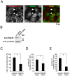
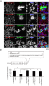
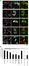
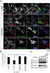
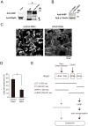
Podosomes are cellular "feet," characterized by F-actin-rich membrane protrusions, which drive cell migration and invasion into the extracellular matrix. Small GTPases that regulate the actin cytoskeleton, such as Cdc42 and Rac are central regulators of podosome formation. The adaptor protein IRSp53 contains an I-BAR domain that deforms membranes into protrusions and binds to Rac, a CRIB motif that interacts with Cdc42, an SH3 domain that binds to many actin cytoskeletal regulators with proline-rich peptides including VASP, and the C-terminal variable region by splicing. However, the role of IRSp53 and VASP in podosome formation had been unclear. Here we found that the knockdown of IRSp53 by RNAi attenuates podosome formation and migration in Src-transformed NIH3T3 (NIH-Src) cells. Importantly, the differences in the IRSp53 C-terminal splicing isoforms did not affect podosome formation. Overexpression of IRSp53 deletion mutants suggested the importance of linking small GTPases to SH3 binding partners. Interestingly, VASP physically interacted with IRSp53 in NIH-Src cells and was essential for podosome formation. These data highlight the role of IRSp53 as a linker of small GTPases to VASP for podosome formation.
Conflict of interest statement
Figures
Similar articles
-
CDC42 switches IRSp53 from inhibition of actin growth to elongation by clustering of VASP.EMBO J. 2013 Oct 16;32(20):2735-50. doi: 10.1038/emboj.2013.208. Epub 2013 Sep 27. EMBO J. 2013. PMID: 24076653 Free PMC article.
-
Characterisation of IRTKS, a novel IRSp53/MIM family actin regulator with distinct filament bundling properties.J Cell Sci. 2007 May 1;120(Pt 9):1663-72. doi: 10.1242/jcs.001776. Epub 2007 Apr 12. J Cell Sci. 2007. PMID: 17430976
-
Cdc42 induces filopodia by promoting the formation of an IRSp53:Mena complex.Curr Biol. 2001 Oct 30;11(21):1645-55. doi: 10.1016/s0960-9822(01)00506-1. Curr Biol. 2001. PMID: 11696321
-
I-BAR domains, IRSp53 and filopodium formation.Semin Cell Dev Biol. 2010 Jun;21(4):350-6. doi: 10.1016/j.semcdb.2009.11.008. Epub 2009 Nov 11. Semin Cell Dev Biol. 2010. PMID: 19913105 Review.
-
Ena/VASP proteins: regulators of the actin cytoskeleton and cell migration.Annu Rev Cell Dev Biol. 2003;19:541-64. doi: 10.1146/annurev.cellbio.19.050103.103356. Annu Rev Cell Dev Biol. 2003. PMID: 14570581 Review.
Cited by
-
Cdc42 and Tks5: a minimal and universal molecular signature for functional invadosomes.Cell Adh Migr. 2014;8(3):280-92. doi: 10.4161/cam.28833. Cell Adh Migr. 2014. PMID: 24840388 Free PMC article.
-
IRSp53 controls plasma membrane shape and polarized transport at the nascent lumen in epithelial tubules.Nat Commun. 2020 Jul 14;11(1):3516. doi: 10.1038/s41467-020-17091-x. Nat Commun. 2020. PMID: 32665580 Free PMC article.
-
BAR domain proteins-a linkage between cellular membranes, signaling pathways, and the actin cytoskeleton.Biophys Rev. 2018 Dec;10(6):1587-1604. doi: 10.1007/s12551-018-0467-7. Epub 2018 Nov 19. Biophys Rev. 2018. PMID: 30456600 Free PMC article. Review.
-
Mechanism of IRSp53 inhibition by 14-3-3.Nat Commun. 2019 Jan 29;10(1):483. doi: 10.1038/s41467-019-08317-8. Nat Commun. 2019. PMID: 30696821 Free PMC article.
-
IRSp53 coordinates AMPK and 14-3-3 signaling to regulate filopodia dynamics and directed cell migration.Mol Biol Cell. 2019 May 15;30(11):1285-1297. doi: 10.1091/mbc.E18-09-0600. Epub 2019 Mar 20. Mol Biol Cell. 2019. PMID: 30893014 Free PMC article.
References
-
- Ridley AJ, Schwartz MA, Burridge K, Firtel RA, Ginsberg MH, et al. (2003) Cell migration: integrating signals from front to back. Science 302: 1704–1709. - PubMed
-
- Yamaguchi H, Wyckoff J, Condeelis J (2005) Cell migration in tumors. Curr Opin Cell Biol 17: 559–564. - PubMed
-
- Linder S, Aepfelbacher M (2003) Podosomes: adhesion hot-spots of invasive cells. Trends Cell Biol 13: 376–385. - PubMed
-
- Buccione R, Orth JD, McNiven MA (2004) Foot and mouth: podosomes, invadopodia and circular dorsal ruffles. Nat Rev Mol Cell Biol 5: 647–657. - PubMed
-
- Rottiers P, Saltel F, Daubon T, Chaigne-Delalande B, Tridon V, et al. (2009) TGFbeta-induced endothelial podosomes mediate basement membrane collagen degradation in arterial vessels. J Cell Sci 122: 4311–4318. - PubMed
Publication types
MeSH terms
Substances
Grants and funding
LinkOut - more resources
Full Text Sources
Other Literature Sources
Miscellaneous

