. 2013 Mar;9(3):e1003354.
doi: 10.1371/journal.pgen.1003354. Epub 2013 Mar 21.

The conserved SKN-1/Nrf2 stress response pathway regulates synaptic function in Caenorhabditis elegans

Affiliations

The conserved SKN-1/Nrf2 stress response pathway regulates synaptic function in Caenorhabditis elegans

Trisha A Staab et al. PLoS Genet. 2013 Mar.

Abstract

The Nrf family of transcription factors plays a critical role in mediating adaptive responses to cellular stress and defends against neurodegeneration, aging, and cancer. Here, we report a novel role for the Caenorhabditis elegans Nrf homolog SKN-1 in regulating synaptic transmission at neuromuscular junctions (NMJs). Activation of SKN-1, either by acute pharmacological treatment with the mitochondrial toxin sodium arsenite or by mutations that cause constitutive SKN-1 activation, results in defects in neuromuscular function. Additionally, elimination of the conserved WD40 repeat protein WDR-23, a principal negative regulator of SKN-1, results in impaired locomotion and synaptic vesicle and neuropeptide release from cholinergic motor axons. Mutations that abolish skn-1 activity restore normal neuromuscular function to wdr-23 mutants and animals treated with toxin. We show that negative regulation of SKN-1 by WDR-23 in the intestine, but not at neuromuscular junctions, is necessary and sufficient for proper neuromuscular function. WDR-23 isoforms differentially localize to the outer membranes of mitochondria and to nuclei, and the effects of WDR-23 on neuromuscular function are dependent on its interaction with cullin E3 ubiquitin ligase. Finally, whole-transcriptome RNA sequencing of wdr-23 mutants reveals an increase in the expression of known SKN-1/Nrf2-regulated stress-response genes, as well as neurotransmission genes not previously implicated in SKN-1/Nrf2 responses. Together, our results indicate that SKN-1/Nrf2 activation may be a mechanism through which cellular stress, detected in one tissue, affects cellular function of a distal tissue through endocrine signaling. These results provide insight into how SKN-1/Nrf2 might protect the nervous system from damage in response to oxidative stress.


Conflict of interest statement

The authors have declared that no competing interests exist.

Figures
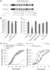
Figure 1
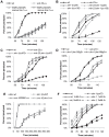
Figure 1. wdr-23 mutants display locomotion defects.
(A) Schematic of WDR-23 isoforms used in this study. Asterisk denotes location of premature stop in the wdr-23(tm1817) allele. The seven WD40 repeat domains and the conservation of the DDB1 binding sequences are identified. The Arg (R) residues which were mutated to His (H) to create WDR-23(DxR) are indicated in bold font. (B) Body bends per minute of the indicated strains. WDR-23 rescue denotes wdr-23 mutants expressing transgenes containing wdr-23a cDNA under control of the wdr-23 promoter. Locomotion was quantified in one minute intervals in the absence of food (Student's t-test). (C) Percent time spent in locomotion of the indicated strains. wdr-23a cDNA was used for rescue with either the endogenous wdr-23 promoter (WDR-23 rescue) or the ges-1 promoter (intestinal rescue). The skn-1 allele zu67 was used for movement analysis. Animals were assayed for three minute intervals in the presence of food. (ANOVA, Tukey's post-hoc). (D) Rates of worm paralysis of the indicated strains when exposed to the acetylcholine esterase inhibitor aldicarb (1.0 mM). Wild type indicated by wt. WDR-23 rescue and over-expression (o/e) were completed using a genomic wdr-23 fragment. WDR-23-GFP rescue denotes co-injection of wdr-23a and wdr-23b cDNA driven by the wdr-23 promoter. (E) Rates of worm paralysis of the indicated strains when exposed to the cholinergic agonist levamisole (200 µM). WDR-23 rescue denotes wdr-23 mutants expressing genomic wdr-23 transgenes. (Error bars indicate ±SEM. *p<0.05, **p<0.01, ***p<0.001.)
Figure 2
Figure 2. Loss of skn-1 suppresses the aldicarb resistance of wdr-23 mutants.
Rates of worm paralysis of the indicated strains when exposed to aldicarb. (A) Expression of WDR-23(DxR) driven by the endogenous wdr-23 promoter does not rescue the aldicarb phenotype of wdr-23 mutants (1.0 mM aldicarb). (B) skn-1(vj24) encodes a R519STOP mutation, and skn-1(zu135) encodes a Q553STOP mutation. Both are in the DNA binding domain and are predicted to affect all three skn-1 isoforms. (C) skn-1(zu67) encodes a R239STOP that specifically affects skn-1a/c. (D) skn-1(gf) alleles lax188 and lax120, which encode E237K and S245L, respectively, are resistant to aldicarb induced paralysis. (E) Loss of skn-1(zu67) does not suppress the aldicarb resistance of unc-10 mutants when exposed to 4.0 mM aldicarb. (F) Exposure to arsenite (Ar) results in skn-1-dependent resistance to aldicarb. The indicated strains were exposed to 0, 1.0, or 2.0 mM arsenite in NGM for 14 hours then allowed to recover for 3 hours prior to assay. (Error bars indicate ±SEM. *p<0.05, **p<0.01, ***p<0.001.)
Figure 3
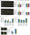
Figure 3. Synaptic vesicle and dense core vesicle secretion defects of wdr-23 mutants.
(A) Left, Representative images of the distribution of the synaptic vesicle protein GFP-RAB-3 and the pro-neuropeptide INS-22-YFP driven by the unc-17/VAChT promoter in cholinergic motor neurons in young adult wild type, wdr-23 mutants and rescued wdr-23 mutants expressing WDR-23a cDNA driven by the endogenous wdr-23 promoter and integrated via Mos insertion. Right, Quantification of the peak fluorescence and the interpunctal interval (Student's t-test). (B) Quantification of the peak fluorescence (left) and interpunctal interval (right) of the synaptic vesicle associated protein GFP-SNB-1/synaptobrevin, the pro-neuropeptide protein NLP-21-YFP, and the active zone protein UNC-10/RIM1-GFP. Control refers to soluble mCherry expressed under the unc-17/VAChT promoter. (C) Left, Representative images of coelomocyte fluorescence in wild type and wdr-23 L4 stage animals expressing NLP-21-YFP. Right, Quantification of NLP-21-YFP in coelomocytes of wild type and wdr-23 mutants. ssGFP refers to GFP tagged to signal sequence which results in a constitutively secreted version of GFP (Student's t-test). (Scale bar represents 10 µm; Error bars indicate ±SEM. *p<0.05, **p<0.01, ***p<0.001.)
Figure 4
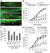
Figure 4. WDR-23 expression pattern and site of action.
(A) Representative images showing fluorescence in the indicated tissues from transgenic animals expressing nuclear-localized gfp from the endogenous 1.3 kb wdr-23a promoter fragment. Arrowheads indicate neuronal cell bodies. (B–D) Paralysis on 1.0 mM aldicarb of the indicated strains. For rescue and knockdown, the following promoters were used: ges-1 (intestine), rab-3 (neuron), col-12 (hypodermis), and myo-3 (muscle). Intestinal and neuronal rescues were independently verified using nlp-40 and snb-1 promoters, respectively (data not shown). (B) Rates of paralysis of tissue-specific rescue of wdr-23 mutants. (C) Hairpin RNAi knockdown of wdr-23 in a sid-1(qt2) background results in tissue-specific aldicarb resistance after 90 minutes on aldicarb (Student's t-test). wdr-23 indicates knockdown with endogenous wdr-23a promoter. (D) RNAi knockdown by feeding of the indicated genes in a wild type or neuronally sensitized (nre-1 lin-15b) strain. Control is the empty expression vector L4440. (Scale bar represents 10 µm; Error bars indicate ±SEM. *p<0.05, **p<0.01.)
Figure 7
Figure 7. Analysis of whole-transcriptome RNA sequencing of wdr-23 mutants.
(A) Correlation plot of RNA-seq reads for wild type versus wdr-23 mutants. Genes significantly different are indicated in blue (up-regulated) and red (down-regulated). (B) Distribution of fold change of genes significantly different in wdr-23 mutants relative to wild type controls. (C) Coverage plot of RNA-seq reads of wdr-23 in wild type and wdr-23(tm1817) mutants.
Figure 5
Figure 5. Subcellular localization and function of WDR-23 isoforms.
(A) Rates of paralysis of the indicated strains on 1.0 mM aldicarb. WDR-23a and WDR-23b rescues denote wdr-23 mutants expressing single copy transgenes containing wdr-23a or wdr-23b cDNA, respectively, driven by the endogenous wdr-23 promoter generated by Mos insertion. (B) Intestinal expression of WDR-23a-GFP and WDR-23b-GFP driven by the ges-1 promoter in a glo-1 background to reduce autofluorescence. Box highlights ring-like pattern of WDR-23a-GFP localization. Arrowheads indicate intestinal nuclei. (C) WDR-23a-GFP co-localization with the inverted outer mitochondrial membrane marker (INVOM-RFP) in muscle cells in wild type and drp-1 mutants. (D) WDR-23b-GFP localization in muscle cells; mitochondria are labeled with INVOM-RFP for reference (Scale bar represents 10 µm; Error bars indicate ±SEM. *p<0.05.)
Figure 6
Figure 6. The WD40 repeat domains are sufficient for WDR-23 function.
(A) Rates of worm paralysis of the indicated strains when exposed to 1.0 mM aldicarb. WDR-23(repeats) rescue denotes wdr-23 mutants expressing transgenes carrying a wdr-23 variant that only contains the seven WD40 repeats driven by the endogenous wdr-23 promoter. (B) WDR-23(repeat) tagged to GFP and expressed in muscle. Dotted circle denotes nucleus. (Scale bar represents 10 µm; Error bars indicate ±SEM. **p<0.01.) (C) Model for SKN-1 regulation of synaptic function. In this model, WDR-23 inhibits SKN-1. Activation of SKN-1 in the intestine, either genetically through loss of wdr-23 or through environmental stress such as arsenite, increases transcription of target genes and may promote the release of a diffusible factor or factors. Endocrine signaling reduces synaptic vesicle and dense core vesicle release, resulting in decreased neurotransmission at neuromuscular junctions.
Figure 8
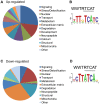
Figure 8. Functional analysis of genes significantly different in wdr-23 mutants.
(A–B) Analysis of 2285 up-regulated genes in wdr-23 mutants. (A) Representation of functional groups of up-regulated genes with known function (1316 genes) in wdr-23 mutants relative to wild type classified by GO terms and protein domain established by DAVID analysis. (B) Canonical SKN-1 binding sequence and enrichment of SKN-1 binding motif identified by RSAT oligonucleotide analysis. One kilobase of intergenic sequence upstream of each ORF for all 2285 up-regulated genes was used for RSAT analysis. RSAT consensus represented by WebLogo. (C–D) Analysis of 134 genes down-regulated in wdr-23 mutants. (C) Representation of functional groups of down-regulated genes with known function (88) in wdr-23 mutants relative to wild type. (D) Canonical SKN-1 binding sequence and enrichment of SKN-1 binding motif identified by RSAT oligo analysis.

Similar articles

Cited by

References

    1. Sykiotis GP, Bohmann D (2010) Stress-activated cap'n'collar transcription factors in aging and human disease. Sci Signal 3: re3. - PMC - PubMed
    1. Halliwell B (2006) Oxidative stress and neurodegeneration: where are we now? J Neurochem 97: 1634–1658. - PubMed
    1. Sykiotis GP, Bohmann D (2008) Keap1/Nrf2 signaling regulates oxidative stress tolerance and lifespan in Drosophila. Dev Cell 14: 76–85. - PMC - PubMed
    1. An JH, Blackwell TK (2003) SKN-1 links C. elegans mesendodermal specification to a conserved oxidative stress response. Genes Dev 17: 1882–1893. - PMC - PubMed
    1. Kobayashi M, Yamamoto M (2006) Nrf2-Keap1 regulation of cellular defense mechanisms against electrophiles and reactive oxygen species. Adv Enzyme Regul 46: 113–140. - PubMed

Publication types

MeSH terms

LinkOut - more resources