. 2013 Mar;9(3):e1003279.
doi: 10.1371/journal.ppat.1003279. Epub 2013 Mar 28.

Pandemic influenza A viruses escape from restriction by human MxA through adaptive mutations in the nucleoprotein

Affiliations

Pandemic influenza A viruses escape from restriction by human MxA through adaptive mutations in the nucleoprotein

Benjamin Mänz et al. PLoS Pathog. 2013 Mar.

Abstract

The interferon-induced dynamin-like MxA GTPase restricts the replication of influenza A viruses. We identified adaptive mutations in the nucleoprotein (NP) of pandemic strains A/Brevig Mission/1/1918 (1918) and A/Hamburg/4/2009 (pH1N1) that confer MxA resistance. These resistance-associated amino acids in NP differ between the two strains but form a similar discrete surface-exposed cluster in the body domain of NP, indicating that MxA resistance evolved independently. The 1918 cluster was conserved in all descendent strains of seasonal influenza viruses. Introduction of this cluster into the NP of the MxA-sensitive influenza virus A/Thailand/1(KAN-1)/04 (H5N1) resulted in a gain of MxA resistance coupled with a decrease in viral replication fitness. Conversely, introduction of MxA-sensitive amino acids into pH1N1 NP enhanced viral growth in Mx-negative cells. We conclude that human MxA represents a barrier against zoonotic introduction of avian influenza viruses and that adaptive mutations in the viral NP should be carefully monitored.


Conflict of interest statement

The authors have declared that no competing interests exist.

Figures
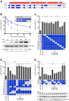
Figure 1
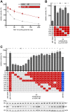
Figure 1. Identification of residues in the NP of the pandemic 1918 influenza A virus responsible for resistance to murine Mx1.
(A) Amino acid differences between NP of H5N1, 1918 and pH1N1. Deviant amino acids of pH1N1 or 1918 NP are highlighted in red and blue, respectively. The 1918/H5N1 chimera (1918*-NP) comprises the N-terminal 365 amino acids of 1918 NP and the C-terminal 133 amino acids of the H5N1 NP and thus lacks 4 1918 NP-specific amino acids. (B) Viral polymerase activity in the presence of increasing concentrations of Mx1. HEK293T cells were transfected with expression plasmids coding for the PB2, PB1 and PA of H5N1, the indicated NP proteins, the firefly luciferase encoding minigenome, increasing amounts of Mx1-coding plasmid and a Renilla-expressing plasmid to normalize variation in transfection efficiency. Polymerase activity (relative activity) in the presence of antivirally inactive Mx1-K49A was used to normalize the data obtained with Mx1. Error bars indicate the standard error of the mean of three independent experiments. Western blot analysis was performed to determine the expression levels of Mx1 and H5N1 NP. (C–E) H5N1 polymerase activity was determined as in (B) after co-transfection of the expression plasmids coding for Mx1 (200 ng) and the indicated NP mutants (100 ng). The polymerase activity (relative activity) observed in the presence of Mx1 was normalized to Mx1-K49A. The resulting relative activity in the presence of either 1918*NP (C–D) or 1918 NP (E) was set to 100%. Western blot analysis shown in panel (E) was performed to determine the expression levels of NP. Error bars indicate the standard error of the mean of three independent experiments. Student's t-test was performed to determine the P value. *P<0.05, **P<0.01, ***P<0.001; NS, not significant.
Figure 2
Figure 2. Identification of residues in the NP of the pandemic 2009 influenza A virus responsible for resistance to murine Mx1.
(A) Reporter activity of H5N1 polymerase in HEK293T cells after co-transfection of expression plasmids coding for the indicated NP proteins (100 ng) and increasing amounts of Mx1. Polymerase activity in the presence of Mx1-K49A was used to normalize the data obtained with Mx1. Error bars indicate the standard error of the mean of three independent experiments. (B) H5N1 polymerase activity (relative activity) was determined as in (A) after co-transfection with expression plasmids coding for Mx1 (200 ng) and the indicated pH1N1 NP mutants (100 ng) harboring single H5N1-derived substitutions. The activity in the presence of Mx1 was normalized to the activity observed with the inactive Mx1 mutant Mx1-K49A. The activity observed in the presence of pH1N1 NP was set to 100%. Error bars indicate the standard error of the mean of three independent experiments. Student's t-test was performed to determine the P value. *P<0.05, **P<0.01, ***P<0.001; NS, not significant. (C) H5N1 polymerase activity was determined as in (B) after co-transfection with expression plasmids coding for Mx1 (200 ng) and the indicated H5N1-NP mutant proteins (100 ng) harboring single or multiple pH1N1-derived substitutions. The activity in the presence of Mx1 was normalized to the activity observed with the inactive Mx1 mutant Mx1-K49A. The activity observed in the presence of 1918 NP was set to 100%. Western blot analysis shown in the lower panel was performed to determine the expression levels of NP and Mx1. Error bars indicate the standard error of the mean of three independent experiments. Student's t-test was performed to determine the P value. *P<0.05, **P<0.01, ***P<0.001; NS, not significant.
Figure 3
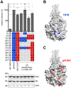
Figure 3. Amino acid clusters in NP of both the 1918 and pH1N1 strain mediate MxA resistance.
(A) H5N1 polymerase activity in HEK293T cells after co-tranfection of the indicated expression plasmids coding for the NP mutants (100 ng) and MxA (200 ng). The activity in the presence of MxA was normalized to the activity observed with the inactive mutant MxA-T103A . The activity observed in the presence of pH1N1 NP was set to 100%. Error bars indicate the standard error of the mean of three independent experiments. Student's t-test was performed to determine the P value. *P<0.05, ***P<0.001, NS, not significant. Western blot analysis was performed to determine the expression levels of MxA and the indicated NPs. (B–C) Amino acid positions of NP mediating Mx resistance. The program PyMOL was used to assign the indicated positions based on the structural model of A/HK/483/97(H5N1) NP (PDB code:2Q06). Positions of adaptive mutations required for Mx resistance of the 1918 NP are marked in blue (B). Amino acids of pH1N1 NP that exhibit only minor contribution to Mx resistance are highlighted in light red, whereas amino acids that strongly increased Mx resistance are indicated in red (C).
Figure 4
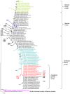
Figure 4. Phylogenetic analysis of representative NP sequences and the presence or loss of Mx-resistance enhancing mutations.
The maximum likelihood tree of 147 aligned representative NP sequences shows four genotypes, i.e., (i) the human seasonal H1N1, H2N2 and H3N2 viruses, (ii) the classical swine H1N1 viruses and pandemic (2009) H1N1 viruses, (iii) the European lineages of swine influenza viruses, and (iv) the North American avian influenza viruses. Strain designations and GenBank acc. nos. are presented. Numbers at nodes indicate bootstrap values obtained after 1,000 replications. Only bootstrap values greater 50% were presented. The bar indicates substitutions per site. Three branches (H2N2/H3N2, recent human H1N1 strains, European lineages of swine influenza viruses) were condensed for clarity. The complete phylogenetic tree is shown in Figure S5. No relevant amino acid substitutions were observed in the condensed branches. Alterations of amino acid positions shown to influence Mx resistance (Figure 3, Figure S9) are highlighted in bold.
Figure 5
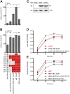
Figure 5. Mx resistance is accompanied by impaired viral growth in cell culture.
(A–B) H5N1 polymerase activity in HEK293T cells after co-transfection of the indicated expression plasmids coding for NP (100 ng) and MxA (200 ng) (A) or porcine Mx1 (poMx1) (200 ng) (B). The activity in the presence of human MxA or poMx1 was normalized to the activity observed with the inactive mutant MxA-T103A. The activity observed with pH1N1 NP was set to 100%. Error bars indicate the standard error of the mean of three independent experiments. Student's t-test was performed to determine the P value. **P<0.01, ***P<0.001; NS, not significant. (C) Expression levels of human MxA and poMx1 in HEK293T cells after reconstitution of the H5N1 polymerase complex using an Mx-specific antibody. (D–E) MDCKII cells were infected with an MOI of 0.001 of wild-type or the indicated pH1N1 (D) or H5N1 mutant viruses (E) and incubated at 37°C. At the indicated time points post infection (p.i.), virus titers were determined by plaque assay. Error bars indicate the standard error of the mean of three independent experiments.
Figure 6
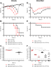
Figure 6. Mx resistance-enhancing mutations in NP increase the virulence of the H5N1 isolate KAN-1 in Mx1-positive mice.
(A–C) BALB/c mice were inoculated intranasally with the indicated amount of viruses. Changes in body weight (A) or survival (B) (n = 6/group) were monitored daily for 14 days. Lungs from infected mice were collected 2 days p.i., homogenized and virus titers were determined by plaque assay (C). (D–F) Mx1-positive BALB/c mice were inoculated intranasally with the indicated amount of viruses. Changes in body weight (D) or survival (E) (n = 6/group) were monitored daily for 14 days. Lungs from infected mice were collected 2, 4, and 6 days p.i., homogenized and virus titers were determined by plaque assay (F). Student's t-test was performed to determine the P value. *P<0.05, **P<0.01, ***P<0.001; NS, not significant.
Figure 7
Figure 7. Temporal appearance of MxA resistance enhancing amino acids in NP of human and swine influenza A viruses.
Bold letters indicate amino acids in NP shown to increase significantly MxA resistance, whereas amino acids highlighted in grey are minor contributors. Adaptive mutations that newly emerged with the appearance of the 2009 pandemic pH1N1 strain are depicted in red. 53D got partially lost after re-introduction of pH1N1 into the swine host.

Similar articles

Cited by

References

    1. Fouchier RA, Schneeberger PM, Rozendaal FW, Broekman JM, Kemink SA, et al. (2004) Avian influenza A virus (H7N7) associated with human conjunctivitis and a fatal case of acute respiratory distress syndrome. Proc Natl Acad Sci U S A 101: 1356–1361. - PMC - PubMed
    1. Tweed SA, Skowronski DM, David ST, Larder A, Petric M, et al. (2004) Human illness from avian influenza H7N3, British Columbia. Emerg Infect Dis 10: 2196–2199. - PMC - PubMed
    1. Lin YP, Shaw M, Gregory V, Cameron K, Lim W, et al. (2000) Avian-to-human transmission of H9N2 subtype influenza A viruses: relationship between H9N2 and H5N1 human isolates. Proc Natl Acad Sci U S A 97: 9654–9658. - PMC - PubMed
    1. Malik Peiris JS (2009) Avian influenza viruses in humans. Rev Sci Tech 28: 161–173. - PubMed
    1. Wang TT, Parides MK, Palese P (2012) Seroevidence for H5N1 Influenza Infections in Humans: Meta-Analysis. Science 335: 1463. - PMC - PubMed

Publication types

MeSH terms

Grants and funding

This work was supported by the Deutsche Forschungsgemeinschaft, grant Ko 1579/8-1 to GK and the Bundesministerium für Bildung und Forschung (FluResearchNet) to MS and RZ. The funders had no role in study design, data collection and analysis, decision to publish, or preparation of the manuscript.

LinkOut - more resources