. 2013 Apr 25;3(4):1117-27.
doi: 10.1016/j.celrep.2013.03.002. Epub 2013 Mar 28.

Mechanism of origin DNA recognition and assembly of an initiator-helicase complex by SV40 large tumor antigen

Affiliations

Mechanism of origin DNA recognition and assembly of an initiator-helicase complex by SV40 large tumor antigen

Y Paul Chang et al. Cell Rep. .

Abstract

The DNA tumor virus Simian virus 40 (SV40) is a model system for studying eukaryotic replication. SV40 large tumor antigen (LTag) is the initiator/helicase that is essential for genome replication. LTag recognizes and assembles at the viral replication origin. We determined the structure of two multidomain LTag subunits bound to origin DNA. The structure reveals that the origin binding domains (OBDs) and Zn and AAA+ domains are involved in origin recognition and assembly. Notably, the OBDs recognize the origin in an unexpected manner. The histidine residues of the AAA+ domains insert into a narrow minor groove region with enhanced negative electrostatic potential. Computational analysis indicates that this region is intrinsically narrow, demonstrating the role of DNA shape readout in origin recognition. Our results provide important insights into the assembly of the LTag initiator/helicase at the replication origin and suggest that histidine contacts with the minor groove serve as a mechanism of DNA shape readout.


Figures

Figure 1
Figure 1. SV40 LTag Domain Structures and Core Replication Ori DNA Sequence
(A) Representation of the LTag domains. The construct used for cocrystallization is boxed in blue (LTag131-627), which contains the OBD, Zn domain, and AAA+ domain. (B) Core ori DNA sequence of SV40. Each of the four pentanucleotide GAGGC sequences (PEN1–PEN4, labeled in red) is recognized by an OBD, and the EP and AT regions are indicated. The hidden-site sequence (GC, colored in blue) is indicated here and discussed in the text. The EP-half ori DNA (boxed in blue) was used for cocrystallization.
Figure 2
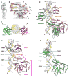
Figure 2. Overall Structure of the LTag Dimer in Complex with EP-Ori
(A) The well-defined electron density map of the EP-ori DNA shows a very clear electron density for the base pairs. (B) The map section corresponding to PEN1 in (A) is enlarged to show the excellent fitting of the DNA model into the density. (C) Top view of the two OBDs (drawn in ribbons) on the opposite faces of the ori DNA (drawn in surface representation) around PEN1. The two DNA strands are shown in the same color scheme as in (B), with yellow for one strand and gray for the other. (D) Overall structure of the dimeric LTag-ori DNA complex. OBD1 binds PEN1 (highlighted), but OBD2 does not bind to PEN2. Instead, OBD2 binds to a hidden site near PEN1 but distal to PEN2. The helicase domains of the two LTag subunits are arranged similarly to two adjacent subunits in LTag hexamers. (E and F) The interactions between EP-ori and the OBD, linker region, and Zn and AAA+ domains are shown along the dsDNA’s consecutive major/minor-groove interfaces (indicated by arrows) in the two subunits (subunit 1 [E] and subunit 2 [F]). Note the difference in the linker conformations and the OBD orientations of the two subunits. See also Figures S1, S2, and S6.
Figure 3
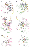
Figure 3. Detailed Interactions between LTag and EP-Ori DNA
In all panels, the leading DNA strand is in gray, the reverse strand is in yellow, subunit 1 is in pink, and subunit 2 is in green. Protein backbone oxygen and nitrogen atoms are represented by red and blue spheres, respectively. (A) Sequence-specific interactions between OBD1 and PEN1. (B) Interactions between OBD2 and the hidden-site sequence 5′-G17′C16′ and 5′-G16C17 (see Figure 1B). (C) Interactions of the Zn domains of both subunits with EP-ori DNA, which is mediated by charge-charge interactions with the DNA backbones. Water molecules are represented by white spheres. (D) Interactions between the AAA+ domains of both subunits and the EP region, which are mainly mediated by residues on the β-hairpin (K512/ H513). Note that the H513 residues of both subunits insert into the minor groove to interact with both strands, whereas the K512 residues of both subunits track only one strand. (E and F) The same interactions shown in (D), except that subunit 1 (E) and subunit 2 (F) are shown separately. See also Figures S2, S3, and S5.
Figure 4
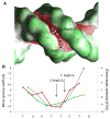
Figure 4. Narrow Minor-Groove Geometry of the 5′-C5T6T7C8T9 Region of EP-Ori DNA where Both β-Hairpin H513 Residues Bind
(A) The shape of the molecular surface is shown with GRASP2 (concave surfaces in dark gray; convex surfaces in green; Petrey and Honig, 2003). The red mesh represents an isopotential surface at −5 kT/e, calculated with DelPhi at a physiologic ionic strength of 0.145 M (Rocchia et al., 2002). The H513 residues from both subunits intrude into the minor groove in a region with enhanced negative electrostatic potential as a result of narrowing the width of the minor groove to 4.5 Å (with an electrostatic potential of −9.4 kT/e) from the normal width of 5.8 Å (with an electrostatic potential of −7.2 kT/e (see B). (B) The minor-groove width of bound ori DNA in our crystal structure (blue) and unbound ori DNA predicted in MC simulations (green), and the electrostatic potential in the center of the minor groove calculated with DelPhi (red) illustrate that the H513 residues bind a region with an intrinsically narrow minor groove. The enhanced negative electrostatic potential in the narrower groove region attracts H513 residues through favorable electrostatic interactions, a mechanism known as shape readout. See also Figure S4.
Figure 5
Figure 5. Helicase Domain Conformations and Dimer Interface
(A) Superimposition of helicase domains in various nucleotide-bound states (white) and the two subunits in the DNA-bound state from this structure (pink and green), which reveals shifting of the AAA+ domains and β-hairpins, illustrating the ability of multiple conformational switches that are critical for a motor protein such as LTag to melt origin and unwind DNA. Note that α10 adopts different orientations for subunits 1 (green) and 2 (pink), which generates a rotation of the AAA+ domains to bring their β-hairpins (and H513 residues) into close proximity to the DNA minor groove (see the box in H here, and Figure 3D). (B–D) Change of contacts between Zn (K281/P311) and AAA+ domains (D367/R371/I374 on α10) in the absence (B: close contact) or presence (C: detached; D: altered contact) of DNA. (E and F) Two views of the protein dimer interface show the tight interface between the two subunits around the β-hairpin regions in the AAA+ domain. (G and H) Close-up views of the extensive interactions between the two AAA+ domains. The close proximity of the two H513 residues is shown in the inset (H).
Figure 6
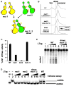
Figure 6. Generation of a Stable LTag Dimer Intermediate and Analysis of its Activities
(A) Strategy to obtain a stable dimer intermediate of LTag through two independent mutations. Mut1 carries V350E/P417D mutations on one side of a subunit, and mut2 has L286Dt/R567E mutations on the opposite surface of LTag131-627. Both mutants are predicted to exist only as monomers when alone, but as a stable dimer when mixed in an equimolar ratio. (B) Superdex-200 column chromatography, showing that mut1 and mut2 alone exist in monomeric form, but when mixed in equimolar ratios, they form stable dimer intermediates. Inset: the two mutants in the dimer peak were detected in a 1:1 ratio by SDS-PAGE, with mut1 being slightly larger due to a C-terminal extension. Note that WT equilibrates between monomeric and hexameric forms; no dimer or any other intermediate oligomeric form can be detected. (C) ATPase activity assay, showing a recovery of ATPase activity only when mut1 and mut2 are mixed, indicating a WT ATP pocket at the dimer interface. (D) Ori DNA unwinding assay, showing that only WT LTag131-627, and not the mutants, can unwind the ori DNA. Lane 1: unboiled blunt-ended ori DNA alone (UB); lane 2: boiled ori DNA (B); lanes 3 and 4: mut1 alone; lanes 5–7: mut2 alone; lanes 8–10: the dimer of mut1 and mut2; lanes 11 and 12: WT LTag131-627. (E) Ori DNA melting assay by KMnO4 reactivity, showing that WT LTag131-627, but not the mutant dimer, has ori-melting activity. Radiolabeled 92 bp ori containing dsDNA was incubated with increasing quantities of LTag protein (lanes 2–5 and lanes 6–9: 100, 200, 400, and 800 ng for WT and mutant dimer, respectively). Lane 1 has no protein added. See also Figure S5.

Similar articles

Cited by

References

    1. Bochkareva E, Martynowski D, Seitova A, Bochkarev A. Structure of the origin-binding domain of simian virus 40 large T antigen bound to DNA. EMBO J. 2006;25:5961–5969. - PMC - PubMed
    1. Borowiec JA, Hurwitz J. Localized melting and structural changes in the SV40 origin of replication induced by T-antigen. EMBO J. 1988;7:3149–3158. - PMC - PubMed
    1. Borowiec JA, Dean FB, Bullock PA, Hurwitz J. Binding and unwinding—how T antigen engages the SV40 origin of DNA replication. Cell. 1990;60:181–184. - PubMed
    1. Deb S, DeLucia AL, Baur CP, Koff A, Tegtmeyer P. Domain structure of the simian virus 40 core origin of replication. Mol Cell Biol. 1986;6:1663–1670. - PMC - PubMed
    1. Deb S, Tsui S, Koff A, DeLucia AL, Parsons R, Tegtmeyer P. The T-antigen-binding domain of the simian virus 40 core origin of replication. J Virol. 1987;61:2143–2149. - PMC - PubMed

Publication types

MeSH terms

Associated data

LinkOut - more resources