. 2013 May 1;190(9):4899-909.
doi: 10.4049/jimmunol.1300271. Epub 2013 Mar 27.

Restoring immune function of tumor-specific CD4+ T cells during recurrence of melanoma

Affiliations

Restoring immune function of tumor-specific CD4+ T cells during recurrence of melanoma

Stephen R Goding et al. J Immunol. .

Abstract

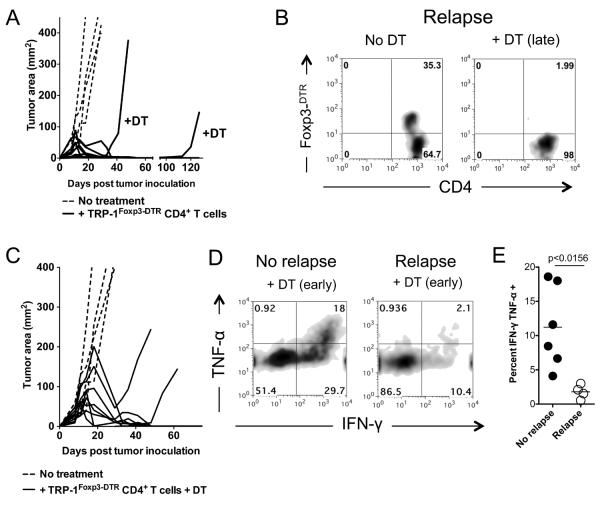
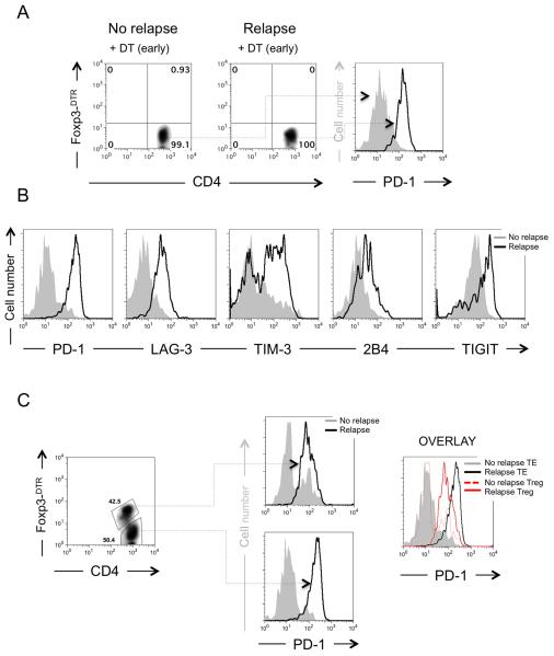
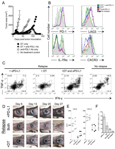
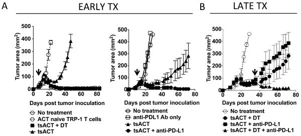
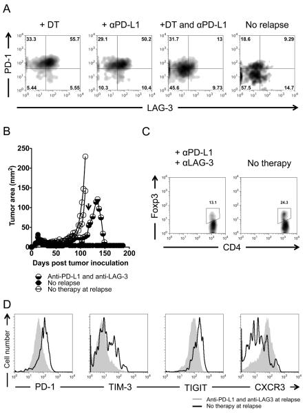
Recurrent solid malignancies are often refractory to standard therapies. Although adoptive T cell transfer may benefit select individuals, the majority of patients succumb to their disease. To address this important clinical dilemma, we developed a mouse melanoma model in which initial regression of advanced disease was followed by tumor recurrence. During recurrence, Foxp3(+) tumor-specific CD4(+) T cells became PD-1(+) and represented >60% of the tumor-specific CD4(+) T cells in the host. Concomitantly, tumor-specific CD4(+) T effector cells showed traits of chronic exhaustion, as evidenced by their high expression of the PD-1, TIM-3, 2B4, TIGIT, and LAG-3 inhibitory molecules. Although blockade of the PD-1/PD-L1 pathway with anti-PD-L1 Abs or depletion of tumor-specific regulatory T cells (Tregs) alone failed to reverse tumor recurrence, the combination of PD-L1 blockade with tumor-specific Treg depletion effectively mediated disease regression. Furthermore, blockade with a combination of anti-PD-L1 and anti-LAG-3 Abs overcame the requirement to deplete tumor-specific Tregs. In contrast, successful treatment of primary melanoma with adoptive cell therapy required only Treg depletion or Ab therapy, underscoring the differences in the characteristics of treatment between primary and relapsing cancer. These data highlight the need for preclinical development of combined immunotherapy approaches specifically targeting recurrent disease.


Figures

Figure 1
Figure 1. Foxp3+ tumor-specific Treg cells increase during melanoma recurrence
(A) After successful tumor-specific CD4+ T cell adoptive cell immunotherapy melanoma recurs. C57BL/6 lymphopenic RAG−/− mice (5 mice/group) were inoculated with B16.F10 melanoma (2 × 10⁵ cells). Tumor-bearing mice were treated with 2 × 10⁵ naïve TRP-1 CD4+ T cells by intravenous tail vein injection on day 10 after tumor inoculation. Tumors were followed until recurrence of melanoma. Experiments repeated at least 10 times. (B) Foxp3+ tumor-specific CD4+ T cells increase during relapsing melanoma. TRP-1 CD4+ T cells were analyzed for Foxp3 expression by flow cytometry from relapsing mice (68.3%) and compared to cells from mice with no tumors (4.4%). Data represent ten independent experiments. (C) Percent Foxp3-eGFP+ cells from ten experiments. Using Foxp3-DTR TRP-1 T cells, which fluoresce with eGFP when expressing Foxp3, similar results were obtained in relapsing and non-relapsing mice (No recurrence, ~5%; Recurrence ~50%). P < 0.0001 for Foxp3 expression between non-relapsing versus relapsing mice. (D) Absolute numbers of tumor-specific CD4+ Treg and Foxp3 CD4+ TE cells during recurrence compared to nonrelapsing mice. Experiments repeated ten times.
Figure 2
Figure 2. Depletion of tumor-specific Treg cells does not prevent or treat relapsing melanoma
(A) Depletion of tumor-specific Foxp3+ Treg cells during recurrence (late) does not retreat melanoma. C57BL/6 lymphopenic RAG−/− mice (5 mice/group) were inoculated with B16.F10 melanoma (2 × 10⁵ cells). Tumor-bearing mice were treated with 2 × 10⁵ naïve TRP-1 Foxp3-DTR CD4+ T cells by intravenous tail vein injection on day 10 after tumor inoculation. Tumors were followed until recurrence of melanoma. DT (diphtheria toxin) was injected i.p. at the prescribed concentration of 50 μg/Kg every other day for three doses total. PBS controls had no effect on Treg cells (not shown). Experiments repeated 5 times. (B) Flow cytometry of Foxp3 eGFP expression in TRP-1 Foxp3-DTR CD4+ T cells from relapsing mice treated with or without DT at time of sacrifice. (No DT, 35.3%; +DT (late), ~2%). Experiments repeated at least five times. (C) Depletion of tumor-specific Foxp3+ Treg cells before recurrence (early) does not prevent melanoma recurrence. C57BL/6 lymphopenic RAG−/− mice (5 mice/group) were inoculated with B16.F10 melanoma (2 × 10⁵ cells). Tumor-bearing mice were treated with 2 × 10⁵ naïve TRP-1 Foxp3-DTR CD4+ T cells by intravenous tail vein injection on day 10 after tumor inoculation. Tumors were followed until recurrence of melanoma. At injection of T cells (day 10), DT (diphtheria toxin) was injected i.p. at the prescribed concentration of 50 μg/Kg every other day for three doses total. PBS controls had no effect on Treg cells (not shown). Experiments repeated five times. (D) Functional characteristics of tumor-specific CD4+ T cells during recurrence with Treg cells depleted early. Flow cytometry of IFN-γ and TNF-α expression in TRP-1 Foxp3-DTR CD4+ T cells from non-relapsing and relapsing mice. Data represent at least three experiments. (E) Percent of TRP-1 CD4+ T cells that are dual producers of IFN-γ and TNF-α during recurrence. P < 0.0156 for non-relapsing and relapsing groups. Experiments repeated four times.
Figure 3
Figure 3. Tumor-specific CD4+ T cells express multiple inhibitory receptors during recurrence of melanoma
(A) Depletion of tumor-specific Treg cells with DT before recurrence does not prevent exhaustion of TRP-1 Foxp3-DTR CD4+ T cells. Flow cytometry of TRP-1 Foxp3-DTR CD4+ T cells showing complete depletion of Treg cells during recurrence compared to non recurring (left two panels) and expression of PD-1 on TRP-1 Foxp3-DTR CD4+ TE cells (right panel) from either group. Experiment repeated five times. (B) Tumor-specific TRP-1 CD4+ TE cells express multiple inhibitory receptors during relapsing melanoma. Foxp3 tumor-specific CD4+ TE cells were analyzed by flow cytometry for expression of inhibitory receptors as indicated. Gray filled histograms represent non-relapsing groups. Open histograms with a black line represent relapsing groups. Representative of three to ten experiments. (C) Foxp3+ tumor-specific Treg cells express PD-1 during relapsing melanoma although at lower levels than tumor-specific TRP-1 CD4+ TE cells. Foxp3+ and Foxp3 tumor-specific CD4+ T cells were analyzed by flow cytometry for expression of the inhibitory receptor PD-1 (left panel). PD-1 is expressed on both CD4+ Treg and TE cells (middle panel). Overlay of flow cytometry shows that PD-1 expression is higher on CD4+ TE cells than on Foxp3+ Treg cells (right panel). Representative of three experiments.
Figure 4
Figure 4. Treatment of relapsing melanoma requires both Treg depletion and blockade of PD-L1
(A) Treatment of relapsing melanoma requires combination Treg depletion and anti-PD-L1 therapy. C57BL/6 lymphopenic RAG−/− mice (5–10/group) were inoculated with B16.F10 melanoma (2 × 10⁵ cells). Tumor-bearing mice were treated with 2 × 10⁵ naïve TRP-1 Foxp3-DTR CD4+ T cells by intravenous tail vein injection on day 7–10 after tumor inoculation. Tumors were followed until recurrence of melanoma. At recurrence, DT (diphtheria toxin) was injected i.p. at the prescribed concentration of 50 mg/Kg every other day for three doses total. At recurrence, anti-PD-L1 was given as a bolus injection i.p. at 500 μg for the first dose and subsequently given every three days thereafter at 200 μg/injection for 5 doses. For combination therapy, both were given at the same time as described above. PBS controls had no effect (not shown). Data shown is representative of more than 5 experiments. (B) Blockade of PD-L1 and depletion of tumor-specific Treg cells reinvigorates tumor-specific CD4+ T cells by reducing inhibitory receptor expression of PD-1 and LAG-3, and increases IL-7R and CXCR3. Flow cytometry shows four groups (No recurrence, DT only, DT plus anti-PD-L1 antibody, and anti-PD-L1 antibody only). Gray filled histograms represent no recurrence, blue solid line represents DT therapy only, red solid line represents anti-PD-L1 only, and green solid line represents therapy with DT and anti-PD-L1 together. All therapies were given at time of recurrence. Flow cytometry is performed on each group 3–5 days after the last dose of therapy. Shown is representative of 5 experiments. (C) Tumor-specific CD4+ T cells regain effector function with dual therapy as defined by re-expression of IFN-γ and TNF-α when compared to relapsing groups with single therapies (anti-PD-L1 (7.3%), DT (3.04%), +DT and anti-PD-L1 (17.4%), no recurrence (18.6%)). Experiment repeated three times. (D) Gross depiction of tumor regression after combination therapy. Days indicate day after therapy was given. All tumors depicted are relapsing tumors that were previously treated as a primary tumor. (E) IFN-γ inducible chemokines (CXCL9 and CXCL10) are highly increased during recurrence and return to treatment levels with combination therapy. Each dot represents and individual mouse. (F) Total number of tumor-specific CD4+ T cells in the spleen stabilizes to non-relapsing levels with combination therapy. Absolute number of TRP-1 CD4+ T cells during different treatments. Each bar graph represents 5–10 individual mice.
Figure 5
Figure 5. Disparate treatment requirements between primary and relapsing melanoma
(A) Early treatment of melanoma with tumor sensitized TRP-1 CD4+ T cells (tsACT) from mice with progressively growing tumors fails in all mice when compared to naïve TRP-1 CD4+ T cell transfer. TRP-1 Foxp3-DTR tyrp1BwRAG−/− transgenic mice (5–10 mice/group) were inoculated with B16.F10 melanoma (1 × 10⁶ cells). When tumors had reached ~400 mm2, tumor sensitized (ts) CD4+ T cells were harvested and sorted and 2 × 10⁵ cells were transferred i.v. into 7 day tumor-bearing lymphopenic mice. tsTRP-1 CD4+ T cells (tsACT) alone fail to control tumor but when depleted of Treg cells (+DT) treated primary tumor (right panel). Anti-PD-L1 alone without T cell transfer fails to treat primary tumors. Anti-PD-L1 with tsTRP-1 CD4+ T cells (tsACT) treated primary tumor (middle panel). (B) Single therapies given late during relapsing tumor fail to treat tumor as in figure (A). Combination therapy with anti-PD-L1 and Treg depletion was still required (right panel). Arrows indicate beginning of therapy with either DT, anti-PD-L1, or both. Experiments repeated three times.
Figure 6
Figure 6. Simultaneous blockade with combination therapy to PD-L1 and LAG-3 inhibitory receptors treats recurring melanoma
(A) PD-1 and LAG-3 are coexpressed on chronically exhausted tumor-specific CD4+ T cells and decrease during anti-PD-L1 and Treg cell depletion therapy or during a potential cure. C57BL/6 lymphopenic RAG−/− mice (5–10 mice/group) were inoculated with B16.F10 melanoma (2 × 10⁵ cells). Tumor-bearing mice were treated with 2 × 10⁵ naïve TRP-1 Foxp3-DTR CD4+ T cells by intravenous tail vein injection on day 7–10 after tumor inoculation. Tumors were followed until recurrence of melanoma. At recurrence, DT (diphtheria toxin) was injected i.p. at the prescribed concentration of 50 mg/Kg every other day for three doses total. At recurrence, Anti-PD-L1 was given as a bolus injection i.p. at 500 μg for the first dose and subsequently given every three days thereafter at 200 μg/injection for 5 doses. For combination therapy, both were given at the same time as described above. Experiment repeated at least five times. (B) Anti-PD-L1 and anti-LAG-3 antibodies given during relapse treat recurring tumor. Arrow indicates the time in which the antibodies were given. Treatment was similar as in (A). However, a 500 μg loading dose of each antibody (anti-PD-L1 and anti-LAG-3) was given initially with 200 μg doses given every 3 days for 5 doses total. (C) Treg cell frequency during treatment with dual antibody therapy in (B). (D) Inhibitory receptor expression on tumor-specific T effector cells during dual antibody therapy. CD4+ T cells were isolated from mice undergoing regression after dual antibody therapy approximately 3–7 days after the last dose of antibody and analyzed by flow cytometry.

Similar articles

Cited by

References

    1. Xie Y, Akpinarli A, Maris C, Hipkiss EL, Lane M, Kwon EK, Muranski P, Restifo NP, Antony PA. Naive tumor-specific CD4(+) T cells differentiated in vivo eradicate established melanoma. J Exp Med. 2010;207:651–667. - PMC - PubMed
    1. Quezada SA, Simpson TR, Peggs KS, Merghoub T, Vider J, Fan X, Blasberg R, Yagita H, Muranski P, Antony PA, Restifo NP, Allison JP. Tumor-reactive CD4(+) T cells develop cytotoxic activity and eradicate large established melanoma after transfer into lymphopenic hosts. J Exp Med. 2010;207:637–650. - PMC - PubMed
    1. Muranski P, Borman ZA, Kerkar SP, Klebanoff CA, Ji Y, Sanchez-Perez L, Sukumar M, Reger RN, Yu Z, Kern SJ, Roychoudhuri R, Ferreyra GA, Shen W, Durum SK, Feigenbaum L, Palmer DC, Antony PA, Chan CC, Laurence A, Danner RL, Gattinoni L, Restifo NP. Th17 cells are long lived and retain a stem cell-like molecular signature. Immunity. 2011;35:972–985. - PMC - PubMed
    1. Landsberg J, Kohlmeyer J, Renn M, Bald T, Rogava M, Cron M, Fatho M, Lennerz V, Wolfel T, Holzel M, Tuting T. Melanomas resist T-cell therapy through inflammation-induced reversible dedifferentiation. Nature. 2012 - PubMed
    1. Jensen SM, Twitty CG, Maston LD, Antony PA, Lim M, Hu HM, Petrausch U, Restifo NP, Fox BA. Increased frequency of suppressive regulatory T cells and T cell-mediated antigen loss results in murine melanoma recurrence. J Immunol. 2012;189:767–776. - PMC - PubMed

Publication types

MeSH terms