. 2013 Apr 17;32(8):1168-82.
doi: 10.1038/emboj.2013.66. Epub 2013 Mar 26.

YY1 controls Igκ repertoire and B-cell development, and localizes with condensin on the Igκ locus

Affiliations

YY1 controls Igκ repertoire and B-cell development, and localizes with condensin on the Igκ locus

Xuan Pan et al. EMBO J. .

Abstract

Conditional knock-out (KO) of Polycomb Group (PcG) protein YY1 results in pro-B cell arrest and reduced immunoglobulin locus contraction needed for distal variable gene rearrangement. The mechanisms that control these crucial functions are unknown. We deleted the 25 amino-acid YY1 REPO domain necessary for YY1 PcG function, and used this mutant (YY1ΔREPO), to transduce bone marrow from YY1 conditional KO mice. While wild-type YY1 rescued B-cell development, YY1ΔREPO failed to rescue the B-cell lineage yielding reduced numbers of B lineage cells. Although the IgH rearrangement pattern was normal, there was a selective impact at the Igκ locus that showed a dramatic skewing of the expressed Igκ repertoire. We found that the REPO domain interacts with proteins from the condensin and cohesin complexes, and that YY1, EZH2 and condensin proteins co-localize at numerous sites across the Ig kappa locus. Knock-down of a condensin subunit protein or YY1 reduced rearrangement of Igκ Vκ genes suggesting a direct role for YY1-condensin complexes in Igκ locus structure and rearrangement.


Conflict of interest statement

The authors declare that they have no conflict of interest.

Figures

Figure 1
Figure 1
The YY1 REPO domain is needed for bone marrow B-cell development. (A) GFP levels correlate with YY1 expression. 38B9 cells were transduced with MigR1-FlagYY1 and 4 days later cells were sorted into low, middle, and high GFP-expressing populations. Cell lysates were evaluated by western blot with anti-YY1 antibody, and blots showed that YY1 expression correlates with the level of GFP expression. (B, C) MigR1, MigR1-FlagYY1, and MigR1-FlagYY1ΔREPO transduced yy1f/f mb1CRE bone marrow was injected into recipient irradiated C57BL/6 mice and bone marrow from reconstituted mice was subject to analyses at 14 weeks post transplantation. (B) FACS analyses of bone marrow B cells. Within the GFP+ cell population, pro-B cells were characterized as B220+CD43+CD19+AA4.1+, pre-B cells as B220+CD43 IgMAA4.1+, immature B cells as B220+CD43IgM+AA4.1+, and mature B cells as B220+CD43IgM+AA4.1. (C) YY1ΔREPO reconstituted mice show decreased pro-B, pre-B, immature B, and mature B sub-populations. Total cell numbers in each bone marrow B-cell sub-population are shown. Mean and standard error of the mean of 4 experiments with 10 mice per cohort are shown. Double asterisks indicate significant differences between YY1 and YY1ΔREPO reconstituted mice at P<0.01. Source data for this figure is available on the online supplementary information page.
Figure 2
Figure 2
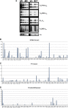
Deletion of the YY1 REPO domain results in skewed Igκ, but not Ig heavy chain, rearrangement patterns. (A) IgH rearrangement in GFP+ splenic B cells from wild-type C57BL/6 and MigR1, YY1, and YY1ΔREPO reconstituted mice. PCR with V family-specific degenerate primers was performed on two-fold serially diluted DNA samples. Results are representative of three experiments. (BD) Deletion of the YY1 REPO domain results in altered Igκ V gene rearrangement pattern. RNA isolated from pre-B cells from C57BL/6 (B), YY1 (C), and YY1ΔREPO (D) mice was subjected to Vκ transcritome sequence analysis. The x axis shows the individual Vκ genes from the most distal (left) to most proximal (right). Percentage of total transcripts representative of each listed Vκ gene is shown on the y axis. The analysis of YY1 mice shows a full representation of numerous Vκ genes that span the Igκ locus, whereas the YY1ΔREPO mice show a dramatically skewed repertoire with reduced Vκ gene usage. Source data for this figure is available on the online supplementary information page.
Figure 3
Figure 3
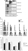
The skewed Vκ repertoire in YY1ΔREPO mice is reproducible. (A) PCR detection of specific Igκ chain rearrangements using DNA from sorted GFP+ splenic B cells from MigR1, YY1, and YY1ΔREPO reconstituted mice. Input DNA was normalized by PCR amplification with primers flanking the Cκ region. Two-fold serially diluted DNA samples were used for PCRs. (B) Real-time PCR was performed to detect specific Igκ chain rearrangements in sorted GFP+ splenic B cells from MigR1, YY1, and YY1ΔREPO reconstituted mice. GAPDH was used as an internal control for normalization and quantitation was determined by the ΔΔCT method. Each bar represents one mouse. (C) Jκ chain gene usage in YY1ΔREPO mice favours Jκ1 and Jκ2, whereas Jκ5 usage is reduced. The percentage of each Jκ segment used in C57BL/6, YY1, and YY1ΔREPO mice is shown.
Figure 4
Figure 4
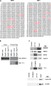
The YY1 REPO domain physically interacts with SMC4 and SMC1. (A) Sequences in red represent peptides identified by MALDI-TOF mass spec analysis of proteins that physically interact with the YY1 REPO domain in co-immunoprecipitation experiments. (B) YY1 interacts with condensin complex protein BRRN1. Retroviral vector or MigR1-FlagYY1 was transduced into 38B9 cells. Lysates were immunoprecipitated with Flag antibody and subjected to western with anti-BRRN1 or anti-YY1 antibodies. (C) YY1 interacts with condensin and PcG proteins. IL-7 cultured B-cell lysates were immunoprecipitated with YY1 antibody or rabbit Ig control. Precipitants then were blotted for BRRN1, SMC4, EZH2, SuZ12, Lamin B1, or β-actin using specific antibodies. Alternatively, lysates from purified primary pro-B cells were precipitated with anti-SMC4 followed by western blot for YY1, Lamin B1, or β-actin with specific antibodies.
Figure 5
Figure 5
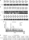
YY1, EZH2, and condensin proteins co-localize across the Vκ locus. (AD) Representative ChIP analyses from IL-7 bone marrow cultures or NIH3T3 cells at YY1 binding sites identified computationally across the Igκ locus for (A) YY1 and EZH2, (B), SMC4, (C) SMC2, (D) YY1, SMC4, and BRRN1. Data in (D) are real-time PCR data presented as percent input. The RP-L30 promoter was used as a positive control for YY1 binding, and actin B was used as a negative control for YY1 binding. The mean and standard error of the mean are shown. (E) ChIP analysis of YY1-negative regions within the Igκ locus showing general loss of EZH2 and SMC4. Primers and antibodies are shown above each lane. Source data for this figure is available on the online supplementary information page.
Figure 6
Figure 6
Location of YY1 binding site clusters within the Igκ locus. Summary of YY1, EZH2, SMC4, SMC2, and BRRN1 (CAP-H) ChIP data. The top panel shows the Vκ rearrangement data for YY1ΔREPO mice. The bottom panel shows the Igκ locus drawn to scale with each Vκ gene represented as a vertical line. YY1, EZH2, and condensin binding sites are shown as black circles on the map. The Vκ genes that continue to rearrange in the YY1ΔREPO background (top panel) are matched to their relative locations on the locus map by arrows. Binding of specific proteins are listed below the map for each site. The YY1-negative region data are summarized in the penultimate lowest panel of the figure. Binding of Pax5 and E47 at the Vκ24-140 and dv-36 regions is summarized in the lowest panel. Presence of YY1, EZH2, SMC4, SMC2, BRRN1, Pax5, or E47 at each site is indicated with a ‘+’. Question marks indicate inconclusive DNA binding.
Figure 7
Figure 7
Condensin subunit or YY1 knock-down inhibits Igκ rearrangement of some Igκ V genes. (A) SMC4 and YY1 knock-down by siRNA in IL-7 cultured B cells. Cell lysates from SMC4 or YY1 knock-down, or scrambled RNA, were subjected to western blot for SMC4 or YY1. β-Actin was used as a loading control. (B) Knock-down of SMC4 impairs recombination of Vκ38-93 to Jκ2 and Jκ5 gene segments. IL-7 cultured B cells electroporated with either SMC4 siRNA or scrambled siRNA were treated with either 5 or 0.1 ng/ml IL-7 right after the electroporation and incubated 72 h. Three-fold serially diluted DNA samples were used for PCRs to detect rearrangement of Vκ38-93 or Vκ12-44 regions to downstream Jκ2 or Jκ5 regions. PCR of the Cκ region was used as an internal control. (C) Knock-down of YY1 results in decreased recombination of Vκ38–93, Vκ21-4, and Vκ2-139, respectively. IL-7 cultured B cells were electroporated with YY1 siRNA or scrambled siRNA and the IL-7 concentration was lowered from 5 to 0.1 ng/ml right after the electroporation. After 72 h, DNA samples were used for PCRs for rearrangement of Vκ38-93, Vκ21-4, Vκ2-139, or Vκ8-27. PCR of the Cκ region was used as an internal control.
Figure 8
Figure 8
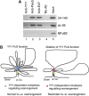
Model for YY1 PcG function and Igκ locus structure. (A) Pax5 and E47 bind to the Vκ24-140 and dv-36 regions. Chromatin from IL-7 cultured bone marrow cells was subjected to ChIP with the antibodies shown above the lanes and the primers shown on the right. (B) Model of Igκ locus loop structure in the presence and absence of YY1 PcG function. Source data for this figure is available on the online supplementary information page.

Similar articles

Cited by

References

    1. Akasaka T, Tsuji K-i, Kawahira H, Kanno M, Harigaya K-i, Hu L, Ebihara Y, Nakahata T, Tetsu O, Taniguchi M, Koseki H (1997) The role of mel-18, a mammalian Polycomb group gene, during IL-7-dependent proliferation of lymphocyte precursors. Immunity 7: 135–146 - PubMed
    1. Alkema MJ, van der Lugt NMT, Bobeldijk RC, Berns A, van Lohuizen M (1995) Transformation of axial skeleton due to overexpression of bmi-1 in transgenic mice. Nature 374: 724–727 - PubMed
    1. Aoki-Ota M, Torkamani A, Ota T, Schork NJ, Nemazee D (2012) Skewed primary Igκ repertoire and V-J joining in C57BL/6 mice: Implications for recombination accessibility and receptor editing. J Immunol 188: 2305–2315 - PMC - PubMed
    1. Atchison L, Ghias A, Wilkinson F, Bonini N, Atchison ML (2003) The YY1 transcription factor functions as a PcG protein in vivo. EMBO J 22: 1347–1358 - PMC - PubMed
    1. Bain G, Romanow WJ, Albers K, Havran WL, Murre C (1999) Positive and negative regulation of V(D)J recombination by the E2A proteins. J Exp Med 189: 289–300 - PMC - PubMed

Publication types

MeSH terms