. 2013 May 15;126(Pt 10):2225-35.
doi: 10.1242/jcs.121327. Epub 2013 Mar 22.

Amotl2 interacts with LL5β, localizes to podosomes and regulates postsynaptic differentiation in muscle

Affiliations

Amotl2 interacts with LL5β, localizes to podosomes and regulates postsynaptic differentiation in muscle

Tomasz J Proszynski et al. J Cell Sci. .

Abstract

Neuromuscular junctions (NMJs) in mammalian skeletal muscle undergo a postnatal topological transformation from a simple oval plaque to a complex branched structure. We previously showed that podosomes, actin-rich adhesive organelles, promote the remodeling process, and demonstrated a key role for one podosome component, LL5β. To further investigate molecular mechanisms of postsynaptic maturation, we purified LL5β-associated proteins from myotubes and showed that three regulators of the actin cytoskeleton--Amotl2, Asef2 and Flii--interact with LL5β. These and other LL5β-interacting proteins are associated with conventional podosomes in macrophages and podosome-like invadopodia in fibroblasts, strengthening the close relationship between synaptic and non-synaptic podosomes. We then focused on Amotl2, showing that it is associated with synaptic podosomes in cultured myotubes and with NMJs in vivo. Depletion of Amotl2 in myotubes leads to increased size of synaptic podosomes and corresponding alterations in postsynaptic topology. Depletion of Amotl2 from fibroblasts disrupts invadopodia in these cells. These results demonstrate a role for Amotl2 in synaptic maturation and support the involvement of podosomes in this process.

Keywords: Acetylcholine receptor; Neuromuscular junction; Podosome.


Figures

Fig. 1.
Fig. 1.
Identification of LL5β binding proteins in myotubes. (A) SDS-PAGE gel of proteins co-purified with TAP–LL5β from C2C12 myotubes. Segments numbered 1–8 show regions analyzed individually by mass spectrometry. M, marker (protein standard); Con, control sample obtained from uninfected myotubes; LL5, sample from myotubes infected with the TAP-LL5β virus. Arrows indicate LL5β. (B) Subset of interacting proteins identified by mass spectrometry; see supplementary material Table S1 for a complete list. Proteins not previously shown to interact with LL5β are in bold. Columns 2–4 show number of detected MS/MS spectra (total number of peptides identified for each protein), coverage of the protein sequence by identified peptides and the number of unique peptides for a given protein. (C–F) Tests of interaction by co-precipitation. Constructs encoding epitope-tagged Amotl2 (C), Asef2 (D), Flii (E) or ELKS (F) were transfected into HEK-293 cells with epitope-tagged LL5β. Cell lysates were used for immunoprecipitation (IP), and precipitates were analyzed by western blotting (WB) with the indicated antibodies. Numbers in parentheses indicate the percentage of the volume of the lysates used for IP that was loaded on the gel as a control. Plus signs at the top of each panel indicate plasmids that were used for co-transfection.
Fig. 2.
Fig. 2.
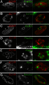
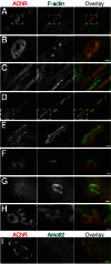
Localization of LL5β-interacting proteins in myotubes. (A,B) C2C12 myotubes were stained with BTX (left panels and red in overlay in right panels) to label AChRs, plus podosome markers (middle panels and green in overlay). Podosomes are characterized by an F-actin-rich core (A) and an LL5β-rich cortex (B). (C,D) ELKS and Plectin are concentrated in a ring at the inside rim of perforations within the AChR-rich plaque. This localization is similar to that of LL5β. ELKS is also present at lower levels in the AChR-rich regions. (E,F) Asef2 and Filamin A are concentrated in the center of perforations, in the actin-rich domain. (G) Amotl2 is present in the actin-rich domain and, at lower levels, at the interface of AChR-rich regions with podosome cortex. Scale bars: 5 µm.
Fig. 3.
Fig. 3.
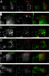
Localization of LL5β and its interacting proteins in RAW 264.7 macrophages. (A) LL5β (green) was concentrated in a region surrounding podosomes, as visualized by phalloidin staining of F-actin (red). (B) ELKS was more broadly distributed but still enriched near podosomes. (C–F) Asef2 (C) and Filamin A (D) have similar localization to LL5β and are concentrated at podosomes. Amotl2 (E) and Flii (F) are concentrated in podosome cores. Scale bar: 5 µm.
Fig. 4.
Fig. 4.
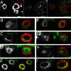
Localization of LL5β and interacting proteins in Src-transformed 3T3 fibroblasts. (A) Src3T3 cells make podosome-related invadopodia that often coalesce into larger, rosette-shaped structures visualized by phalloidin staining of F-actin (red on all images). LL5β (green) is concentrated on the outer and inner side of rosettes. Dashed lines outline the cell. (B–I) ELKS (B), Plectin (C), Asef2 (D), Amotl2 (F), Flii (G), and Paxillin (H) exhibited similar localization, predominantly concentrating in the inner side of rosettes, with some accumulation around the outer edges of actin-rich structures. Filamin A (E) and Talin (I) were associated with the actin-rich rosettes, but some antigen was present beyond the actin-rich region. Scale bar: 5 µm.
Fig. 5.
Fig. 5.
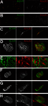
Localization of Amotl2 at the neuromuscular junction. (A) Section of adult skeletal muscle stained with anti-Amotl2 (red) and BTX to label AChRs (green). Amotl2 is concentrated at synaptic sites. (B) As in A, but antibody was absorbed with immunogen peptide before application. (C) High power view of area boxed in A, showing concentration of Amotl2 in the subsynaptic membrane. (D) Whole mount of adult muscle stained with anti-Amotl2 and BTX. (E) High power view of area boxed in D showing highest level of Amotl2 between AChR-rich branches. Note that this is also visible in C. (F–H) Levels of Amotl2 increase at the NMJ during the second and third postnatal weeks, as the AChR-rich postsynaptic membrane acquires its branched morphology (F, P20; G, P14; H, P7). Scale bars: 5 µm.
Fig. 6.
Fig. 6.
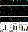
Depletion of Amotl2 leads to enlargement of synaptic podosomes in myotubes. (A–H) Distribution of AChRs (red in overlay) and F-actin (green in overlay) on C2C12 myotubes. (A) Control myotubes form AChR clusters that are perforated by actin-rich podosomes. (B) High power view of area boxed in A. (C) Cells transfected with MuSK siRNA entirely lacked AChR clusters. (D) Depletion of Amotl2 led to a dramatic increase in the size of podosomes and aberrations in the shape of clusters. (E) High power view of area boxed in D. (F) Co-expression of human (RNAi-resistant) Amotl2 rescues the phenotypes shown in D,E. (G) Co-transfection of a control plasmid has no effect on the ability of Amotl2 siRNA to enlarge podosomes. (H) Depletion of Amotl2 with a second interfering RNA (see Materials and Methods) led to increased podosome size. (I) The level of Amotl2 immunoreactivity was dramatically reduced in myotubes transfected with Amotl2 siRNA (compare with Fig. 2G). Scale bars: 10 µm.
Fig. 7.
Fig. 7.
Depletion of Amotl2 affects podosome composition and postsynaptic structure. Distribution of AChRs (red in overlay), F-actin (green in overlay) and indicated antigens (blue in overlay) in C2C12 myotubes. (A,B) LL5β is concentrated at the cortex of podosomes in both control (A) and Amotl2-depleted cells (B), and is also present at lower levels in actin-rich regions of Amotl2-depleted cells. (C,D) ELKS is concentrated at the cortex of podosomes in control myotubes (C) but diffusely distributed in Amotl2-depleted cells (D). (E,F) Average area of actin-rich regions (E) associated with AChR aggregates and average aggregate area (F) (measured as area within cluster outline) in control myotubes and in myotubes treated with Amotl2 RNAi or control RNAi. (G) Percentage of aggregate area occupied by actin, AChRs or neither of them (Empty). *P<0.001; NS, not significant. n = 31 clusters and 121 podosomes for ‘untransfected’; n = 41 and 81 for Amotl2 KD; n = 34 and 53 for non-silencing siRNA. Scale bars: 5 µm.
Fig. 8.
Fig. 8.
Amotl2 is required for the formation of invadopodia. (A) HEK-293 cells were co-transfected with plasmids encoding Amotl2 alone, Amotl2 plus shRNA complementary to Amotl2 or Asef2, or an ‘empty’ vector. Amotl2 levels in lysates were evaluated by western blotting. (B) Control Src3T3 cells stained for F-actin (phalloidin), showing multiple actin-rich puncta and rosettes. (C) Amotl2-depleted Src3T3 cells stained for actin show actin cables but few rosettes or puncta. (D–F) Percentage of control and transfected Src3T3 cells containing rosettes (D), containing no rosettes but ≥5 actin-rich puncta (E), or no rosettes and ≤4 actin-rich puncta (F). n = 243 for untreated cells, 226 for Src3T3 cells expressing Amotl2 shRNA, 100 for cells expressing Amotl2 shRNA plus human (shRNA-resistant) Amotl2 and 142 for cells expressing Amotl2 shRNA plus GFP. (G,H) Filamin A (G) and ELKS (H) immunostaining showing that in the absence of actin concentrated into rosettes other components of invadopodia are also dispersed. Scale bar: 5 µm.

Similar articles

Cited by

References

    1. Adams D. H., Ruzehaji N., Strudwick X. L., Greenwood J. E., Campbell H. D., Arkell R., Cowin A. J. (2009). Attenuation of Flightless I, an actin-remodelling protein, improves burn injury repair via modulation of transforming growth factor (TGF)-beta1 and TGF-beta3. Br. J. Dermatol. 161, 326–336 10.1111/j.1365-2133.2009.09296.x - DOI - PubMed
    1. Archer S. K., Behm C. A., Claudianos C., Campbell H. D. (2004). The flightless I protein and the gelsolin family in nuclear hormone receptor-mediated signalling. Biochem. Soc. Trans. 32, 940–942 10.1042/BST0320940 - DOI - PubMed
    1. Bratt A., Wilson W. J., Troyanovsky B., Aase K., Kessler R., Van Meir E. G., Holmgren L. (2002). Angiomotin belongs to a novel protein family with conserved coiled-coil and PDZ binding domains. Gene 298, 69–77 10.1016/S0378-1119(02)00928-9 - DOI - PubMed
    1. Bristow J. M., Sellers M. H., Majumdar D., Anderson B., Hu L., Webb D. J. (2009). The Rho-family GEF Asef2 activates Rac to modulate adhesion and actin dynamics and thereby regulate cell migration. J. Cell Sci. 122, 4535–4546 10.1242/jcs.053728 - DOI - PMC - PubMed
    1. Buccione R., Orth J. D., McNiven M. A. (2004). Foot and mouth: podosomes, invadopodia and circular dorsal ruffles. Nat. Rev. Mol. Cell Biol. 5, 647–657 10.1038/nrm1436 - DOI - PubMed

Publication types

MeSH terms