Combining technologies to create bioactive hybrid scaffolds for bone tissue engineering
- PMID: 23507924
- PMCID: PMC3749798
- DOI: 10.4161/biom.23705
Combining technologies to create bioactive hybrid scaffolds for bone tissue engineering
Abstract
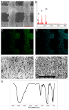
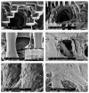
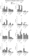
Combining technologies to engineer scaffolds that can offer physical and chemical cues to cells is an attractive approach in tissue engineering and regenerative medicine. In this study, we have fabricated polymer-ceramic hybrid scaffolds for bone regeneration by combining rapid prototyping (RP), electrospinning (ESP) and a biomimetic coating method in order to provide mechanical support and a physico-chemical environment mimicking both the organic and inorganic phases of bone extracellular matrix (ECM). Poly(ethylene oxide terephthalate)-poly(buthylene terephthalate) (PEOT/PBT) block copolymer was used to produce three dimensional scaffolds by combining 3D fiber (3DF) deposition, and ESP, and these constructs were then coated with a Ca-P layer in a simulated physiological solution. Scaffold morphology and composition were studied using scanning electron microscopy (SEM) coupled to energy dispersive X-ray analyzer (EDX) and Fourier Tranform Infrared Spectroscopy (FTIR). Bone marrow derived human mesenchymal stromal cells (hMSCs) were cultured on coated and uncoated 3DF and 3DF + ESP scaffolds for up to 21 d in basic and mineralization medium and cell attachment, proliferation, and expression of genes related to osteogenesis were assessed. Cells attached, proliferated and secreted ECM on all the scaffolds. There were no significant differences in metabolic activity among the different groups on days 7 and 21. Coated 3DF scaffolds showed a significantly higher DNA amount in basic medium at 21 d compared with the coated 3DF + ESP scaffolds, whereas in mineralization medium, the presence of coating in 3DF+ESP scaffolds led to a significant decrease in the amount of DNA. An effect of combining different scaffolding technologies and material types on expression of a number of osteogenic markers (cbfa1, BMP-2, OP, OC and ON) was observed, suggesting the potential use of this approach in bone tissue engineering.
Keywords: biomimetic coating; bone tissue engineering; calcium-phosphate; electrospinning; polymer; rapid prototyping.
Figures


Similar articles
-
Monolithic and assembled polymer-ceramic composites for bone regeneration.Acta Biomater. 2013 Mar;9(3):5708-17. doi: 10.1016/j.actbio.2012.10.044. Epub 2012 Nov 7. Acta Biomater. 2013. PMID: 23142480
-
Nano-ceramic composite scaffolds for bioreactor-based bone engineering.Clin Orthop Relat Res. 2013 Aug;471(8):2422-33. doi: 10.1007/s11999-013-2859-0. Clin Orthop Relat Res. 2013. PMID: 23436161 Free PMC article.
-
Osteogenic Differentiation of Mesenchymal Stem Cells with Silica-Coated Gold Nanoparticles for Bone Tissue Engineering.Int J Mol Sci. 2019 Oct 16;20(20):5135. doi: 10.3390/ijms20205135. Int J Mol Sci. 2019. PMID: 31623264 Free PMC article.
-
The Synergetic Effect of 3D Printing and Electrospinning Techniques in the Fabrication of Bone Scaffolds.Ann Biomed Eng. 2024 Jun;52(6):1518-1533. doi: 10.1007/s10439-024-03500-5. Epub 2024 Mar 26. Ann Biomed Eng. 2024. PMID: 38530536 Review.
-
Hierarchically designed bone scaffolds: From internal cues to external stimuli.Biomaterials. 2019 Oct;218:119334. doi: 10.1016/j.biomaterials.2019.119334. Epub 2019 Jul 3. Biomaterials. 2019. PMID: 31306826 Free PMC article. Review.
Cited by
-
3D-Printed Scaffolds and Biomaterials: Review of Alveolar Bone Augmentation and Periodontal Regeneration Applications.Int J Dent. 2016;2016:1239842. doi: 10.1155/2016/1239842. Epub 2016 Jun 5. Int J Dent. 2016. PMID: 27366149 Free PMC article. Review.
-
The Role of Bioceramics for Bone Regeneration: History, Mechanisms, and Future Perspectives.Biomimetics (Basel). 2024 Apr 12;9(4):230. doi: 10.3390/biomimetics9040230. Biomimetics (Basel). 2024. PMID: 38667241 Free PMC article. Review.
-
Biomimetic Hybrid Systems for Tissue Engineering.Biomimetics (Basel). 2020 Oct 9;5(4):49. doi: 10.3390/biomimetics5040049. Biomimetics (Basel). 2020. PMID: 33050136 Free PMC article. Review.
-
Open-source three-dimensional printing of biodegradable polymer scaffolds for tissue engineering.J Biomed Mater Res A. 2014 Dec;102(12):4326-35. doi: 10.1002/jbm.a.35108. J Biomed Mater Res A. 2014. PMID: 25493313 Free PMC article.
-
Engineering biomimetic periosteum with β-TCP scaffolds to promote bone formation in calvarial defects of rats.Stem Cell Res Ther. 2017 Jun 5;8(1):134. doi: 10.1186/s13287-017-0592-4. Stem Cell Res Ther. 2017. PMID: 28583167 Free PMC article.
References
-
- Hutmacher DW, Schantz T, Zein I, Ng KW, Teoh SH, Tan KC. Mechanical properties and cell cultural response of polycaprolactone scaffolds designed and fabricated via fused deposition modeling. J Biomed Mater Res. 2001;55:203–16. doi: 10.1002/1097-4636(200105)55:2<203::AID-JBM1007>3.0.CO;2-7. - DOI - PubMed
MeSH terms
Substances
LinkOut - more resources
Full Text Sources
Other Literature Sources
Miscellaneous
