. 2013 Mar 21;38(3):437-49.
doi: 10.1016/j.immuni.2012.11.018. Epub 2013 Mar 14.

Dephosphorylation of the RNA sensors RIG-I and MDA5 by the phosphatase PP1 is essential for innate immune signaling

Affiliations

Dephosphorylation of the RNA sensors RIG-I and MDA5 by the phosphatase PP1 is essential for innate immune signaling

Effi Wies et al. Immunity. .

Abstract

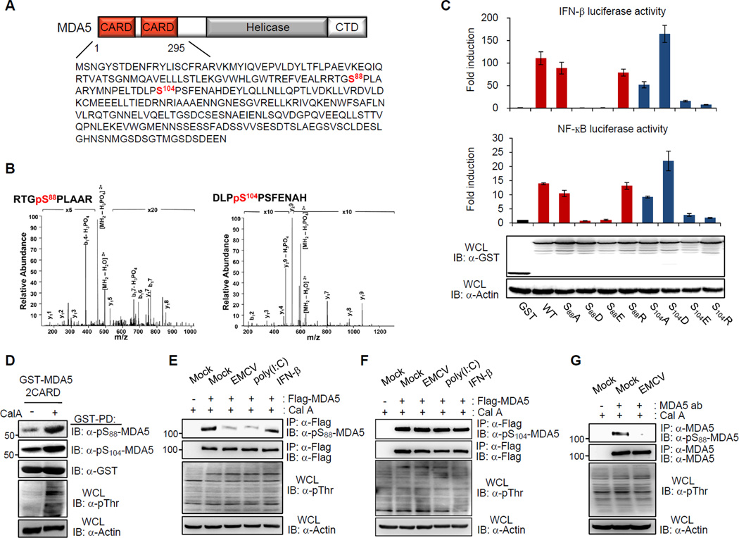
RIG-I and MDA5 have emerged as key cytosolic sensors for the detection of RNA viruses and lead to antiviral interferon (IFN) production. Recent studies have highlighted the importance of posttranslational modifications for controlling RIG-I antiviral activity. However, the regulation of MDA5 signal-transducing ability remains unclear. Here, we show that MDA5 signaling activity is regulated by a dynamic balance between phosphorylation and dephosphorylation of its caspase recruitment domains (CARDs). Employing a phosphatome RNAi screen, we identified PP1α and PP1γ as the primary phosphatases that are responsible for MDA5 and RIG-I dephosphorylation and that lead to their activation. Silencing of PP1α and PP1γ enhanced RIG-I and MDA5 CARD phosphorylation and reduced antiviral IFN-β production. PP1α- and PP1γ-depleted cells were impaired in their ability to induce IFN-stimulated gene expression, which resulted in enhanced RNA virus replication. This work identifies PP1α and PP1γ as regulators of antiviral innate immune responses to various RNA viruses, including influenza virus, paramyxovirus, dengue virus, and picornavirus.


Figures

Figure 1
Figure 1. Phosphorylation of the MDA5 CARDs
(A) MDA5 domain structure and amino acid sequence of the CARDs. Phosphorylation sites identified by MS are highlighted in red. CTD, C-terminal domain. (B) MS/MS spectra of tryptic phosphopeptides RTGpSPLAAR and DLPpSPSFENAH, which identified phosphorylation sites at S88 and S104, respectively. p, phosphorylation. b- and y-ion designations are shown (Aebersold and Goodlett, 2001). (C) GST, GST-MDA5(2CARD) WT or the indicated mutants were expressed together with IFN-β or NF-κB luciferase and constitutive β-gal expressing pGK-β-gal in HEK293T cells. At 48 h posttransfection, values for luciferase and β-galactosidase were determined. The results are expressed as means +/− s.d. (n=3). Whole-cell lysates (WCLs) were used for immunoblotting (IB) with anti-GST and anti-Actin antibodies. (D) Detection of S88 and S104 phosphorylations of MDA5 in vivo by using phospho-specific pS88-MDA5 and pS104-MDA5 antibodies. At 48 h posttransfection with GST-MDA5(2CARD), HEK293T cells were mock-treated or treated with Calyculin A (Cal A). WCLs were subjected to GST pull-down (GST-PD), followed by IB with the indicated antibodies. WCLs were further used for IB with anti-phosphothreonine (anti-pThr) and anti-Actin antibodies. (E and F) HEK293T cells transfected with Flag-MDA5, were either mock-treated, infected with EMCV (MOI 0.5) for 3 h, transfected with 5 µg/ml poly(I:C) or treated with 1,000 U/ml IFN-β for 20 h. WCLs were subjected to immunoprecipitation (IP) with anti-Flag antibody, followed by IB with anti-pS88-MDA5 (E) or anti-pS104-MDA5 (F), and anti-Flag antibodies (E and F). (G) HEK293T cells were either mock-infected or infected with EMCV (MOI 0.5) for 3 h. WCLs were used for IP with anti-MDA5 antibody, followed by IB. See also Figure S1.
Figure 2
Figure 2. MDA5 phosphorylation at S88 inhibits its downstream signaling, whereas S88 dephosphorylation is required for optimal MDA5 activity
(A) Vector, Myc-tagged MDA5 WT or mutants together with IFN-β luciferase and pGK-β-gal were transfected into HEK293T cells. 24 h after transfection, cells were either mock-treated or transfected with 0.1 µg/ml poly(I:C). 20 h later, luciferase and β-galactosidase activity were determined. The results are expressed as means +/− s.d. (n=3). Protein expressions were determined by IB. (B) HEK293T cells were transfected with MAVS-CARD-PRD-Flag together with GST or GST-MDA5(2CARD) constructs. WCLs were subjected to GST-PD. (C) 48 h after transfection with Flag-IRF3 and GST or GST-MDA5(2CARD) fusion constructs, WCLs of HEK293T cells were subjected to native PAGE, followed by IB with anti-Flag antibody. WCLs were also used for SDS-PAGE, followed by IB with anti-GST and anti-Flag antibodies. (D) HEK293T cells were transfected with GST, GST-MDA5(2CARD) WT or the indicated mutants and IRF3-eGFP. 48 h later, cells were stained for GST (red) and imaged by confocal microscopy. (E) MDA5 S88D mutant inhibits the poly(I:C)-induced IFN-β promoter activation. HEK293T were transfected with vector or increasing amounts of Flag-tagged MDA5 S88D or S88A mutant together with IFN-β luciferase and pGK-β-gal. Cells were either mock-treated or co-transfected with 3 µg/ml poly(I:C). 48 h later, luciferase activity was determined as described in Fig.1C. The results, expressed as means +/− s.d. (n=2), are representative of 3 independent experiments. (F) Mda5-deficient (Ifih1−/−) MEFs were transfected with vector, MDA5 WT or mutants. At 24 h posttransfection, cells were either mock-treated or transfected with 0.1 µg/ml poly(I:C). 20 h later, IFN-β production in the supernatant was measured by ELISA. The results are expressed as means +/− s.d. (n=3). ND, non-detectable.
Figure 3
Figure 3. Identification of PP1α and PP1γ as critical regulators of MDA5 and RIG-I signal transducing activities
(A) Identified candidate phosphatases using a siRNA-based screen. HEK293T cells were transfected with GST-MDA5(2CARD) together with siRNA pools specific for one of 257 human phosphatases, as well as IFN-β luciferase and pGK-β-gal. 48 h posttransfection, luciferase and β-gal activities were determined. Results of the top 9 siRNA hits as well as of the siRNA specific for IRF3 (positive control) are shown. Activation by GST-MDA5(2CARD) upon co-transfection of a non-targeting control siRNA (si.C) was set to 100%. (B) Flag-MDA5 was co-transfected with the indicated candidate phosphatases together with IFN-β luciferase and pGK-β-gal. Luciferase and β-gal activities were determined 48 h after transfection. The results are expressed as means +/− s.d. (n=3). (C) Flag-MDA5 was transfected together with increasing amounts of HA-tagged PP1α, PP1β, or PP1γ, and IFN-β luciferase and pGK-β-gal. Luciferase and β-gal activities were measured 48 h after transfection. The results, expressed as means +/− s.d. (n=2), are representative of 3 independent experiments. (D and E) HEK293T were transfected with GST or GST-MDA5(2CARD) together with IFN-β or NF-κB luciferase and pGK-β-gal as well as non-targeting control siRNA (si.C), or siRNAs specific for PP1α, PP1β, PP1γ, or IRF3 (control). 48 h later, luciferase and β-gal activities were determined. The results are expressed as means +/− s.d. (n=3). (F and G) HEK293T cells, transfected with IFN-β luciferase, pGK-β-gal, and the indicated siRNAs, were either co-transfected with GST or GST-RIG-I(2CARD) (F), or infected with SeV (5 HA units/ml) for 14 h (G). Luciferase and β-gal activities were determined. The results are expressed as means +/− s.d. (n=3). See also Figure S2.
Figure 4
Figure 4. PP1α and PP1γ interact with RIG-I and MDA5, leading to their CARD dephosphorylation
(A) WCLs of HEK293T cells, transfected with Flag-tagged MDA5 and HA-tagged phosphatase constructs, were used for IP with anti-Flag, followed by IB with anti-pS88-MDA5 or anti-Flag. (B and C) HEK293T cells were transfected with GST-MDA5(2CARD) (B) or GST-RIG-I(2CARD) (C), together with control siRNA (si.C) or siRNA specific for PP1α and PP1γ. WCLs were subjected to GST-PD, followed by IB with the indicated antibodies. (D) HEK293T cells were transfected with vector or Flag-MDA5. At 45 h posttransfection, cells were mock-treated or infected with EMCV (MOI 0.5) for 3 h. WCLs were subjected to IP with anti-Flag. (E and F) HEK293T (E) or NHLF (F) cells were either mock-treated, infected with SeV (50 HA units/ml) for 18 h (E), or infected with ΔNS1 PR8 influenza virus (MOI 2) for the indicated time points (F). WCLs were subjected to IP with anti-RIG-I antibody. n.s., non-specific band. (G and H) HEK293T (G) or NHLF (H) cells were either mock-treated, transfected with poly(I:C) for 36 h (G), or infected with EMCV (MOI 0.5) for 3 h (H). WCLs were subjected to IP with anti-MDA5 antibody. See also Figures S3 and S4.
Figure 5
Figure 5. PP1-binding deficient mutants of RIG-I and MDA5 exhibit enhanced CARD phosphorylation levels and abolished downstream signaling abilities
(A) (upper) Identified PP1-binding motifs in MDA5 and RIG-I. (lower) Flag-tagged MDA5 WT or mutants (left), or RIG-I WT or mutants (right) together with IFN-β luciferase and pGK-β-gal were transfected into HEK293T cells. 48 h later, luciferase and β-gal activities were measured. The results are expressed as means +/− s.d. (n=3). (B) HEK293T cells were transfected with vector, Flag-MDA5, Flag-RIG-I or the indicated mutants together with HA-tagged PP1α or PP1γ, followed by infection with EMCV (MOI 0.5) (left panel) or SeV (50 HA units/ml) (right panel) for 3 h. WCLs were used for IP with anti-Flag. (C) WCLs of HEK293T cells transfected with Flag-MDA5, Flag-RIG-I or the indicated mutants were used for IP with anti-Flag. (D) Ifih1−/− and Ddx58−/− MEFs were complemented with MDA5 WT or mutants and RIG-I WT or mutants, respectively. IFN-β production upon mock-treatment, poly(I:C) (0.1 µg/ml) transfection (left), or SeV infection (50 HAU/ml) (right) was determined by ELISA. The results are expressed as means +/− s.d. (n=3). ND, non-detectable. (E) WCLs of HEK293T cells, transfected with GST-RIG-I(2CARD) WT or MI, were subjected to GST-PD. Arrows, ubiquitinated bands. (F) HEK293T cells were transfected with GST, GST-RIG-I(2CARD) WT or MI mutant together with IFN-β luciferase and pGK-β-gal. 48 h after transfection, luciferase and β-gal activities were determined. The results are expressed as means +/− s.d. (n=3). See also Figure S5.
Figure 6
Figure 6. Depletion of PP1α and PP1γ decreases antiviral IFN production and enhances viral replication
(A) NHLF cells were transfected with the indicated siRNAs. 48 h later, cells were either mock-transfected or transfected with poly(I:C) or Vero-EMCV-RNA, or infected with ΔNS1 A/PR/8/34 influenza virus or SeV. 24 h later, IFN-β production was measured by ELISA. PP1 knockdown was confirmed by IB. The results are expressed as means +/− s.d. (n=3). *p < 0.05; **p < 0.005. ND, non-detectable. (B and C) NHLF cells were transfected with the indicated siRNAs. 48 h later, cells were infected with VSV-eGFP (MOI 0.05). 24 h after infection, eGFP expression was determined by microscopy and WB, and virus titers were determined by plaque assay. PP1 knockdown was confirmed by IB (C). Pfu, plaque forming unit. (D and E) THP-1 cells were transfected with control siRNA (si.C), or with PP1α and PP1γ specific siRNAs, followed by mock-infection or infection with EMCV (MOI 1) for 5 h. Total RNA was isolated and subjected to RT-PCR using gene-specific primers for ISG15, ISG54 and ISG56 (D). PP1 knockdown was confirmed by IB (E). See also Figure S6.
Figure 7
Figure 7. PP1α and PP1γ are critical for ISG induction and host antiviral activity to Dengue virus
(A) At 30 h posttransfection with vector, PP1α, or PP1γ, HEK293T cells were infected with DenV (MOI 0.5). 42 h later, cells were stained for viral envelope protein (red). Nuclei were stained with DAPI (blue). Quantification of infection is presented as relative infection compared to control. The results are expressed as means +/− s.d. (n=3). *p < 0.05; **p < 0.005. (B) At 48 h posttransfection with the indicated siRNAs, HeLa cells were infected with DenV (MOI 0.01). 42 h later, cells were stained for viral envelope protein (red). Nuclei were stained with DAPI (blue). Quantification of infection is presented as relative infection compared to control. The results are expressed as means +/− s.d. (n=3). *p < 0.05; **p < 0.005. (C and D) NHLF cells were either mock-treated or infected with DenV (MOI 0.1) for the indicated time points. WCLs were used for IP with anti-MDA5 (C) or anti-RIG-I (D) antibody. (E) NHLF cells were transfected with the indicated siRNAs. 48 h later, cells were either mock-infected or infected with DenV (MOI 0.25) for 24 h. WCLs were subjected to IB with the indicated antibodies. See also Figure S7.

Comment in

Similar articles

Cited by

References

    1. Aebersold R, Goodlett DR. Mass spectrometry in proteomics. Chem Rev. 2001;101:269–295. - PubMed
    1. Andrejeva J, Childs KS, Young DF, Carlos TS, Stock N, Goodbourn S, Randall RE. The V proteins of paramyxoviruses bind the IFN-inducible RNA helicase, mda-5, and inhibit its activation of the IFN-beta promoter. Proc Natl Acad Sci U S A. 2004;101:17264–17269. - PMC - PubMed
    1. Arimoto K, Takahashi H, Hishiki T, Konishi H, Fujita T, Shimotohno K. Negative regulation of the RIG-I signaling by the ubiquitin ligase RNF125. Proc Natl Acad Sci U S A. 2007;104:7500–7505. - PMC - PubMed
    1. Barbalat R, Ewald SE, Mouchess ML, Barton GM. Nucleic acid recognition by the innate immune system. Annu Rev Immunol. 2011;29:185–214. - PubMed
    1. Cohen PT. Protein phosphatase 1--targeted in many directions. J Cell Sci. 2002;115:241–256. - PubMed

Publication types

MeSH terms