. 2013 Mar 12;6(266):ra16.
doi: 10.1126/scisignal.2003706.

Signaling by p38 MAPK stimulates nuclear localization of the microprocessor component p68 for processing of selected primary microRNAs

Affiliations

Signaling by p38 MAPK stimulates nuclear localization of the microprocessor component p68 for processing of selected primary microRNAs

Sungguan Hong et al. Sci Signal. .

Abstract

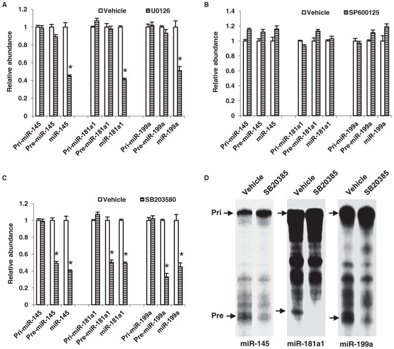
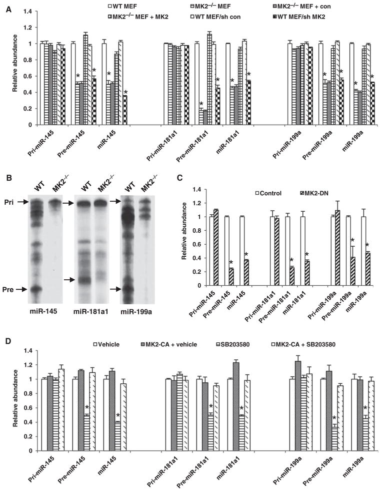
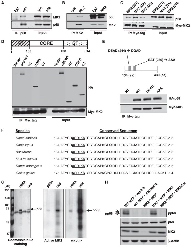
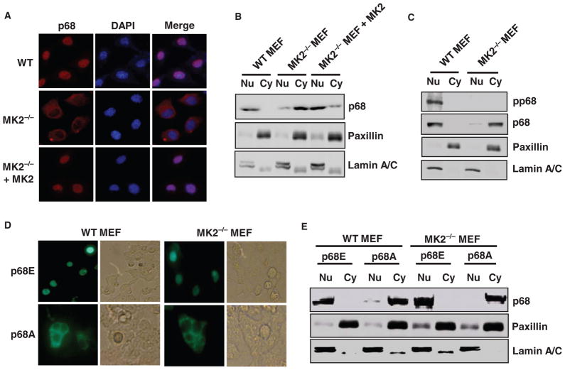
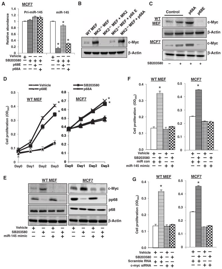
The importance of microRNAs (miRNAs) in biological and disease processes necessitates a better understanding of the mechanisms that regulate miRNA abundance. We showed that the activities of the mitogen-activated protein kinase (MAPK) p38 and its downstream effector kinase MAPK-activated protein kinase 2 (MK2) were necessary for the efficient processing of a subset of primary miRNAs (pri-miRNAs). Through yeast two-hybrid screening, we identified p68 (also known as DDX5), a key component of the Drosha complex that processes pri-miRNAs, as an MK2-interacting protein, and we found that MK2 phosphorylated p68 at Ser(197) in cells. In wild-type mouse embryonic fibroblasts (MEFs) treated with a p38 inhibitor or in MK2-deficient (MK2(-/-)) MEFs, expression of a phosphomimetic mutant p68 fully restored pri-miRNA processing, suggesting that MK2-mediated phosphorylation of p68 was essential for this process. We found that, whereas p68 was present in the nuclei of wild-type MEFs, it was found mostly in the cytoplasm of MK2(-/-) MEFs. Nuclear localization of p68 depended on MK2-mediated phosphorylation of Ser(197). In addition, inhibition of p38 MAPK promoted the growth of wild-type MEFs and breast cancer MCF7 cells by enhancing the abundance of c-Myc through suppression of the biogenesis of the miRNA miR-145, which targets c-Myc. Because pri-miRNA processing occurs in the nucleus, our findings suggest that the p38 MAPK-MK2 signaling pathway promotes miRNA biogenesis by facilitating the nuclear localization of p68.


Conflict of interest statement

Competing interests: The authors declare that they have no competing interests.

Figures

Fig. 1
Fig. 1
The activity of p38 MAPK is essential for pri-miRNA processing. (A to C) qRT-PCR analysis of the amounts of the pri-, pre-, and mature forms of miR-145, miR-181a1, and miR-199a in wild-type (WT) MEFs treated with (A to C) dimethyl sulfoxide (DMSO; vehicle) or (A) 10 μM U0126, (B) 10 μM SP600125, or (C) 10 μM SB203580 for 1 day. Data are means ± SE from three experiments. *P < 0.005 versus vehicle-treated cells. (D) In vitro pri-miRNA processing in whole-cell extracts of WT MEFs treated with vehicle or 10 μM SB203580. Data are representative of two independent experiments.
Fig. 2
Fig. 2
MK2 is required for pri-miRNA processing. (A) qRT-PCR analysis of the amounts of the pri-, pre-, and mature forms of miR-145, miR-181a1, and miR-199a in WT MEFs, MK2−/− MEFs, MK2−/− MEFs transfected with empty vector (con), MK2−/− MEFs transfected with plasmid encoding MK2, WT MEFs transfected with luciferase-specific shRNA (sh Luc), and WT MEFs in which MK2 was knocked down. Data are means ± SE from three experiments. *P < 0.005 versus WT MEFs. (B) In vitro pri-miRNA processing with whole-cell extracts of WT and MK2−/− MEFs. Data are representative of two independent experiments. (C) qRT-PCR analysis of the amounts of the pri-, pre-, and mature forms of miR-145, miR-181a1, and miR-199a in WT MEFs transfected with empty vector(control) or with plasmid encoding MK2-DN. Data are means ± SE from three experiments. *P < 0.005 versus control. (D) WT MEFs were transfected with plasmid encoding MK2-CA and then were treated for 1 day with solvent (vehicle) or 10 μM SB203580. Total RNA was isolated and subjected to qRT-PCR to analyze the amounts of the pri-, pre-, and mature forms of miR-145, miR-181a1, and miR-199a. Data are means ± SE from three experiments. *P < 0.005 versus vehicle alone.
Fig. 3
Fig. 3
MK2 physically interacts with p68 and phosphorylates it at Ser197. (A) WT MEFs were subjected to immunoprecipitation with mouse immunoglobulin G (IgG) or monoclonal antibody against p68, and immunoprecipitates were subjected to Western blotting analysis to detect MK2. Data are representative of two independent experiments. (B) WT MEFs were subjected to immunoprecipitation with rabbit IgG or polyclonal antibody against MK2, and immunoprecipitates were subjected to Western blotting analysis to detect p68. Data are representative of two independent experiments. (C) WT MEFs were transfected with plasmid encoding Myc-tagged MK2, MK2-CA, or MK2-DN and then subjected to coimmunoprecipitations to detect their interaction with p68. Data are representative of three independent experiments. (D) WT MEFs were transfected with empty plasmid (vector) or with plasmids encoding HA-tagged full-length p68 (FL) or the N-terminal (NT), core, or C-terminal (CT) regions of p68, and cell lysates were subjected to coimmunoprecipitations to detect interactions between the p68 proteins and MK2. Data are representative of two independent experiments. (E) WT MEFs were transfected with plasmids encoding HA-tagged WT p68 or p68 mutants, and then cell lysates were subjected to coimmunoprecipitation experiments to detect interactions between the p68 proteins and MK2. Data are representative of two independent experiments. aa, amino acid. (F) Alignment of p68 protein sequences from different species. The conserved MK2 phosphorylation target motif is underlined. Amino acid residue numbers are to the left and right of the sequences. (G) Left panel: Coomassie blue staining of recombinant p68 and p68A. Right panel: In vitro kinase assay with either recombinant active MK2 or MK2 immunoprecipitates (MK2-IP) to determine the ability of MK2 to phosphorylate p68. Data are representative of three independent experiments. (H) Western blotting analysis to detect Ser197-phosphorylated p68, total p68, MK2, and β-actin in WT MEFs treated with solvent (vehicle) or 10 μM SB203580 for 1 day; MK2−/− MEFs; and MK2−/− MEFs transduced to express either WT MK2 or MK2-DN. Data are representative of two independent experiments.
Fig. 4
Fig. 4
Phosphorylation of Ser197 of p68 is essential for pri-miRNA processing. (A) qRT-PCR analysis of the amounts of the pri-, pre-, and mature forms of miR-145, miR-181a1, and miR-199a in WT MEFs that were transduced with lentiviruses to express luciferase-specific shRNA (control) or p68-specific shRNA. Data are means ± SE from three experiments. *P< 0.005 versus control. (B) WT MEFs were transfected with plasmids encoding WT p68 or the p68E or p68A mutants and then were treated for 1 day with solvent (vehicle) or 10 μM SB203580. Total RNA was isolated and subjected to qRT-PCR analysis to determine the amounts of the pri-, pre-, and mature forms of miR-145, miR-181a, and miR-199a1. Data are means ± SE from three experiments. *P < 0.005 versus vehicle. (C) qRT-PCR analysis of the amounts of the pri-, pre-, and mature forms of miR-145, miR-181a1, and miR-199a in WT MEFs, MK2−/− MEFs, and MK2−/− MEFs transfected with plasmids encoding either p68E or p68A. Data are means ± SE from three experiments. *P < 0.005 versus WT MEFs. (D) WT MEFs, MK2−/− MEFs, and WT MEFs treated with 10 μM SB203580 for 1 day were subjected to immunoprecipitation with polyclonal antibody against Drosha, and immunoprecipitates were subjected to Western blotting analysis with antibodies specific for total p68 and p68 phosphorylated at Ser197 (pp68). Data are representative of three independent experiments. (E) Lysates of WT MEFs were precleared with or without polyclonal antibody against phosphorylated p68 followed by immunoprecipitation with polyclonal antibody against Drosha. Immunoprecipitates were subjected to Western blotting analysis with an antibody against p68. As a control, MEF lysates were subjected to immunoprecipitation with rabbit IgG. Data are representative of two independent experiments. (F) WT MEFs were transfected with plasmids encoding HA-tagged WT p68 or the p68E or p68A mutants, and then cell lysates were subjected to immunoprecipitation with polyclonal antibody against Drosha. Immunoprecipitates were analyzed by Western blotting with antibody against HA-tagged p68. As a control, WT MEFs were transfected with empty vector. Data are representative of two independent experiments. (G) WT MEFs were transfected with plasmids encoding HA-tagged WT p68 or the p68E or p68A mutants, and cell lysates were subjected to immunoprecipitation with monoclonal antibody against the HA tag. Immunoprecipitates were analyzed by Western blotting with an antibody against Drosha. As a control, WT MEFs were transfected with empty vector. Data are representative of two independent experiments.
Fig. 5
Fig. 5
Phosphorylation of Ser197 of p68 is essential for its nuclear localization. (A) WT MEFs, MK2−/− MEFs, and MK2−/− MEFs transfected to express MK2 were subjected to immunofluorescence staining to detect the subcellular localization of p68. Cell nuclei were visualized with DAPI (4′,6-diamidino-2-phenylindole). Data are representative of four independent experiments. (B) Nuclear and cytoplasmic fractions of WT MEFs, MK2−/− MEFs, and MK2−/− MEFs transfected to express MK2 were analyzed by Western blotting to detect p68. The cytoplasmic (Cy) and nuclear fractions (Nu) were verified with antibodies specific for paxillin and lamin A/C, respectively. Data are representative of two independent experiments. (C) The nuclear and cytoplasmic fractions of WT and MK2−/− MEFs were analyzed by Western blotting with antibodies specific for total p68 and for p68 phosphorylated at Ser197 (pp68). Data are representative of two independent experiments. (D) WT or MK2−/− MEFs were transfected with plasmids encoding GFP-fused p68E or p68A mutants. One day later, live cells were monitored with a fluorescence microscope. Data are representative of three independent experiments. (E) WT and MK2−/− MEFs were transfected with plasmids encoding either HA-tagged p68E or HA-tagged p68A. One day later, nuclear and cytoplasmic fractions were prepared and subjected to Western blotting analysis to detect HA-tagged p68E or p68A. Data are representative of three independent experiments.
Fig. 6
Fig. 6
SB203580 enhances cell growth by suppressing miR-145 production and increasing the abundance of c-Myc protein. (A) Untransfected MCF7 cells and MCF7 cells transfected with plasmids encoding the p68E or p68A mutants were treated with 10 μM SB203580 for 1 day and then were analyzed by qRT-PCR to measure the amounts of the pri- and mature forms of miR-145. Data are means ± SE from three experiments. *P < 0.005 versus vehicle. (B) WT MEFs, MK2−/− MEFs, and MK2−/− MEFs transfected with plasmids encoding MK2, p68E, or p68A were subjected to Western blotting analysis with an antibody against c-Myc. We used β-actin as a loading control. Data are representative of two independent experiments. (C) WT MEFs and MCF7 cells were transfected with plasmids encoding p68E or p68A. Cells were treated with SB203580 for 1 day before being analyzed by Western blotting to detect c-Myc. We used β-actin as a loading control. Data are representative of three independent experiments. (D) WT MEFs and MCF7 cells were transfected with plasmids encoding p68E or p68A. Cells were then treated with SB203580 for the indicated times before being analyzed by the MTT assay to determine the extent of cell proliferation. Data are means ± SE from three experiments. *P < 0.005 versus vehicle. (E) WT MEFs and MCF7 cells were transfected with the miR-145 mimic (33 nM). After 3 days, the cells were treated with SB203580 for 1 day. Cells were then analyzed by Western blotting to detect c-Myc, total p68, p68 phosphorylated at Ser197 (pp68), and β-actin. Data are representative of three independent experiments. (F) WT MEFs and MCF7 cells were transfected with the miR-145 mimic (33 nM). Three days later, the cells were treated with 10 μM SB203580 for 2 days. The cells were then analyzed with the MTT to determine cell proliferation. Data are means ± SE from three experiments. *P < 0.005 versus vehicle. (G) WT MEFs and MCF7 cells were transfected with a c-Myc–specific siRNA pool (33 nM). Three days later, the cells were treated with 10 μM SB203580 for 2 days. The cells were then analyzed with the MTT assay to determine the extent of cell proliferation. Data are means ± SE from three experiments. *P < 0.005 versus vehicle.

Similar articles

Cited by

References

    1. Ambros V. The functions of animal microRNAs. Nature. 2004;431:350–355. - PubMed
    1. Lewis BP, Burge CB, Bartel DP. Conserved seed pairing, often flanked by adenosines, indicates that thousands of human genes are microRNA targets. Cell. 2005;120:15–20. - PubMed
    1. Bartel DP. MicroRNAs: Genomics, biogenesis, mechanism, and function. Cell. 2004;116:281–297. - PubMed
    1. Ventura A, Jacks T. MicroRNAs and cancer: Short RNAs go a long way. Cell. 2009;136:586–591. - PMC - PubMed
    1. Murchison EP, Hannon GJ. miRNAs on the move: miRNA biogenesis and the RNAi machinery. Curr Opin Cell Biol. 2004;16:223–229. - PubMed

Publication types

Substances

LinkOut - more resources