

The role of hepatic expression of STAT1, SOCS3 and PIAS1 in the response of chronic hepatitis C patients to therapy
- PMID: 23472246
- PMCID: PMC3731121
- DOI: 10.1155/2013/562765
The role of hepatic expression of STAT1, SOCS3 and PIAS1 in the response of chronic hepatitis C patients to therapy
Abstract
Background: The underlying mechanisms of hepatitis C virus (HCV) resistance to treatment are unknown. Signal transducers and activators of transcription (STAT) proteins play a critical role in antiviral defense.
Objective: To explore some of the mechanisms of HCV resistance to interferon, the expression of STAT1 and its negative regulators, protein inhibitor of activated STAT (PIAS1) and suppressor of cytokine signalling (SOCS3), in liver tissues of both inteferon responders and nonresponders in chronic HCV patients.
Methods: Sixty patients were divided into the following groups: group 1a comprised 38 treatment-responder chronic HCV patients; group 1b consisted of 22 treatment-nonresponder chronic HCV patients; and group 2 consisted of six control subjects. Liver biopsies were examined for histological scoring; STAT1, SOCS3 and PIAS1 expression was analyzed using Western blotting methods.
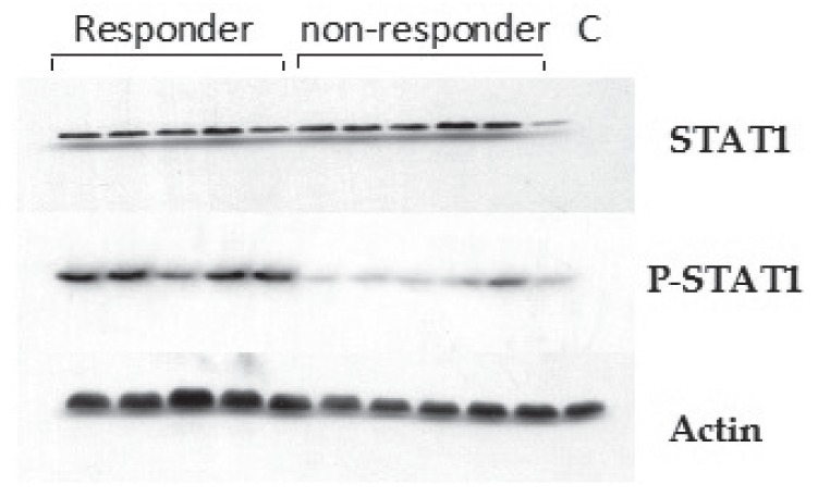
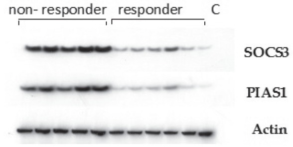
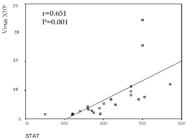
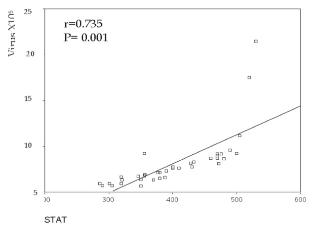
Results: STAT1 expression in the liver tissue of patients in group 1 was significantly increased compared with group 2 patients (P=0.001), while no significant difference in expression was observed between group 1a and group 1b patients (P=0.747). However, phosphorylated STAT1 protein was expressed at a significantly higher level in liver tissue of patients in group 1a compared with patients in group 1b (P=0.001). Western blot analysis of PIAS1 and SOCS3 protein expression in liver tissues from groups 1 and 2 revealed significantly increased expression in group 1 compared with group 2 (P=0.001). In addition, PIAS1 and SOCS3 protein expression was significantly higher in the liver tissues of patients in group 1b compared with patients in group 1a.
Conclusion: Levels of STAT1 and⁄or the protein expression of its negative regulators, PIAS1 and SOCS3, may be a good predictor of response to therapy. These could be used as biomarkers that are easily detected by Western blotting or immunostaining during standard histopathological liver biopsy analysis.
HISTORIQUE :: On ne connaît pas les mécanismes sous-jacents de la résistance au traitement du virus de l’hépatite C (VHC). Les protéines des transducteurs de signal et des activateurs de transcription (STAT) jouent un rôle essentiel dans la défense antivirale.
OBJECTIF :: Explorer certains mécanismes de la résistance du VHC à l’interféron, de l’expression de la STAT1 et de ses régulateurs négatifs, de l’inhibiteur protéique de la STAT activée (PIAS1) et du suppresseur de la signalisation des cytokines (SOCS3) dans les tissus hépatiques de patients atteints du VHC chronique répondant ou non à l’ interféron.
MÉTHODOLOGIE :: Soixante patients ont été répartis dans les groupes suivants : le groupe 1a se composait de 38 patients atteints du VHC chronique qui répondaient au traitement, le groupe 1b, de 22 patients atteints du VHC chronique qui ne répondaient pas au traitementet le groupe 2, de six sujets témoins. Les chercheurs ont examiné leur biopsie hépatique pour établir leur indice histologique et ont analysé l’expression des protéines STAT1, SOCS3 et PIAS1 au moyen du transfert Western.
RÉSULTATS :: L’expression de la STAT1 dans le tissu hépatique des patients du groupe 1 était significativement plus élevée que celle des patients du groupe 2 (P=0,001), mais il n’y avait pas de différence significative de l’expression entre les patients du groupe 1a et ceux du groupe 1b (P=0,747). Cependant, la protéine STAT1 phosphorylée était exprimée à un taux considérablement plus élevé dans les tissus hépatiques des patients du groupe 1a que dans ceux du groupe 1b (P=0,001). L’analyse de l’expression des protéines PIAS1 et SOCS3 des tissus hépatiques des groupes 1 et 2 par transfert Western a révélé une expression considérablement plus élevée dans le groupe 1 que dans le groupe 2 (P=0,001). En outre, l’expression des protéines PIAS1 et SOCS3 était considérablement plus élevée dans les tissus hépatiques des patients du groupe 1b que dans ceux des patients du groupe 1a.
CONCLUSION :: Le titrage de la STAT1 phosphorylée ou de l’expression protéique de ses régulateurs négatifs, les protéines PIAS1 et SOCS3, pour-rait être un bon prédicteur de la réponse au traitement. On pourrait l’utiliser comme biomarqueur facilement décelé par transfert Western ou par immunocoloration pendant l’analyse histopatologique standard de la biopsie hépatique.
Figures
Similar articles
-
Liver expression of proteins controlling interferon-mediated signalling as predictive factors in the response to therapy in patients with hepatitis C virus infection.J Pathol. 2007 Nov;213(3):347-55. doi: 10.1002/path.2214. J Pathol. 2007. PMID: 17940994
-
Hepatic SOCS3 expression is strongly associated with non-response to therapy and race in HCV and HCV/HIV infection.J Hepatol. 2009 Apr;50(4):705-11. doi: 10.1016/j.jhep.2008.12.021. Epub 2009 Feb 14. J Hepatol. 2009. PMID: 19231005 Free PMC article.
-
Suppressor of cytokine signaling 3 (SOCS3) expression and hepatitis C virus-related chronic hepatitis: Insulin resistance and response to antiviral therapy.Hepatology. 2007 Oct;46(4):1009-15. doi: 10.1002/hep.21782. Hepatology. 2007. PMID: 17668875
-
Hepatitis C virus inhibits interferon signaling through up-regulation of protein phosphatase 2A.Gastroenterology. 2004 Jan;126(1):263-77. doi: 10.1053/j.gastro.2003.10.076. Gastroenterology. 2004. PMID: 14699505
-
Defective hepatic response to interferon and activation of suppressor of cytokine signaling 3 in chronic hepatitis C.Gastroenterology. 2007 Feb;132(2):733-44. doi: 10.1053/j.gastro.2006.11.045. Epub 2006 Nov 29. Gastroenterology. 2007. PMID: 17258724 Free PMC article.
Cited by
-
Hepatitis C virus induces a prediabetic state by directly impairing hepatic glucose metabolism in mice.J Biol Chem. 2017 Aug 4;292(31):12860-12873. doi: 10.1074/jbc.M117.785030. Epub 2017 May 30. J Biol Chem. 2017. PMID: 28559285 Free PMC article.
-
The IFN-λ4 Conundrum: When a Good Interferon Goes Bad.J Interferon Cytokine Res. 2019 Oct;39(10):636-641. doi: 10.1089/jir.2019.0044. Epub 2019 Jun 26. J Interferon Cytokine Res. 2019. PMID: 31241411 Free PMC article. Review.
-
Involvement of the Interleukin-23/Interleukin-17 Axis in Chronic Hepatitis C Virus Infection and Its Treatment Responses.Int J Mol Sci. 2016 Jul 15;17(7):1070. doi: 10.3390/ijms17071070. Int J Mol Sci. 2016. PMID: 27428948 Free PMC article.
-
HCV infection causes cirrhosis in human by step-wise regulation of host genes involved in cellular functioning and defense during fibrosis: Identification of bio-markers.Genes Dis. 2019 May 8;6(3):304-317. doi: 10.1016/j.gendis.2019.04.007. eCollection 2019 Sep. Genes Dis. 2019. PMID: 32042870 Free PMC article.
-
PIAS1 Regulates Hepatitis C Virus-Induced Lipid Droplet Accumulation by Controlling Septin 9 and Microtubule Filament Assembly.Pathogens. 2021 Oct 15;10(10):1327. doi: 10.3390/pathogens10101327. Pathogens. 2021. PMID: 34684276 Free PMC article.
References
-
- Zhu H, Nelson DR, Crawford JM, Liu C. Defective JaK-Stat activation in hepatoma cells is associated with hepatitis C viral INF- α resistance. J Interferon and Cytokine Res. 2005;25:528–39. - PubMed
-
- Mohamed MK. Epidemiology of HCV in Egypt. Afro-Arab Liver J. 2004;3:41–52.
-
- Abdel-Hamid M, El-Daly M, Molnegren V, et al. Genetic diversity in hepatitis C virus in Egypt and possible association with hepatocellular carcinoma. J Gen Virol. 2007;88:1526–31. - PubMed
-
- El-Zayadi A, Simmonds P, Dabbous H, Prescott L, Selim O, Andy A. Response to interferon α of Egyptian patients infected with hepatitis C virus genotype 4. J Viral Hepat. 2007;1:261–4. - PubMed
MeSH terms
Substances
LinkOut - more resources
Full Text Sources
Other Literature Sources
Research Materials
Miscellaneous