

A new insect-specific flavivirus from northern Australia suppresses replication of West Nile virus and Murray Valley encephalitis virus in co-infected mosquito cells
- PMID: 23460804
- PMCID: PMC3584062
- DOI: 10.1371/journal.pone.0056534
A new insect-specific flavivirus from northern Australia suppresses replication of West Nile virus and Murray Valley encephalitis virus in co-infected mosquito cells
Abstract
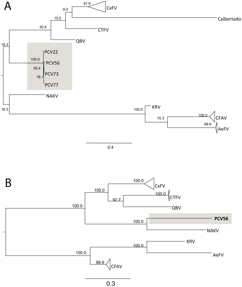
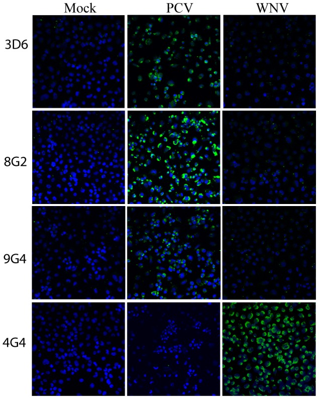
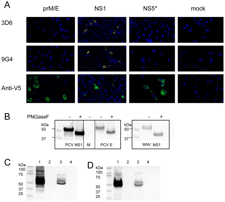
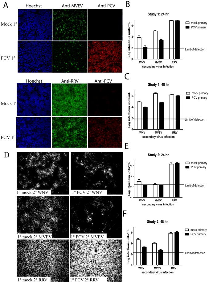
Recent reports of a novel group of flaviviruses that replicate only in mosquitoes and appear to spread through insect populations via vertical transmission have emerged from around the globe. To date, there is no information on the presence or prevalence of these insect-specific flaviviruses (ISFs) in Australian mosquito species. To assess whether such viruses occur locally, we used reverse transcription-polymerase chain reaction (RT-PCR) and flavivirus universal primers that are specific to the NS5 gene to detect these viruses in mosquito pools collected from the Northern Territory. Of 94 pools of mosquitoes, 13 were RT-PCR positive, and of these, 6 flavivirus isolates were obtained by inoculation of mosquito cell culture. Sequence analysis of the NS5 gene revealed that these isolates are genetically and phylogenetically similar to ISFs reported from other parts of the world. The entire coding region of one isolate (designated 56) was sequenced and shown to have approximately 63.7% nucleotide identity and 66.6% amino acid identity with its closest known relative (Nakiwogo virus) indicating that the prototype Australian ISF represents a new species. All isolates were obtained from Coquillettidia xanthogaster mosquitoes. The new virus is tentatively named Palm Creek virus (PCV) after its place of isolation. We also demonstrated that prior infection of cultured mosquito cells with PCV suppressed subsequent replication of the medically significant West Nile and Murray Valley encephalitis viruses by 10-43 fold (1 to 1.63 log) at 48 hr post-infection, suggesting that superinfection exclusion can occur between ISFs and vertebrate-infecting flaviviruses despite their high level of genetic diversity. We also generated several monoclonal antibodies (mAbs) that are specific to the NS1 protein of PCV, and these represent the first ISF-specific mAbs reported to date.
Conflict of interest statement
Figures
Similar articles
-
Antigenic Characterization of New Lineage II Insect-Specific Flaviviruses in Australian Mosquitoes and Identification of Host Restriction Factors.mSphere. 2020 Jun 17;5(3):e00095-20. doi: 10.1128/mSphere.00095-20. mSphere. 2020. PMID: 32554715 Free PMC article.
-
The insect-specific Palm Creek virus modulates West Nile virus infection in and transmission by Australian mosquitoes.Parasit Vectors. 2016 Jul 25;9(1):414. doi: 10.1186/s13071-016-1683-2. Parasit Vectors. 2016. PMID: 27457250 Free PMC article.
-
Co-circulation of West Nile virus and distinct insect-specific flaviviruses in Turkey.Parasit Vectors. 2017 Mar 20;10(1):149. doi: 10.1186/s13071-017-2087-7. Parasit Vectors. 2017. PMID: 28320443 Free PMC article.
-
Insect-specific flaviviruses: a systematic review of their discovery, host range, mode of transmission, superinfection exclusion potential and genomic organization.Viruses. 2015 Apr 10;7(4):1927-59. doi: 10.3390/v7041927. Viruses. 2015. PMID: 25866904 Free PMC article. Review.
-
Unleashing Nature's Allies: Comparing the Vertical Transmission Dynamics of Insect-Specific and Vertebrate-Infecting Flaviviruses in Mosquitoes.Viruses. 2024 Sep 23;16(9):1499. doi: 10.3390/v16091499. Viruses. 2024. PMID: 39339975 Free PMC article. Review.
Cited by
-
Uncovering the Worldwide Diversity and Evolution of the Virome of the Mosquitoes Aedes aegypti and Aedes albopictus.Microorganisms. 2021 Aug 3;9(8):1653. doi: 10.3390/microorganisms9081653. Microorganisms. 2021. PMID: 34442732 Free PMC article.
-
Characterization of Three New Insect-Specific Flaviviruses: Their Relationship to the Mosquito-Borne Flavivirus Pathogens.Am J Trop Med Hyg. 2018 Feb;98(2):410-419. doi: 10.4269/ajtmh.17-0350. Epub 2017 Aug 31. Am J Trop Med Hyg. 2018. PMID: 29016330 Free PMC article.
-
Detection of DENV-2 and Insect-Specific Flaviviruses in Mosquitoes Collected From Jeddah, Saudi Arabia.Front Cell Infect Microbiol. 2021 Feb 25;11:626368. doi: 10.3389/fcimb.2021.626368. eCollection 2021. Front Cell Infect Microbiol. 2021. PMID: 33718273 Free PMC article.
-
Viral RNA intermediates as targets for detection and discovery of novel and emerging mosquito-borne viruses.PLoS Negl Trop Dis. 2015 Mar 23;9(3):e0003629. doi: 10.1371/journal.pntd.0003629. eCollection 2015 Mar. PLoS Negl Trop Dis. 2015. PMID: 25799391 Free PMC article.
-
Abundance of Phasi-Charoen-like virus in Aedes aegypti mosquito populations in different states of India.PLoS One. 2022 Dec 9;17(12):e0277276. doi: 10.1371/journal.pone.0277276. eCollection 2022. PLoS One. 2022. PMID: 36490242 Free PMC article.
References
-
- Mackenzie JS, Lindsay MD, Coelen RJ, Broom AK, Hall RA, et al. (1994) Arboviruses causing human disease in the Australasian zoogeographic region. Arch Virol 136: 447–467. - PubMed
-
- Mackenzie JS, Gubler DJ, Petersen LR (2004) Emerging flaviviruses: the spread and resurgence of Japanese encephalitis, West Nile and dengue viruses. Nat Med 10: S98–109. - PubMed
-
- Stollar V, Thomas VL (1975) An agent in the Aedes aegypti cell line (Peleg) which causes fusion of Aedes albopictus cells. Virology 64: 367–377. - PubMed
-
- Crabtree MB, Sang RC, Stollar V, Dunster LM, Miller BR (2003) Genetic and phenotypic characterization of the newly described insect flavivirus, Kamiti River virus. Arch Virol 148: 1095–1118. - PubMed
-
- Cook S, Bennett SN, Holmes EC, De Chesse R, Moureau G, et al. (2006) Isolation of a new strain of the flavivirus cell fusing agent virus in a natural mosquito population from Puerto Rico. J Gen Virol 87: 735–748. - PubMed
Publication types
MeSH terms
Substances
Grants and funding
LinkOut - more resources
Full Text Sources
Other Literature Sources
Medical
Molecular Biology Databases
Research Materials
