. 2013 Mar 11;210(3):517-34.
doi: 10.1084/jem.20120572. Epub 2013 Mar 4.

IL-27 inhibits HIV-1 infection in human macrophages by down-regulating host factor SPTBN1 during monocyte to macrophage differentiation

Affiliations

IL-27 inhibits HIV-1 infection in human macrophages by down-regulating host factor SPTBN1 during monocyte to macrophage differentiation

Lue Dai et al. J Exp Med. .

Abstract

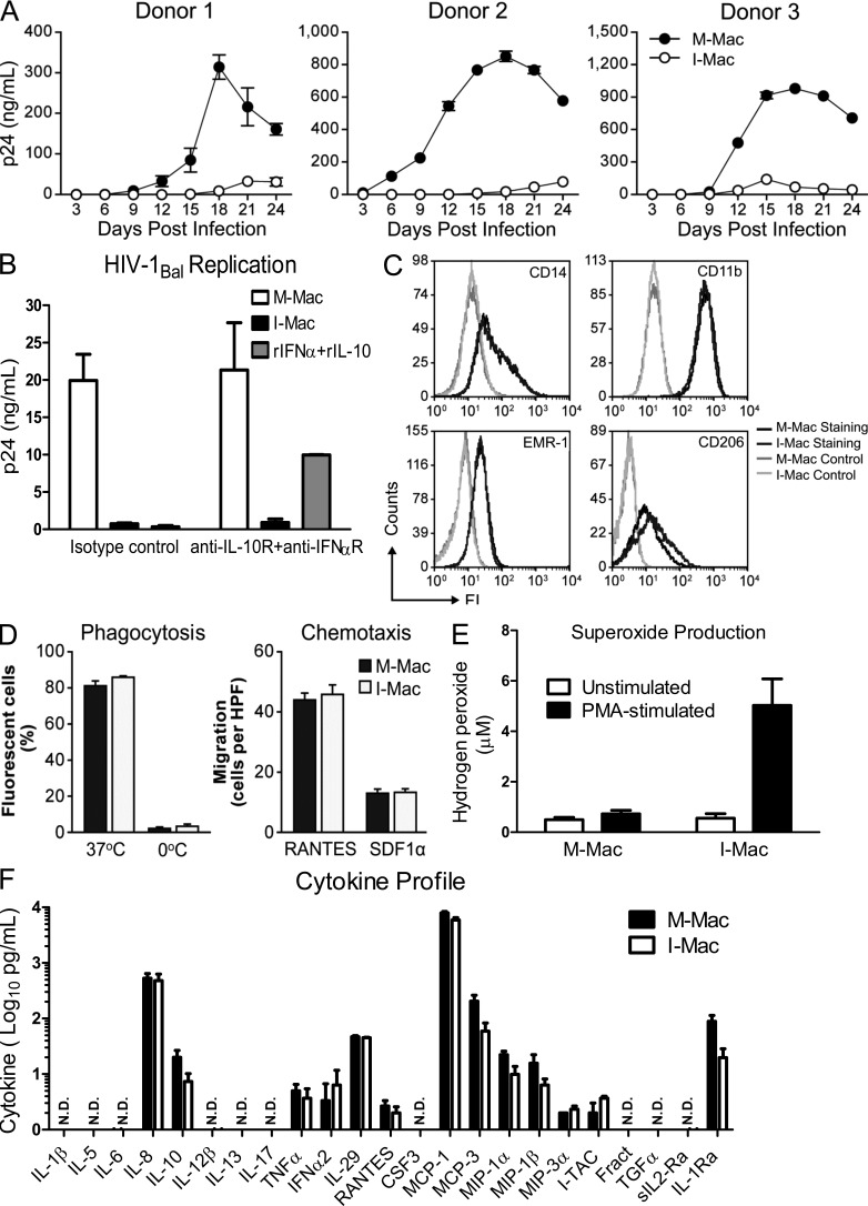
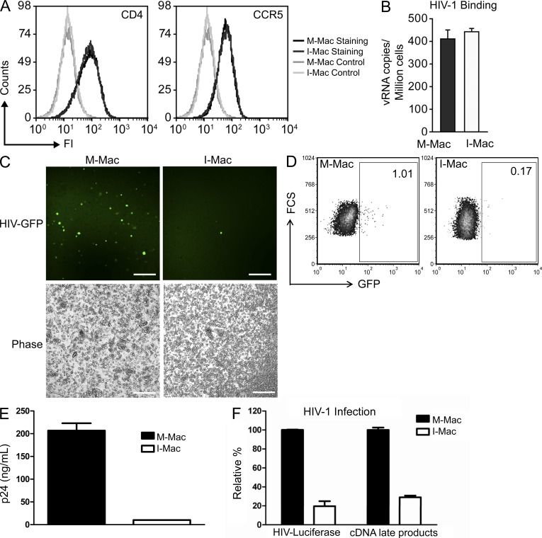
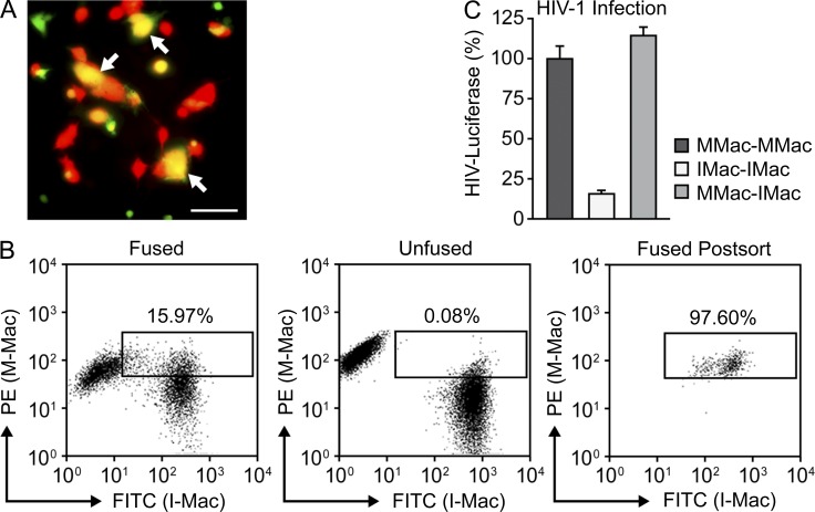
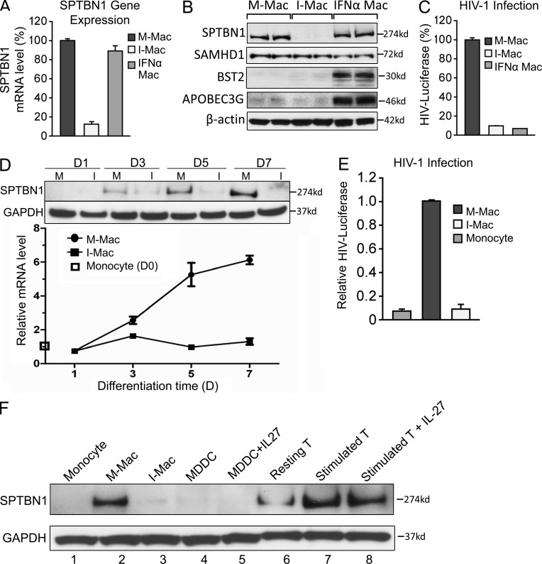
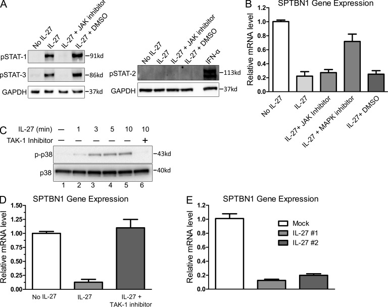
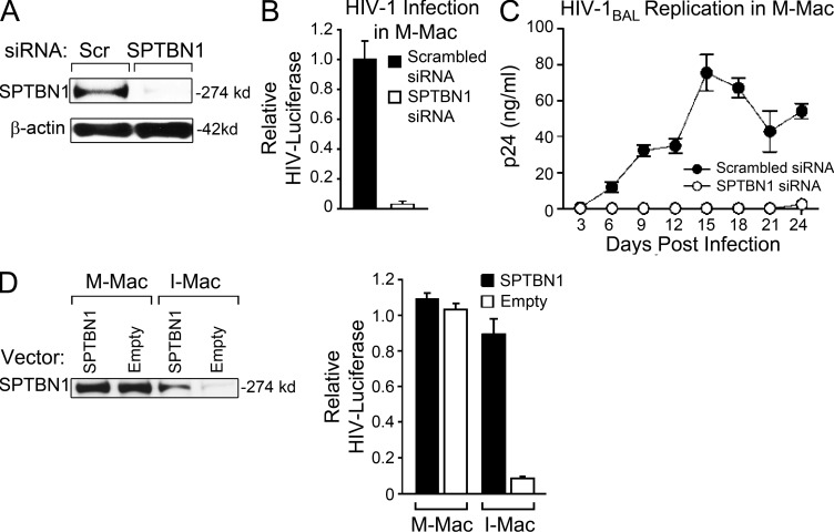
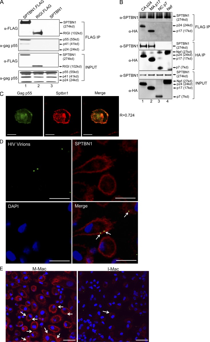
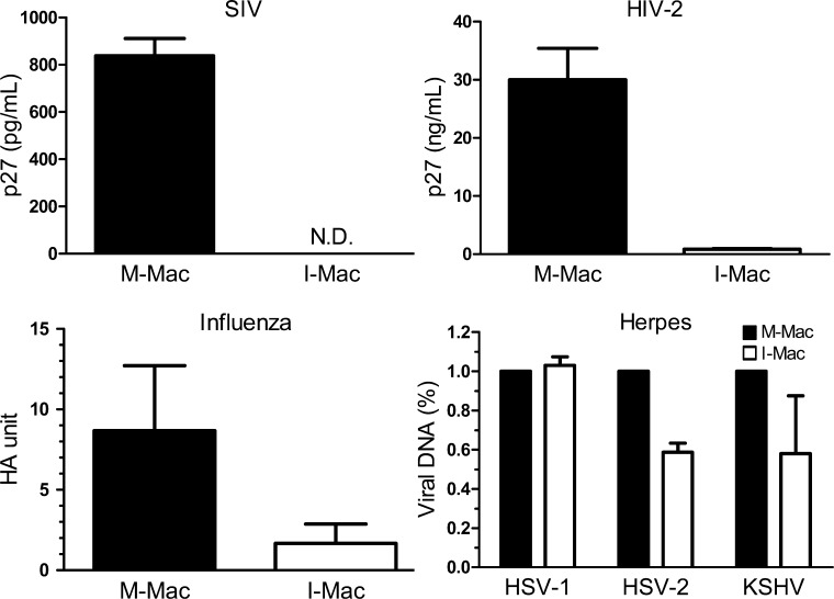
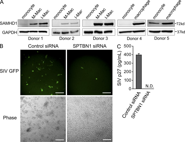
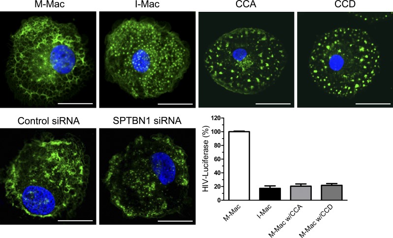
The susceptibility of macrophages to HIV-1 infection is modulated during monocyte differentiation. IL-27 is an anti-HIV cytokine that also modulates monocyte activation. In this study, we present new evidence that IL-27 promotes monocyte differentiation into macrophages that are nonpermissive for HIV-1 infection. Although IL-27 treatment does not affect expression of macrophage differentiation markers or macrophage biological functions, it confers HIV resistance by down-regulating spectrin β nonerythrocyte 1 (SPTBN1), a required host factor for HIV-1 infection. IL-27 down-regulates SPTBN1 through a TAK-1-mediated MAPK signaling pathway. Knockdown of SPTBN1 strongly inhibits HIV-1 infection of macrophages; conversely, overexpression of SPTBN1 markedly increases HIV susceptibility of IL-27-treated macrophages. Moreover, we demonstrate that SPTBN1 associates with HIV-1 gag proteins. Collectively, our results underscore the ability of IL-27 to protect macrophages from HIV-1 infection by down-regulating SPTBN1, thus indicating that SPTBN1 is an important host target to reduce HIV-1 replication in one major element of the viral reservoir.


Figures

Figure 1.
Figure 1.
IL-27 induces macrophages resistant to HIV-1 infection while retaining normal functions and differentiation markers. (A) M-Mac and I-Mac were infected with HIV-1BAL. Viral replication was monitored by measuring p24 antigen in culture supernatants. Results are shown for macrophages cultures obtained from three independent donors and data shown represent mean ± SD of triplicate infection samples. (B) Antibodies that neutralize human IFN-α receptor (10 µg/ml) and IL-10 receptors (10 µg/ml) were kept in culture during the 7-d differentiation of M-Mac and I-Mac. M-Mac and I-Mac differentiated in the presence of neutralizing antibodies were infected with HIV-1Bal, and p24 amount in culture supernatants was measured 14 d after infection. As an additional control to indicate the neutralizing effect, M-Mac differentiated in the presence of neutralizing antibodies were also incubated with recombinant human IFN-α (10 u/ml) and IL-10 (1 ng/ml) for 1 h before HIV-1 infection. Data shown represent means ± SE of three independent experiments. (C) M-Mac and I-Mac were analyzed for CD14, CD11b, EMR-1, and CD206 expression by FACS before infection. (D, left) Phagocytic activities of M-Mac and I-Mac were assessed using a pHrodo dye phagocytosis assay. As a negative control, macrophages were placed on ice to avoid phagocytosis. (right) the migration of M-Mac and I-Mac to RANTES (10 ng/ml) and SDF-1α (100 ng/ml) were assessed by counting cells migrating across filters of microchemotaxis chambers. Data shown represent mean ± SE of three independent experiments. (E) M-Mac and I-Mac were analyzed for superoxide production with or without PMA stimulation. Data shown represent means ± SE of three independent experiments. (F) Supernatants of M-Mac and I-Mac were analyzed for cytokine concentrations using the Multiplex Cytokine assay. Data are shown on a log scale. N.D., not detected. Data shown represent means ± SE of three independent experiments.
Figure 2.
Figure 2.
HIV-1 infection of I-Mac is blocked after entry and before reverse transcription of viral cDNA. (A) M-Mac and I-Mac were analyzed for CD4 and CCR5 expression by FACS. (B) M-Mac and I-Mac were incubated with HIV-1BAL on ice for 1 h to allow virus binding. Virus attachment was determined by measuring the copy number of HIV-1 RNA bound to the cells. Data shown represent means ± SE of three independent experiments. (C) M-Mac and I-Mac were infected with HIV-EGFP-V. (left) Infected cells were examined with fluorescent microscopy. Bar, 200 µm. (D) GFP+ cells were analyzed by FACS 4 d after infection. (E) p24 antigen amount was measured by ELISA. Data shown represent mean ± SD of triplicate infection samples. (F) M-Mac and I-Mac were infected with HIV-LUC-V. Viral cDNA copy number and luciferase activity was measured 48 h after infection. Data shown represent mean ± SE of three independent experiments.
Figure 3.
Figure 3.
I-Mac lacks a cellular host factor to support HIV-1 infection. (A) Heterokaryons were formed between M-Mac and I-Mac. M-Mac was stained with CellTracker Green and I-Mac was stained with CellTracker Red. Double-stained heterokaryons were yellow as indicated by arrows. Bar, 50 µm. (B, left) Double-stained heterokaryons between M-Mac and I-Mac were sorted by FACS with the indicated gate. (middle) M-Mac and I-Mac were mixed without fusion. (right) Heterokaryons were reanalyzed by FACS to confirm purity after sorting. (C) Sorted heterokaryons were infected with HIV-LUC-V. HIV-1 infection of heterokaryons between M-Mac and I-Mac was compared with infection levels of M-Mac or I-Mac homokaryons. Data shown represent mean ± SE of three independent experiments.
Figure 4.
Figure 4.
IL-27 down-regulates SPTBN1 during monocyte differentiation. (A) M-Mac, I-Mac, and macrophages treated with IFN-α (1 ng/ml) for 24 h (IFN-α Mac) were analyzed for gene expression levels of SPTBN1 by RT-PCR. (B) Whole-cell lysates of M-Mac, I-Mac, and IFN-α Mac were used for Western blotting. Protein expression levels of SPTBN1, SAMHD1, BST-2, and APOBEC3G were examined with specific antibodies. Samples were loaded in duplicate and β-actin served as an internal loading control. (C) M-Mac, I-Mac, and IFN-α Mac were infected with HIV-LUC-V. Data shown represent mean ± SE of three independent experiments. (D) M-Mac and I-Mac were analyzed for SPTBN1 expression on day 1, 3, 5, and 7 during differentiation. (top) Whole-cell lysates were subjected to Western blotting to examine protein levels of SPTBN1. (bottom) Gene expression levels of SPTBN1 were examined by RT-PCR. Relative mRNA levels of M-Mac and I-Mac were compared with that of undifferentiated monocyte on day 0, which is equal to 1. (E) Monocytes, M-Mac, and I-Mac derived from the same donor were infected with HIV-LUC-V. Values of luciferase activity were expressed relative to those obtained for M-Mac. Data shown represent mean ± SE of three independent experiments. (F) Monocytes, macrophages, dendritic cells, and CD4+ T cells obtained from the same donor were analyzed for SPTBN1 expression by Western blotting. MDDCs were induced by G-MCSF (50 ng/ml) and IL-4 (50 ng/ml) with or without IL-27 (50 ng/ml). CD4+ T cells were stimulated with PHA (5 µg/ml) and IL-2 (20 U/ml) with or without IL-27 (50 ng/ml).
Figure 5.
Figure 5.
IL-27 down-regulates SPTBN1 through a TAK-1–mediated p38 MAPK signaling pathway. On day 4 during differentiation, macrophages were stimulated with IL-27 (50 ng/ml), with or without the pretreatment of a JAK inhibitor or its solvent control DMSO. (A) Whole-cell lysates were harvested 15 min after IL-27 stimulation, and the phosphorylation of STAT1, STAT2, and STAT3 was examined by Western blotting using specific antibodies against p-STATs (Cell Signaling Technology). IFN-α–treated macrophage lysate was included to validate the antibody against phosphorylated STAT2. (B) Macrophages were pretreated with a JAK inhibitor (EMD), a p38 MAPK inhibitor (Cell Signaling Technology), or the solvent control DMSO. After 2 h, macrophages were stimulated with IL-27 (50 ng/ml). Gene expression of SPTBN1 was examined by RT-PCR 24h after IL-27 stimulation. Data shown represent means ± SE of three independent experiments. (C) Macrophages were pretreated with a TAK-1 inhibitor (Cell Signaling Technology) or mock treated for 2 h. Cells were then stimulated with IL-27 (50 ng/ml) and harvested at different time points as indicated. The phosphorylation of p38 MAPK was examined by Western blotting using an anti–p-p38 antibody (Cell Signaling Technology). The expression of unphosphorylated p38 was shown as an internal control. (D) Macrophages were pretreated with a TAK-1 inhibitor (Cell Signaling Technology) or mock treated. After 2 h, macrophages were stimulated with IL-27 (50 ng/ml). Gene expression of SPTBN1 was examined by RT-PCR 24h after IL-27 stimulation. Data shown represent means ± SE of three independent experiments. (E) Recombinant human IL-27 #1 and IL-27 #2 were obtained from R&D Systems and HumanZyme, respectively. On day 4 during differentiation, macrophages were stimulated with IL-27 #1 and IL-27 #2 (50 ng/ml). Gene expression of SPTBN1 was examined by RT-PCR 24 h after IL-27 stimulation. Data shown represent means ± SD of three independent samples.
Figure 6.
Figure 6.
SPTBN1 is required to support HIV-1 infection in primary macrophages. (A) M-Mac was mock transfected or transfected with siRNA targeting SPTBN1. 48 h after transfection, whole-cell lysates were used to examine SPTBN1 expression by Western blotting. (B) M-Mac was infected with HIV-LUC-V 48 h after siRNA transfection. Data shown represent mean ± SE of three independent experiments. (C) M-Mac was infected with HIV-1BAL. HIV-1 replication was monitored by measuring p24 antigen levels in culture supernatants for 24 d after infection. Data shown represent mean ± SD of triplicate infection samples. (D) M-Mac and I-Mac were transfected with an SPTBN1 expression vector or an empty vector for 48 h. (left) Whole-cell lysates were used to examine SPTBN1 expression by Western blotting. (right) Transfected cells were infected with HIV-LUC-V. Relative luciferase activity was shown, and data shown represent mean ± SE of three independent experiments.
Figure 7.
Figure 7.
SPTBN1 associates with HIV-1 gag proteins. (A) 293T cells were transfected to express FLAG-tagged SPTBN1 and gag p55. Whole-cell lysates from transfected cells were subjected to a pull-down assay using anti-FLAG agarose. Unrelated FLAG-tagged RIG-I and untagged SPTBN1 were negative controls for nonspecific binding. Immunoprecipitates were analyzed by immunoblotting using anti-FLAG tag and anti-gag p55 antibodies. (B) 293T cells were transfected to express FLAG-tagged SPTBN1 and HA-tagged CA p24, MA p17, and NC p7. Whole-cell lysates from transfected cells were subjected to a pull-down assay using anti-FLAG agarose or anti-HA agarose. Immunoprecipitates were analyzed by immunoblotting using anti-SPTBN1 and anti-HA antibodies. Nef was a negative control for nonspecific binding. (C) Macrophages were transfected to express GFP-tagged Gag p55 (green). After 24 h, macrophages were fixed and labeled with antibody against SPTBN1 (red). A representative merged image of Gag p55 and SPTBN1 shown on the right (yellow) indicates the colocalization between Gag and SPTBN1. R, Pearson coefficient of correlation. Bars, 10 µm. (D) Macrophages were infected with VSV-G–pseudotyped Vpr-GFP-packaged HIV-1 virions (green). After 30-min incubation, cells were fixed and labeled with antibody against SPTBN1 (red) or DAPI (blue). White arrows on the merged image indicate the colocalization of SPTBN1 and incoming HIV-1 virions (yellow). Bars, 30 µm. (E) M-Mac and I-Mac were infected with VSV-G-pseudotyped Vpr-GFP-packaged HIV-1 virions (green). After 30-min incubation, cells were fixed and labeled with antibody against SPTBN1 (red) or DAPI (blue). White arrows on the merged image indicate the colocalization of SPTBN1 and incoming HIV-1 virions (yellow). Bars, 30 µm.
Figure 8.
Figure 8.
IL-27 inhibits various viral infections. M-Mac and I-Mac were infected with SIV, HIV-2, Influenza A, HSV-1, HSV-2, and KSHV. Infection of SIV and HIV-2 was monitored by measuring p27 antigen amount in supernatants. N.D., not detected. Detection limit of SIV p27 was 15.625 pg/ml. Infection of Influenza A was determined using HA assay. Infection of herpes viruses was gauged by measuring viral DNA copies with real-time PCR. Data shown represent means ± SE of three independent experiments.
Figure 9.
Figure 9.
SPTBN1 affects macrophage susceptibility to HIV-1 infection independently of SAMHD1. (A) Whole-cell lysates of monocytes, M-Mac, and I-Mac (donor 1–3) or whole-cell lysates of monocytes and macrophages (donor 4 and 5) were analyzed for protein expression levels of SPTBN1. GAPDH served as an internal loading control. (B) Macrophages were infected with SIV-GFP after transfection with control siRNA or SPTBN1 specific siRNA (50 pmol). A representative field of macrophages for SIV-GFP expression was shown. Bars, 200 µm. (C) SIV p27 level in culture supernatants was measured. Data shown represent means ± SD of triplicate infection samples.
Figure 10.
Figure 10.
Actin disarrangement impacts HIV-1 infection. Macrophages were treated with IL-27 (50 ng/ml), cytochalasin A (5 µM), or cytochalasin D (5 µM), or transfected with control or SPTBN1 siRNA (50 pmol). F-actin was labeled with fluorescein phalloidin and nuclei were stained with DAPI. Bars, 10 µm. Cells were infected with HIV-LUC-V and luciferase activity was measured 4 d after infection. Data shown represent means ± SE of three independent experiments.

Similar articles

Cited by

References

    1. Andrei G., Fiten P., De Clercq E., Snoeck R., Opdenakker G. 2000. Monitoring drug resistance for herpesviruses. Methods Mol. Med. 24:151–169 - PubMed
    1. Arhel N. 2010. Revisiting HIV-1 uncoating. Retrovirology. 7:96 10.1186/1742-4690-7-96 - DOI - PMC - PubMed
    1. Arhel N., Genovesio A., Kim K.A., Miko S., Perret E., Olivo-Marin J.C., Shorte S., Charneau P. 2006. Quantitative four-dimensional tracking of cytoplasmic and nuclear HIV-1 complexes. Nat. Methods. 3:817–824 10.1038/nmeth928 - DOI - PubMed
    1. Baldauf H.M., Pan X., Erikson E., Schmidt S., Daddacha W., Burggraf M., Schenkova K., Ambiel I., Wabnitz G., Gramberg T., et al. 2012. SAMHD1 restricts HIV-1 infection in resting CD4(+) T cells. Nat. Med. 18:1682–1687 10.1038/nm.2964 - DOI - PMC - PubMed
    1. Bender H., Wiesinger M.Y., Nordhoff C., Schoenherr C., Haan C., Ludwig S., Weiskirchen R., Kato N., Heinrich P.C., Haan S. 2009. Interleukin-27 displays interferon-gamma-like functions in human hepatoma cells and hepatocytes. Hepatology. 50:585–591 10.1002/hep.22988 - DOI - PubMed

Publication types

MeSH terms