

Dissociating memory networks in early Alzheimer's disease and frontotemporal lobar degeneration - a combined study of hypometabolism and atrophy
- PMID: 23457466
- PMCID: PMC3573064
- DOI: 10.1371/journal.pone.0055251
Dissociating memory networks in early Alzheimer's disease and frontotemporal lobar degeneration - a combined study of hypometabolism and atrophy
Abstract
Introduction: We aimed at dissociating the neural correlates of memory disorders in Alzheimer's disease (AD) and frontotemporal lobar degeneration (FTLD).
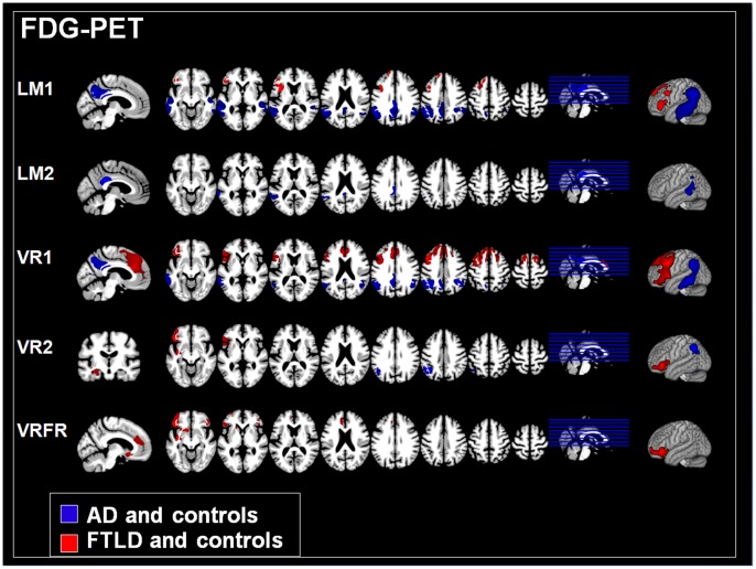
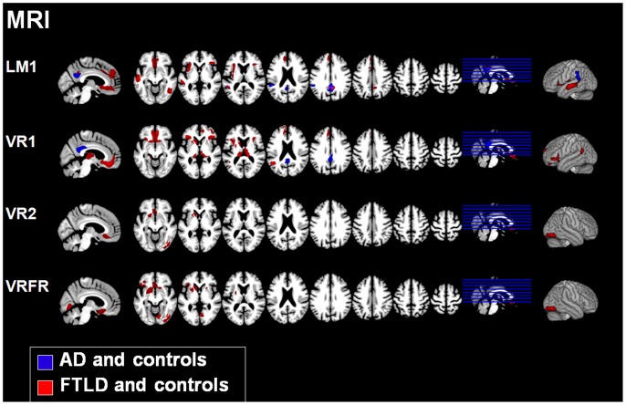
Methods: We included patients with AD (n = 19, 11 female, mean age 61 years) and FTLD (n = 11, 5 female, mean age 61 years) in early stages of their diseases. Memory performance was assessed by means of verbal and visual memory subtests from the Wechsler Memory Scale (WMS-R), including forgetting rates. Brain glucose utilization was measured by [18F]fluorodeoxyglucose positron emission tomography (FDG-PET) and brain atrophy by voxel-based morphometry (VBM) of T1-weighted magnetic resonance imaging (MRI) scans. Using a whole brain approach, correlations between test performance and imaging data were computed separately in each dementia group, including a group of control subjects (n = 13, 6 female, mean age 54 years) in both analyses. The three groups did not differ with respect to education and gender.
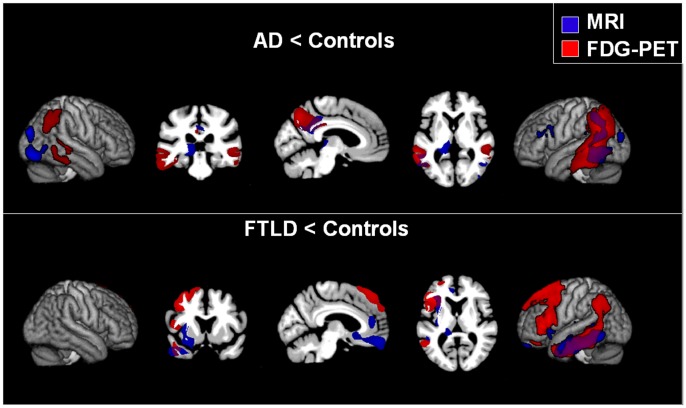
Results: Patients in both dementia groups generally performed worse than controls, but AD and FTLD patients did not differ from each other in any of the test parameters. However, memory performance was associated with different brain regions in the patient groups, with respect to both hypometabolism and atrophy: Whereas in AD patients test performance was mainly correlated with changes in the parieto-mesial cortex, performance in FTLD patients was correlated with changes in frontal cortical as well as subcortical regions. There were practically no overlapping regions associated with memory disorders in AD and FTLD as revealed by a conjunction analysis.
Conclusion: Memory test performance may not distinguish between both dementia syndromes. In clinical practice, this may lead to misdiagnosis of FTLD patients with poor memory performance. Nevertheless, memory problems are associated with almost completely different neural correlates in both dementia syndromes. Obviously, memory functions are carried out by distributed networks which break down in brain degeneration.
Conflict of interest statement
Figures
Similar articles
-
Temporoparietal hypometabolism in frontotemporal lobar degeneration and associated imaging diagnostic errors.Arch Neurol. 2011 Mar;68(3):329-37. doi: 10.1001/archneurol.2010.295. Epub 2010 Nov 8. Arch Neurol. 2011. PMID: 21059987 Free PMC article.
-
Removing outliers from the normative database improves regional atrophy detection in single-subject voxel-based morphometry.Neuroradiology. 2024 Apr;66(4):507-519. doi: 10.1007/s00234-024-03304-3. Epub 2024 Feb 21. Neuroradiology. 2024. PMID: 38378906 Free PMC article.
-
A visual [18F]FDG-PET rating scale for the differential diagnosis of frontotemporal lobar degeneration.Eur Arch Psychiatry Clin Neurosci. 2011 Sep;261(6):433-46. doi: 10.1007/s00406-010-0184-0. Epub 2011 Jan 5. Eur Arch Psychiatry Clin Neurosci. 2011. PMID: 21207049
-
[Brain functional imaging of frontotemporal lobar degeneration].Brain Nerve. 2009 Nov;61(11):1275-84. Brain Nerve. 2009. PMID: 19938684 Review. Japanese.
-
[Neuroimaging in corticobasal syndrome].Rinsho Shinkeigaku. 2013;53(11):1029-32. doi: 10.5692/clinicalneurol.53.1029. Rinsho Shinkeigaku. 2013. PMID: 24291869 Review. Japanese.
Cited by
-
Update on Major Neurocognitive Disorders.Focus (Am Psychiatr Publ). 2021 Jul;19(3):271-281. doi: 10.1176/appi.focus.20210004. Epub 2021 Jul 9. Focus (Am Psychiatr Publ). 2021. PMID: 34690593 Free PMC article.
-
How cognitive neuroscience could be more biological-and what it might learn from clinical neuropsychology.Front Hum Neurosci. 2014 Jul 21;8:541. doi: 10.3389/fnhum.2014.00541. eCollection 2014. Front Hum Neurosci. 2014. PMID: 25100981 Free PMC article.
-
MRI signatures of brain macrostructural atrophy and microstructural degradation in frontotemporal lobar degeneration subtypes.J Alzheimers Dis. 2013;33(2):431-44. doi: 10.3233/JAD-2012-121156. J Alzheimers Dis. 2013. PMID: 22976075 Free PMC article. Clinical Trial.
-
Papez Circuit Gray Matter and Episodic Memory in Amyotrophic Lateral Sclerosis and Behavioural Variant Frontotemporal Dementia.Brain Imaging Behav. 2021 Apr;15(2):996-1006. doi: 10.1007/s11682-020-00307-5. Brain Imaging Behav. 2021. PMID: 32734436
-
Genuine Memory Deficits as Assessed by the Free and Cued Selective Reminding Test (FCSRT) in the Behavioural Variant of Frontotemporal Dementia. A Systematic Review and Meta-analysis Study.Neuropsychol Rev. 2024 Sep;34(3):823-837. doi: 10.1007/s11065-023-09613-3. Epub 2023 Sep 22. Neuropsychol Rev. 2024. PMID: 37736861 Free PMC article. Review.
References
-
- Neary D, Snowden JS, Gustafson L, Passant U, Stuss D, et al. (1998) Frontotemporal lobar degeneration: a consensus on clinical diagnostic criteria. Neurology 51: 1546–54. - PubMed
-
- Neary D, Snowden J, Mann D (2005) Frontotemporal dementia. Lancet Neurol 4: 771–80. - PubMed
-
- Raczka KA, Becker G, Seese A, Frisch S, Heiner S, et al. (2010) Executive and behavioral deficits share common neural substrates in frontotemporal lobar degeneration - a pilot FDG-PET study. Psychiatry Res 182: 274–80. - PubMed
Publication types
MeSH terms
Substances
Grants and funding
LinkOut - more resources
Full Text Sources
Other Literature Sources
Medical
