D-series resolvin attenuates vascular smooth muscle cell activation and neointimal hyperplasia following vascular injury
- PMID: 23407709
- PMCID: PMC3659350
- DOI: 10.1096/fj.12-225615
D-series resolvin attenuates vascular smooth muscle cell activation and neointimal hyperplasia following vascular injury
Abstract
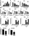
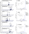
Recent evidence suggests that specialized lipid mediators derived from polyunsaturated fatty acids control resolution of inflammation, but little is known about resolution pathways in vascular injury. We sought to determine the actions of D-series resolvin (RvD) on vascular smooth muscle cell (VSMC) phenotype and vascular injury. Human VSMCs were treated with RvD1 and RvD2, and phenotype was assessed by proliferation, migration, monocyte adhesion, superoxide production, and gene expression assays. A rabbit model of arterial angioplasty with local delivery of RvD2 (10 nM vs. vehicle control) was employed to examine effects on vascular injury in vivo. Local generation of proresolving lipid mediators (LC-MS/MS) and expression of RvD receptors in the vessel wall were assessed. RvD1 and RvD2 produced dose-dependent inhibition of VSMC proliferation, migration, monocyte adhesion, superoxide production, and proinflammatory gene expression (IC50≈0.1-1 nM). In balloon-injured rabbit arteries, cell proliferation (51%) and leukocyte recruitment (41%) were reduced at 3 d, and neointimal hyperplasia was attenuated (29%) at 28 d by RvD2. We demonstrate endogenous biosynthesis of proresolving lipid mediators and expression of receptors for RvD1 in the artery wall. RvDs broadly reduce VSMC responses and modulate vascular injury, suggesting that local activation of resolution mechanisms expedites vascular homeostasis.
Keywords: inflammation; intracellular signaling; resolvin.
Figures


Similar articles
-
17R/S-Benzo-RvD1, a synthetic resolvin D1 analogue, attenuates neointimal hyperplasia in a rat model of acute vascular injury.PLoS One. 2022 Feb 28;17(2):e0264217. doi: 10.1371/journal.pone.0264217. eCollection 2022. PLoS One. 2022. PMID: 35226675 Free PMC article.
-
Systemic delivery of proresolving lipid mediators resolvin D2 and maresin 1 attenuates intimal hyperplasia in mice.FASEB J. 2015 Jun;29(6):2504-13. doi: 10.1096/fj.14-265363. Epub 2015 Mar 16. FASEB J. 2015. PMID: 25777995 Free PMC article.
-
Perivascular delivery of resolvin D1 inhibits neointimal hyperplasia in a rat model of arterial injury.J Vasc Surg. 2017 Jan;65(1):207-217.e3. doi: 10.1016/j.jvs.2016.01.030. Epub 2016 Mar 29. J Vasc Surg. 2017. PMID: 27034112 Free PMC article.
-
Resolvin E1 attenuates injury-induced vascular neointimal formation by inhibition of inflammatory responses and vascular smooth muscle cell migration.FASEB J. 2018 Oct;32(10):5413-5425. doi: 10.1096/fj.201800173R. Epub 2018 May 3. FASEB J. 2018. PMID: 29723062
-
Perivascular delivery of resolvin D1 inhibits neointimal hyperplasia in a rabbit vein graft model.J Vasc Surg. 2018 Dec;68(6S):188S-200S.e4. doi: 10.1016/j.jvs.2018.05.206. Epub 2018 Jul 29. J Vasc Surg. 2018. PMID: 30064835 Free PMC article.
Cited by
-
Biomimetic Nanocarriers of Pro-Resolving Lipid Mediators for Resolution of Inflammation in Atherosclerosis.Adv Healthc Mater. 2024 Jan;13(3):e2302238. doi: 10.1002/adhm.202302238. Epub 2023 Nov 8. Adv Healthc Mater. 2024. PMID: 37852632 Free PMC article.
-
Pro-resolving lipid mediators are leads for resolution physiology.Nature. 2014 Jun 5;510(7503):92-101. doi: 10.1038/nature13479. Nature. 2014. PMID: 24899309 Free PMC article. Review.
-
Resolvins and omega three polyunsaturated fatty acids: Clinical implications in inflammatory diseases and cancer.World J Clin Cases. 2016 Jul 16;4(7):155-64. doi: 10.12998/wjcc.v4.i7.155. World J Clin Cases. 2016. PMID: 27458590 Free PMC article. Review.
-
17R/S-Benzo-RvD1, a synthetic resolvin D1 analogue, attenuates neointimal hyperplasia in a rat model of acute vascular injury.PLoS One. 2022 Feb 28;17(2):e0264217. doi: 10.1371/journal.pone.0264217. eCollection 2022. PLoS One. 2022. PMID: 35226675 Free PMC article.
-
Aspirin enhances protective effect of fish oil against thrombosis and injury-induced vascular remodelling.Br J Pharmacol. 2015 Dec;172(23):5647-60. doi: 10.1111/bph.12986. Epub 2015 Jan 12. Br J Pharmacol. 2015. PMID: 25339093 Free PMC article.
References
-
- Hansson G. K. (2005) Inflammation, atherosclerosis, and coronary artery disease. N. Engl. J. Med. 352, 1685–1695 - PubMed
-
- Libby P. (2002) Inflammation in atherosclerosis. Nature 420, 868–874 - PubMed
-
- Conte M. S., Bandyk D. F., Clowes A. W., Moneta G. L., Seely L., Lorenz T. J., Namini H., Hamdan A. D., Roddy S. P., Belkin M., Berceli S. A., DeMasi R. J., Samson R. H., Berman S. S. (2006) Results of PREVENT III: a multicenter, randomized trial of edifoligide for the prevention of vein graft failure in lower extremity bypass surgery. J. Vasc. Surg. 43, 742–751; discussion 751 - PubMed
-
- Norgren L., Hiatt W. R., Dormandy J. A., Nehler M. R., Harris K. A., Fowkes F. G. (2007) Inter-society consensus for the management of peripheral arterial disease (TASC II). J. Vasc. Surg. 45(Suppl. S), S5–S67 - PubMed
-
- Rocha-Singh K. J., Jaff M. R., Crabtree T. R., Bloch D. A., Ansel G. (2007) Performance goals and endpoint assessments for clinical trials of femoropopliteal bare nitinol stents in patients with symptomatic peripheral arterial disease. Catheter Cardiovasc. Interv. 69, 910–919 - PubMed
Publication types
MeSH terms
Substances
Grants and funding
LinkOut - more resources
Full Text Sources
Other Literature Sources

