. 2013 Mar 22;288(12):8238-8249.
doi: 10.1074/jbc.M112.438119. Epub 2013 Feb 6.

RIAM and vinculin binding to talin are mutually exclusive and regulate adhesion assembly and turnover

Affiliations

RIAM and vinculin binding to talin are mutually exclusive and regulate adhesion assembly and turnover

Benjamin T Goult et al. J Biol Chem. .

Abstract

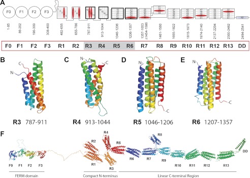
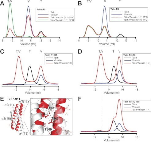
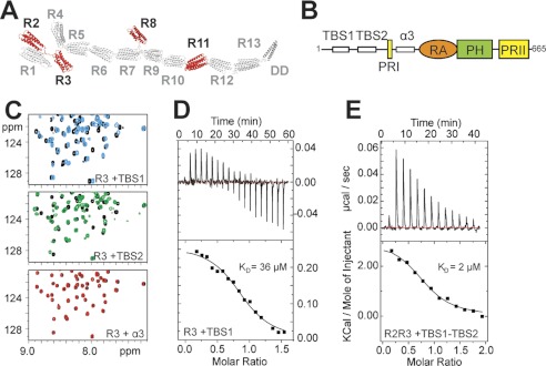
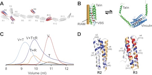
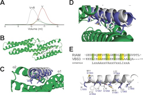
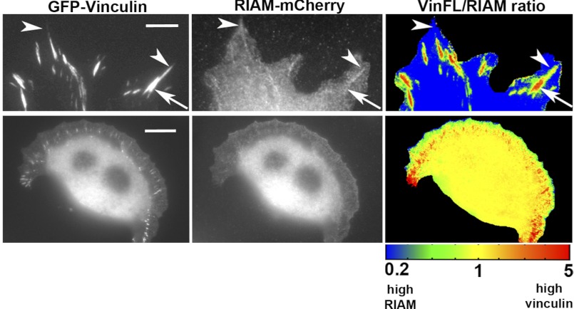
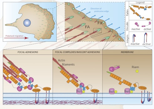
Talin activates integrins, couples them to F-actin, and recruits vinculin to focal adhesions (FAs). Here, we report the structural characterization of the talin rod: 13 helical bundles (R1-R13) organized into a compact cluster of four-helix bundles (R2-R4) within a linear chain of five-helix bundles. Nine of the bundles contain vinculin-binding sites (VBS); R2R3 are atypical, with each containing two VBS. Talin R2R3 also binds synergistically to RIAM, a Rap1 effector involved in integrin activation. Biochemical and structural data show that vinculin and RIAM binding to R2R3 is mutually exclusive. Moreover, vinculin binding requires domain unfolding, whereas RIAM binds the folded R2R3 double domain. In cells, RIAM is enriched in nascent adhesions at the leading edge whereas vinculin is enriched in FAs. We propose a model in which RIAM binding to R2R3 initially recruits talin to membranes where it activates integrins. As talin engages F-actin, force exerted on R2R3 disrupts RIAM binding and exposes the VBS, which recruit vinculin to stabilize the complex.


Figures

FIGURE 1.
FIGURE 1.
Domain structure of talin. A, schematic diagram of talin showing the N-terminal FERM domain (F0, F1, F2, and F3 domains) linked via an unstructured region to the 13 amphipathic helical bundles of the talin rod, which terminates in a single helix, the dimerization domain (DD). Residue numbers for each domain (R1–R13) are shown. Helices are numbered and vinculin-binding sites are colored red. Domains corresponding to the new structures reported here are shaded. B–E, NMR structures of the talin R3, R4, R5, and R6 rod domains. Ribbon diagrams of representative low-energy structures show the overall topology of each bundle. F, model of talin showing the structures of all 18 domains.
FIGURE 2.
FIGURE 2.
Vinculin binding to the talin rod. A and B, vinculin Vd1 (V) was incubated at a 1:1 molar ratio with talin (T) R2 (A) and R3 polypeptides (B) at 25 or 37 °C as indicated, and complex (T/V) formation was analyzed on a Superdex-75 (10/300) GL gel filtration column. C and D, vinculin Vd1 was incubated (25 °C) with talin R1–R5 (C) and talin R1–R3 (D) at a molar ratio of 4:1, and complex formation was analyzed by gel filtration on a Superdex-200 (10/300). The results show that the VBS in the N-terminal region of talin are active even in larger fragments. E, structure of R3; the two vinculin-binding helices are shown in red. The enlarged region shows the four threonine residues embedded within the hydrophobic core. F, mutation of the four threonines in R3 to hydrophobic residues (T809I/T833V/T867V/T901I; the IVVI mutation) abolished Vd1 binding to the R1–R3 IVVI talin polypeptide at 25 °C.
FIGURE 3.
FIGURE 3.
Characterization of the RIAM binding sites in the talin rod. A, schematic of the talin1 rod. Domains that interact with a RIAM(6–30) peptide as determined by NMR are in red. B, schematic of RIAM showing the two N-terminal talin-binding helices (TBS1 and TBS2), the proline-rich region (PRI), the additional helix (α3), the Ras association (RA) domain, the pleckstrin homology (PH) domain, and the second proline-rich region (PRII). C, 1H,15N HSQC spectra of 100 μm 15N-labeled talin R3 rod domain in the absence (black) or presence of RIAM TBS1 (6-30, blue), RIAM TBS2 (45–127; green) or RIAM α3 (147–174; red) at a ratio of 1:3. D and E, ITC characterization of RIAM binding to talin rod domains at 25°C. D, talin R3 titration with RIAM(1–32) (TBS1): 500 μm R3 in the cell and 5 mm TBS1 in the syringe. The lower panel shows fitting to a single-site binding model; KD, 36 μm. E, talin R2R3 titration with RIAM(1–127) (TBS1-TBS2); 18 μm R2R3 in the cell and 180 μm TBS1-TBS2 in the syringe. The lower panel shows fitting to a single-site binding model; KD, 2 μm.
FIGURE 4.
FIGURE 4.
RIAM and vinculin binding to talin is mutually exclusive. A, schematic of the talin1 rod. Domains that interact with a RIAM(6–30) peptide as determined by NMR are shown in red. VBS within these domains are shown in blue. B, left, model of single RIAM helix (orange) bound to a talin rod helical bundle (green). A single vinculin binding helix is shown in white, and residues that interact with vinculin are in red. Right, model of the Vd1 domain of vinculin (blue) bound to the VBS in a talin helical bundle illustrating that binding involves domain unfolding. C, analytical gel filtration (Superdex 75/10 300 GL) of talin R2R3 1:1 with vinculin Vd1 (V+T; cyan); talin R2R3 1:1 with RIAM 1–127 (T+R; orange); talin R2R3 1:1:1 with RIAM(1–127) and vinculin Vd1 (V+T+R; purple); Vd1 alone (V; Red); R2R3 alone (T; black). D, effects of RIAM(6–30) binding to R2 and R3 as detected by NMR. Weighted chemical shift differences, determined as described previously (16), are shown on ribbon representations of the R2 and R3 structures; peaks that broaden are shown in red, shifts > 0.13 ppm are in dark blue, and shifts > 0.07 ppm are in light blue. The VBS helices are shown in orange.
FIGURE 5.
FIGURE 5.
RIAM binding to vinculin. A, analytical gel filtration (Superdex 75/10 300 GL) of RIAM(1–127) mixed with vinculin Vd1. Protein samples are vinculin Vd1 1:1 with RIAM(1–127) (V+R; gray); RIAM1–127 alone (R; green); and Vd1 alone (V; red). All proteins were loaded at 50 μm. B, crystal structure of RIAM TBS1(1–32) (white) bound to vinculin Vd1 (green). C, top down view of B. The position of the talin R10 VBS (VBS3; blue) complexed to Vd1 (Protein Data Bank code 1XWJ) is shown for comparison. D, close up showing the similarity in binding of RIAM TBS1 and talin VBS3 to vinculin Vd1 and the overall displacement of RIAM TBS1 helix relative to talin VBS. E, sequence alignment of RIAM(1–32) and talin VBS3 plus the consensus VBS sequence. Lower panel, overlay of RIAM(1–32) and the VBS3 in talin R10. Side chains of residues contacting vinculin in the complex are shown in stick representation and labeled.
FIGURE 6.
FIGURE 6.
Differential localization of vinculin and RIAM in FA. Vinculin (−/−) cells were transfected with full-length GFP-vinculin and RIAM-mCherry. Upper panel, RIAM and vinculin localize to FAs. Ratio images displayed in a spectral color scale ranging from 0.2 (blue, high in RIAM) to 5 (red, high in vinculin), reveal that FA areas with high vinculin intensity (arrows) have low amounts of RIAM, and areas with high RIAM intensity (arrowheads) have low amounts of vinculin. Scale bar, 3 μm. Lower panel, in a migrating cell, RIAM localizes strongly to the leading edge in front of developing FAs, which are high in vinculin. Scale bar,10 μm.
FIGURE 7.
FIGURE 7.
Talin R1–R3 are essential for FA assembly. Human endothelial cells were electroporated with a talin1 siRNA or control RNA (Ctrl.) plus constructs encoding either GFP alone, full-length GFP-talin1, or GFP-mini-talin1 constructs (mouse) containing the rod domain deletions shown in A. Cells were grown on collagen-coated plastic for 72 h and then replated on uncoated glass coverslips and fixed/stained 4 h later. B, quantitative analysis shows that knockdown of endogenous talin1 markedly reduced FA numbers/cell and that the phenotype is rescued by full-length talin (FL-Tln) and in a large part by the mini-talin1 (GFP-ΔR4–R10) containing the R2R3 rod domains. However, the GFP-ΔR1-R10 construct lacking R2R3 failed to do so. This is unlikely to be due to incorrect folding because the expressed protein was stable (supplemental Fig. 12C) and both the isolated talin head (10) and rod fragment (35) contained in this construct are biologically active when expressed in cells. A two-tailed paired Student's t test was performed to test for significance. Results significantly different from control + GFP are indicated; t test p < 0.05 (*) and p < 0.001 (***). C–E, epifluorescence images showing the localization of GFP-talin1 constructs indicated (GFP), paxillin (Pax), and vinculin (Vin). Bar, 15 microns.
FIGURE 8.
FIGURE 8.
Role of RIAM and vinculin binding to talin in the assembly of focal adhesions at the front of a migrating cell. Small dynamic cell-matrix contacts (focal complexes; FX) form at the protrusive edge of cells; some disassemble, whereas others engage cytoskeletal actin and mature into larger streak-shaped FAs. Our data support a model in which Rap1-GTP recruits RIAM to the plasma membrane. RIAM binds synergistically to the R2R3 domains (blue) of the talin rod recruiting talin to the membrane. Here, talin binds to and activates integrins and also engages the actin cytoskeleton, which exerts force on talin. This changes the conformation of the talin R2R3 rod domains reducing their affinity for RIAM and increasing their affinity for vinculin. Vinculin also binds F-actin re-enforcing the link between talin and the acto-myosin contractile apparatus, promoting integrin clustering and the maturation of focal complexes into FAs.

Similar articles

Cited by

References

    1. Zaidel-Bar R., Geiger B. (2010) The switchable integrin adhesome. J. Cell Sci. 123, 1385–1388 - PMC - PubMed
    1. Legate K. R., Fässler R. (2009) Mechanisms that regulate adaptor binding to β-integrin cytoplasmic tails. J. Cell Sci. 122, 187–198 - PubMed
    1. Critchley D. R. (2009) Biochemical and structural properties of the integrin-associated cytoskeletal protein talin. Annu. Rev. Biophys. 38, 235–254 - PubMed
    1. Saltel F., Mortier E., Hytönen V. P., Jacquier M. C., Zimmermann P., Vogel V., Liu W., Wehrle-Haller B. (2009) New PI(4,5)P2- and membrane proximal integrin-binding motifs in the talin head control β3-integrin clustering. J. Cell Biol. 187, 715–731 - PMC - PubMed
    1. Shattil S. J., Kim C., Ginsberg M. H. (2010) The final steps of integrin activation: the end game. Nat. Rev. Mol. Cell Biol. 11, 288–300 - PMC - PubMed

Publication types

MeSH terms