. 2013;11(1):e1001474.
doi: 10.1371/journal.pbio.1001474. Epub 2013 Jan 29.

Strigolactone can promote or inhibit shoot branching by triggering rapid depletion of the auxin efflux protein PIN1 from the plasma membrane

Affiliations

Strigolactone can promote or inhibit shoot branching by triggering rapid depletion of the auxin efflux protein PIN1 from the plasma membrane

Naoki Shinohara et al. PLoS Biol. 2013.

Abstract

Plants continuously extend their root and shoot systems through the action of meristems at their growing tips. By regulating which meristems are active, plants adjust their body plans to suit local environmental conditions. The transport network of the phytohormone auxin has been proposed to mediate this systemic growth coordination, due to its self-organising, environmentally sensitive properties. In particular, a positive feedback mechanism termed auxin transport canalization, which establishes auxin flow from active shoot meristems (auxin sources) to the roots (auxin sinks), has been proposed to mediate competition between shoot meristems and to balance shoot and root growth. Here we provide strong support for this hypothesis by demonstrating that a second hormone, strigolactone, regulates growth redistribution in the shoot by rapidly modulating auxin transport. A computational model in which strigolactone action is represented as an increase in the rate of removal of the auxin export protein, PIN1, from the plasma membrane can reproduce both the auxin transport and shoot branching phenotypes observed in various mutant combinations and strigolactone treatments, including the counterintuitive ability of strigolactones either to promote or inhibit shoot branching, depending on the auxin transport status of the plant. Consistent with this predicted mode of action, strigolactone signalling was found to trigger PIN1 depletion from the plasma membrane of xylem parenchyma cells in the stem. This effect could be detected within 10 minutes of strigolactone treatment and was independent of protein synthesis but dependent on clathrin-mediated membrane trafficking. Together these results support the hypothesis that growth across the plant shoot system is balanced by competition between shoot apices for a common auxin transport path to the root and that strigolactones regulate shoot branching by modulating this competition.


Conflict of interest statement

The authors have declared that no competing interests exist.

Figures

Figure 1
Figure 1. Genetic interactions between max2, gn, and tir3.
(A) Micrographs and (B) their quantitative analysis of longitudinal sections from inflorescence stems of 6-wk-old soil-grown Arabidopsis plants harbouring PIN1:PIN1–GFP in either the wild-type, gn, or tir3 genetic background, with or without max2. (C) Stem polar auxin transport levels and (D) the number of rosette branches in plants of the above genotypes grown on soil. In (A), green shows the PIN1–GFP signal, predominantly localised to xylem parenchyma cells, and magenta shows autofluorescence of chloroplasts; scale bar: 20 µm. In (B), the whole-cell signal (light grey) and signal localised to the basal PM (dark grey) are shown as means ± s.e.m. of nine xylem parenchyma cells in three to four plants as a percentage to the whole-cell signal of the wild-type; samples were compared by Tukey's test. In (C), stem segments were excised from 6-wk-old plants and incubated in liquid medium containing 1 µM [14C] IAA, and the amount of radiolabelled auxin transported over a period of 6 h was measured and converted to the percentage of wild-type; means ± s.e.m. of 16 segments are shown; samples were compared by Tukey's test. (D) shows means ± s.e.m. of 16 8-wk-old plants; samples were compared by Steel–Dwass test. Results presented are typical of at least two independent experiments.
Figure 2
Figure 2. Landscapes of simulated polar auxin transport and shoot branching.
(A) Simulated polar auxin transport levels and (B) shoot branching levels shown as heights on the μ–ρ plane, where μ is the removal constant and ρ is the insertion constant for PM PIN1 . Cross-sectional views of (A) and (B) at the simulated wild-type ρ value (C) and at the simulated wild-type μ value (D). Relative positions (red marks) of simulated genotypes in the μ–ρ plane are shown.
Figure 3
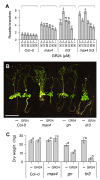
Figure 3. Combinatorial effect of GR24 and tir3 on shoot branching and growth.
(A) The number of rosette branches of wild-type, max4, tir3, and max4 tir3 Arabidopsis plants grown for 8 wk in glass jars on agar medium supplemented with the indicated concentrations of GR24. (B) Images and (C) dry weights of wild-type, max4, gn, and tir3 Arabidopsis plants grown for 8 wk in glass jars on agar medium supplemented with the vehicle control or 5 µM GR24. In (A), means ± s.e.m. of 18 plants are shown; samples in each genotype were compared by Steel–Dwass test. In (B), scale bar: 5 cm. In (C), means ± s.e.m. of 15 plants are shown; samples were compared by Tukey's test.
Figure 4
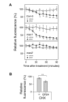
Figure 4. Effect of GR24 on PIN1 protein dynamics in inflorescence stems.
(A) Real-time monitoring of GR24-induced PIN1 depletion from the basal PM in wild-type, max1, or max2. (B) Effect of the protein synthesis inhibitor cycloheximide (CHX) on GR24-induced PIN1 instability in wild-type. The GFP signal in longitudinal sections from inflorescence stems of 6-wk-old soil-grown plants harbouring PIN1:PIN1–GFP in either the wild-type, max1, or max2 genetic background was monitored. Sections were mounted with the vehicle control or 5 µM GR24. For (B), sections were pretreated with 50 µM CHX for 30 min before addition of the vehicle control or 5 µM GR24 for 60 min. Means ± s.e.m. of the basal PM region of 7–9 cells are shown as a percentage of the value just after mounting. The vehicle control versus GR24-treated samples were compared by one-tailed Student's test. Results presented are typical of three independent experiments.
Figure 5
Figure 5. Characterisation of strigolactone-induced PIN1 protein instability in inflorescence stems.
(A) Effect of the vesicle trafficking inhibitor brefeldin A (BFA) on PIN1 protein. (B) Lack of recovery of PIN1 signal after photobleaching. (C) Effect of the clathrin-dependent endocytosis inhibitor A23 on GR24-induced instability of PIN1 protein. (D) Effect of GR24 on stability of a PM-localised protein other than PIN1. In (A–C), the GFP signal in longitudinal sections from inflorescence stems of 6-wk-old soil-grown plants harbouring PIN1:PIN1–GFP was monitored up to 180 min after mounting with 50 µM BFA for (A), before and up to 180 min after photobleaching for (B), or up to 90 min after mounting prior to 30-min pretreatment of 50 µM A23 or its inactive analogue A51 before addition of the vehicle control or 5 µM GR24 (indicated by the arrow) for (C); means ± s.e.m. of the basal PM region of 5–9 cells are shown as a percentage to the value just after mounting; comparison was performed between samples just after mounting and either 60, 120, or 180 min after mounting by one-tailed paired t test for (A), between samples 10 min and 90 min after photobleaching by one-tailed paired t test for (B), or between the vehicle control and GR24-treated samples by one-tailed Student's test at each time point for (C). In micrographs of (A) and (B), green shows the PIN1–GFP signal, and magenta shows autofluorescence of chloroplasts; scale bar: 20 µm; an arrowhead indicates PIN1-rich compartment in (A), or a bleached region of the basal PM in (B). In (D) the YFP signal in longitudinal sections from inflorescence stems of 6-wk-old soil-grown plants harbouring UBQ10:PIP1–YFP, which encodes a fluorescence-tagged aquaporin protein, was monitored up to 90 min after mounting with the vehicle control or 5 µM GR24; means ± s.e.m. of the basal PM region of seven cells are shown as a percentage of the value just after mounting; the vehicle control versus GR24-treated samples were compared by Student's test at each time point. Results presented are typical of at least two independent experiments.
Figure 6
Figure 6. Effects of GR24 on development and PIN1 localisation in roots.
(A) Dose–response of gravitropic root growth to GR24 in wild-type seedlings. (B) Gravitropic root growth in wild-type and max2 seedlings treated with the vehicle control or 100 µM GR24. (C) Dose–response of root growth to GR24 in wild-type and max2 seedlings. (D) Root growth inhibition by 0.1 µM 2,4-D and 5 µM GR24 in seedlings of various genotypes. (E, G) Micrographs and (F, H) their quantitative analysis of longitudinal optical sections from the primary root. In (A–D), all seedlings were grown for 3 d on hormone-free medium and were preincubated for 24 h on medium containing the vehicle control or relevant hormone before the observation of root growth; means ± s.e.m. of 8–12 seedlings are shown. In (A), the vehicle control versus GR24-treated samples were compared by Shirley–Williams test. In (B), the vehicle control versus GR24-treated samples were compared by Wilcoxon's test in each genotype. In (C), wild-type versus max2 samples were compared by Student's test at each concentration of GR24; the significant effect of GR24 at 1 µM or higher in both wild-type and max2 was detected by Williams test at p<0.01 (not shown in the graph). In (D), the percentage of each vehicle control-treated sample is shown; wild-type versus mutant samples were compared by Dunnett's test. In (E–H), 4-d-old Arabidopsis seedlings harbouring PIN1:PIN1–GFP in either the wild-type, gn, or tir3 genetic backgrounds were treated with the vehicle control or 10 µM GR24 for 12 h (E, F) or 48 h (G, H). In (E) and (G), green colour shows the PIN1–GFP signal, and magenta colour shows cell wall counterstained with propidium iodide; scale bar: 20 µm. In (F) and (H), average intensity of the PIN1–GFP signal in the stele region was measured for each seedling; means ± s.e.m. of three seedlings are shown; in each genotype, the vehicle control versus GR24-treated samples were compared by Student's test. In (F), for each treatment, there was no significant difference (p>0.05) between wild-type and either gn or tir3 samples compared by Dunnett's test (not shown in the graph).
Figure 7
Figure 7. Schematic representation of PIN1 protein regulation by strigolactone and its effect on bud activity.
Strigolactone, signalling via MAX2, depletes PIN1 from the PM of cells in the shoot, for example by promoting clathrin-mediated endocytosis. Strigolactone acts systemically, influencing PM PIN1 levels throughout the shoot. In the main stem, PIN1 on the PM is at steady state. In an activating bud, canalization is underway, with rapid PIN1 insertion, outstripping PIN1 removal. In an inhibited bud, PIN1 insertion is slower than PIN1 depletion, such that PIN1 does not accumulate on the PM ,. Systemically higher strigolactone levels will reduce the number of active buds and the steady-state levels of PM PIN1. Systemically lower strigolactone will have the opposite effect.

Comment in

  • Transforming a stem into a bush.
    Coombs A. Coombs A. PLoS Biol. 2013;11(1):e1001476. doi: 10.1371/journal.pbio.1001476. Epub 2013 Jan 29. PLoS Biol. 2013. PMID: 23382653 Free PMC article. No abstract available.

Similar articles

Cited by

References

    1. Leyser O (2009) The control of shoot branching: an example of plant information processing. Plant Cell Environ 32: 694–703. - PubMed
    1. Petrášek J, Mravec J, Bouchard R, Blakeslee JJ, Abas M, et al. (2006) PIN proteins perform a rate-limiting function in cellular auxin efflux. Science 312: 914–918. - PubMed
    1. Wiśniewska J, Xu J, Seifertová D, Brewer PB, Růžička K, et al. (2006) Polar PIN localization directs auxin flow in plants. Science 312: 883. - PubMed
    1. Okada K, Ueda J, Komaki MK, Bell CJ, Shimura Y (1991) Requirement of the auxin polar transport system in early stages of Arabidopsis floral bud formation. Plant Cell 3: 677–684. - PMC - PubMed
    1. Gälweiler L, Guan C, Müller A, Wisman E, Mendgen K, et al. (1998) Regulation of polar auxin transport by AtPIN1 in Arabidopsis vascular tissue. Science 282: 2226–2230. - PubMed

Publication types

MeSH terms

LinkOut - more resources