Comment
. 2013 May;3(5):520-33.
doi: 10.1158/2159-8290.CD-12-0531. Epub 2013 Jan 29.

Relief of feedback inhibition of HER3 transcription by RAF and MEK inhibitors attenuates their antitumor effects in BRAF-mutant thyroid carcinomas

Affiliations
Comment

Relief of feedback inhibition of HER3 transcription by RAF and MEK inhibitors attenuates their antitumor effects in BRAF-mutant thyroid carcinomas

Cristina Montero-Conde et al. Cancer Discov. 2013 May.

Abstract

The RAF inhibitor vemurafenib (PLX4032) increases survival in patients with BRAF-mutant metastatic melanoma, but has limited efficacy in patients with colorectal cancers. Thyroid cancer cells are also comparatively refractory to RAF inhibitors. In contrast to melanomas, inhibition of mitogen-activated protein kinase (MAPK) signaling by PLX4032 is transient in thyroid and colorectal cancer cells. The rebound in extracellular signal-regulated kinase (ERK) in thyroid cells is accompanied by increased HER3 signaling caused by induction of ERBB3 (HER3) transcription through decreased promoter occupancy by the transcriptional repressors C-terminal binding protein 1 and 2 and by autocrine secretion of neuregulin-1 (NRG1). The HER kinase inhibitor lapatinib prevents MAPK rebound and sensitizes BRAF-mutant thyroid cancer cells to RAF or MAP-ERK kinase inhibitors. This provides a rationale for combining ERK pathway antagonists with inhibitors of feedback-reactivated HER signaling in this disease. The determinants of primary resistance to MAPK inhibitors vary between cancer types, due to preferential upregulation of specific receptor tyrosine kinases, and the abundance of their respective ligands.


Conflict of interest statement

Conflict of interest: JAF is a consultant for Novo Nordisk and receives grant support from AstraZeneca. NR is on the Scientific Advisory Board for Astrazeneca and Chugai, is a consultant for Novartis and receives laboratory funding from Astrazeneca and Chugai.

Figures

Figure 1
Figure 1
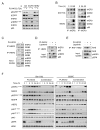
Differential effects of the RAF inhibitor PLX4032 in BRAF mutant melanoma, thyroid and colorectal cancer cell lines. A, bars represent IC50 values for PLX4032 in 15 BRAF (+) cell lines. The majority of thyroid and colorectal cell lines were comparatively refractory to PLX4032, whereas melanoma lines were uniformly sensitive. A set of BRAF wild type thyroid cancer cell lines were resistant to growth inhibition by the compound. B, western blots of cell lines from (A) treated with 2 μM PLX4032 immunoblotted for pMEK1/2 (Ser217/221) and pERK1/2 (Thr202/Tyr204). Melanoma cell lines had a sustained inhibition of pERK1/2 after exposure to the compound, whereas most thyroid and colorectal cancer cell lines had a rebound of pERK1/2 as early as 6 h post-treatment. As expected, BRAF wild type thyroid cancer cell lines were resistant to MAPK pathway inhibition by PLX4032. C, SW1736 and 8505C were treated as in (B) and collected at the indicated times. 72 h post-treatment cells were re-treated with fresh PLX4032 for 1 h (lane 72 + 1). Immunoblots of pMEK1/2 and pERK1/2 demonstrated that re-addition of PLX4032 did not fully re-inhibit MAPK signaling. D, SW1736 and SK-MEL-28 cells were treated as in (B) and collected at 0, 24 and 48 h post-treatment. Lysates were immunoprecipitated with the RAS binding domain of CRAF (RBD) and immunoblotted for pan-RAS. Activation of RAS increased at 24 and 48 h of treatment in SW1736 cells.
Figure 2
Figure 2
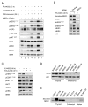
Phospho-ERK inhibition promotes expression and activation of RTKs in BRAF mutant thyroid cancer cells. A, SW1736 cells were left untreated or exposed for 72 h to 2 μM PLX4032 and lysates incubated with phospho-RTK arrays. Spots are in duplicate, with each pair corresponding to a specific pRTK. The pair spots in the corners are positive controls. Comparison between treated and untreated cells demonstrates increased phosphorylation of several RTKs by PLX4032, with pHER3 being the most prominently induced. Normalized data from densitometry analysis of the arrays are listed in the table. B, western blots of SW1736 cells treated with 2 μM PLX4032 and collected at the indicated times. Rebound in phospho-ERK and pAKT is associated with induction of total and pHER3, and total HER2. C, a panel of 6 thyroid cancer, 3 melanoma and 4 colorectal cell lines with BRAFV600E mutation were treated with or without PLX4032 for 72 h. Immunoblots show an increase of pHER3 in 5/6 thyroid cancer cell lines (SW1736, Hth104, 8505C, BCPAP and T235, see boxes). By contrast, EGFR phosphorylation was lower in 4/6 thyroid cell lines, and unchanged in the others. No comparable induction of pHER3 was observed in melanoma or colorectal cell lines. Lysates of SW1736 were used as an inter-blot control (*). D, western immunoblots of thyroid cancer tissue lysates of TPO-Cre/LSL-BrafV600E mice treated with a single 25 mg/kg dose of the MEK inhibitor PD0325901 for 6 h. Each lane corresponds to lysates from one mouse thyroid cancer tissue.
Figure 3
Figure 3
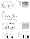
PLX4032 induces HER2/HER3 heterodimers, recruitment of p85 and activation of RAS. A, 8505C cells were transfected with siRNAs against EGFR or HER2, or with a scrambled siRNA control. After 16 h cells were treated with 2 μM PLX4032 for 72 h. Western blotting with the indicated antibodies shows that knockdown of HER2, but not EGFR, reduces PLX4032-induced HER3 phosphorylation. As HER4 was not expressed, RNAi experiments for this RTK were not done. B, SW1736 and 8505C cells were treated with 2 μM PLX4032 and collected at 24 and 48 h post-treatment. Lysates were immunoprecipitated with either anti-HER2 or anti-HER3 antibodies and Western blotted for HER3, HER2 or p85. C, 8505C cells were treated with 2 μM PLX4032 and collected 72 h post-treatment. Lysates were immunoprecipitated with either anti-HER3 or anti-HER2 antibodies and Western blotted for HER3, HER2, pHER2 or GRB2. PLX4032-treated cells show increased association of GRB2 to the HER2-HER3 immunocomplex. D, 8505C cells were treated with PLX4032 for 22 h and then treated with or without 1 μM lapatinib for 2 h. Lysates were IP with antiHER3 and blotted with the indicated antibodies. E, 8505C cells were treated with PLX4032 for 70 h and then incubated with or without 1 μM lapatinib for 2 h. Lysates were immunoprecipitated with RAF-RBD and blotted with the indicated antibodies. PLX4032 treatment induced RAS activation that is blocked by lapatinib. Immunoblotting of the input lysates demonstrates inhibition of pHER3 and pERK1/2 by lapatinib. F, western blot of lysates of SW1736 and 8505C cells treated with 2 μM PLX4032 with or without 1 μM lapatinib for the indicated times. Lapatinib blocked the PLX4032-induced HER3 and AKT phosphorylation and the pERK1/2 rebound. EGFR phosphorylation, which was not induced by vemurafenib, was also blocked by lapatinib.
Figure 4
Figure 4
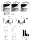
PLX4032-induced HER2/HER3 activation is dependent on autocrine secretion of Neuregulin-1. A, 8505C cells were grown in 10% FBS for 72 h, or for 48 h followed by 24 h in serum free medium, and treated with or without 2 μM PLX4032 for 72 h. Cells in lane 7 were also incubated with 1 μM lapatinib for 24 h. Starved cells were stimulated with the HER3 ligand neuregulin-1 for 5 min. B, western blot of lysates or concentrated media of 8505C cells incubated with control or NRG1 siRNA for 60 h and with 2 μM PLX4032 for the final 48 h. Knockdown of NRG1 inhibited the PLX4032-induced HER3 activation. C, effects of short term incubation with pertuzumab on PLX4032-induced signaling in 8505C. Addition of 10 μg/ml pertuzumab for 2 h decreased pHER3, pMEK and pERK, with more subtle effects on pT308AKT. Similar findings were seen in SW1736 and Hth104 cells. D, endogenous expression of NRG1 in protein lysates of the indicated thyroid cancer and melanoma cell lines. E, western immunoblotting for NRG1 of concentrated serum-free media conditioned by the indicated cell lines.
Figure 5
Figure 5
CtBPs modulate PLX4032 induction of HER3 gene transcription. A, a panel of BRAF mutant thyroid cells was treated with 2 μM PLX4032 for 1, 6 or 48 h and cell lysates analyzed for expression of HER3 and HER2 by Q-RT-PCR. Points represent fold-change of HER3/GAPDH Q-RT-PCR values of triplicate assays (mean +/− SD) over untreated controls. B, 8505C cells were treated with increasing concentrations of AZD6244. Lysates were extracted at 24 h post-treatment and immunoblotted with the indicated antibodies. C, luciferase assays of 8505C cells transfected with plasmids containing HER3 promoter-reporter constructs (−992/+63; −730/+63; −401/+63 or −42/+63, relative to transcriptional start site) and pRenilla-CMV, used as transfection normalizing control plasmid. Twelve hours post-transfection, complete media containing 2 μM PLX4032 or 0.5 μM AZD6244 was added to cells. Lysates were obtained at different time points post-treatment (0, 6, 24 and 48 h), and luciferase activity measured. Promoter activity was determined as the ratio between luciferase and renilla, relative to untreated cells. The results shown are the mean +/− SD of triplicate samples. * p< 0.05, **p < 0.01, ***p < 0.001. D, 8505C cells were transfected with control, CtBP1 or CtBP2 siRNAs and treated with or without 2 μM PLX4032 for 24 h. Lysates were analyzed for expression of HER3 and HER2 by RT-PCR. Bars represent mean +/− SD of triplicate assays of HER/GAPDH Q-RT-PCR values relative to untreated controls. E, panels show protein levels of HER3, HER2, CtBP1, CtBP2 and p85 (loading control) in cells transfected with control, CtBP1 and CtBP2 siRNAs and grown in either absence or presence of 2 μM PLX4032 for 24 h. F, CtBP1 and CtBP2 chromatin immunoprecipitation assays were performed in 8505C cells treated with or without 2 μM PLX4032 for 24 (left) or 48 h (right). A fragment (–246 to –162) of the HER3 promoter that includes known interacting sites for CtBP proteins was amplified by means of RT-PCR for both conditions. Graph shows normalized RT-PCR data of HER3 fragment of the immunoprecipitated complex compared to input lysate. Data represent mean +/− SD of two independent biological replicates performed in quadruplicate.
Figure 6
Figure 6
Lapatinib cooperates with PLX4032 to inhibit BRAF mutant thyroid cancer cell growth. A, bars represent growth inhibition of 8505C, SW1736 and Hth104 cells by lapatinib, PLX4032 or their combination. Growth was measured 4 days after addition of the indicated concentration of PLX4032 in the absence (open bars) or presence of 1 μM lapatinib (black), or after addition of the indicated concentration of lapatinib in the absence (hatched) or presence of 2 μM PLX4032 (grey). Bars represent percent change (mean +/− SD) in cell count of triplicate wells compared to untreated cells. B, SW1736 and 8505 cells were treated with 2 μM PLX4032, 0.1 μM AZD6244 or 1 μM lapatinib alone or in combination for 48 h. FACS shows a significant increase in G1 phase in cells exposed to PLX4032 or AZD6244 when combined with lapatinib. C, Thyroid volumes of TPO-Cre/LSL-BrafV600E mice treated with lapatinib (150 mg/kg five times a week), PLX4720-impregnated chow, or their combination at days 0 and 21 of treatment. The fold-change of thyroid volume was significantly lower in mice treated with the combination compared to vehicle (p= 0.02), and PLX4720 alone (p= 0.02). D, Bars represent average number of Ki67 positive cells per high power field in mice treated as described above for two weeks, except that lapatinib was given three times a week alone or in combination with PLX4720. At least 8 higher powered fields/section on 4 separate sections were counted for each mouse (2–3 mice per treatment group). **p < 0.01.
Figure 7
Figure 7
Model of HER2/HER3-induced primary resistance to MAPK inhibitors in BRAF mutant thyroid cancer cells. RAF or MEK inhibitors release transcription repressor CTBP protein/s from the HER3 promoter and induce HER3 gene expression. Autocrine-secreted NRG-1 binds to HER3, triggers HER3/HER2 heterodimerization and receptor phosphorylation, inducing PI3K and reactivating MAPK signaling, thus promoting resistance to growth inhibition.

Comment on

Similar articles

Cited by

References

    1. Davies H, Bignell GR, Cox C, Stephens P, Edkins S, Clegg S, et al. Mutations of the BRAF gene in human cancer. Nature. 2002;417:949–54. - PubMed
    1. Kimura ET, Nikiforova MN, Zhu Z, Knauf JA, Nikiforov YE, Fagin JA. High prevalence of BRAF mutations in thyroid cancer: genetic evidence for constitutive activation of the RET/PTC-RAS-BRAF signaling pathway in papillary thyroid carcinoma. Cancer Res. 2003;63:1454–7. - PubMed
    1. Xing M. BRAF mutation in papillary thyroid cancer: pathogenic role, molecular bases, and clinical implications. Endocr Rev. 2007;28:742–62. - PubMed
    1. Paik PK, Arcila ME, Fara M, Sima CS, Miller VA, Kris MG, et al. Clinical characteristics of patients with lung adenocarcinomas harboring BRAF mutations. J Clin Oncol. 2011;29:2046–51. - PMC - PubMed
    1. Yang H, Higgins B, Kolinsky K, Packman K, Go Z, Iyer R, et al. RG7204 (PLX4032), a selective BRAFV600E inhibitor, displays potent antitumor activity in preclinical melanoma models. Cancer Res. 2010;70:5518–27. - PubMed

Publication types

MeSH terms