. 2013 Mar;24(6):858-69.
doi: 10.1091/mbc.E12-10-0747. Epub 2013 Jan 30.

Regulation of apoptosis by Bcl-2 cysteine oxidation in human lung epithelial cells

Affiliations

Regulation of apoptosis by Bcl-2 cysteine oxidation in human lung epithelial cells

Sudjit Luanpitpong et al. Mol Biol Cell. 2013 Mar.

Abstract

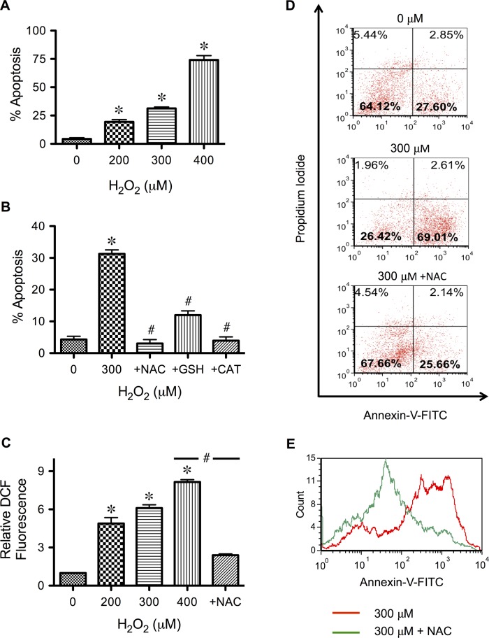
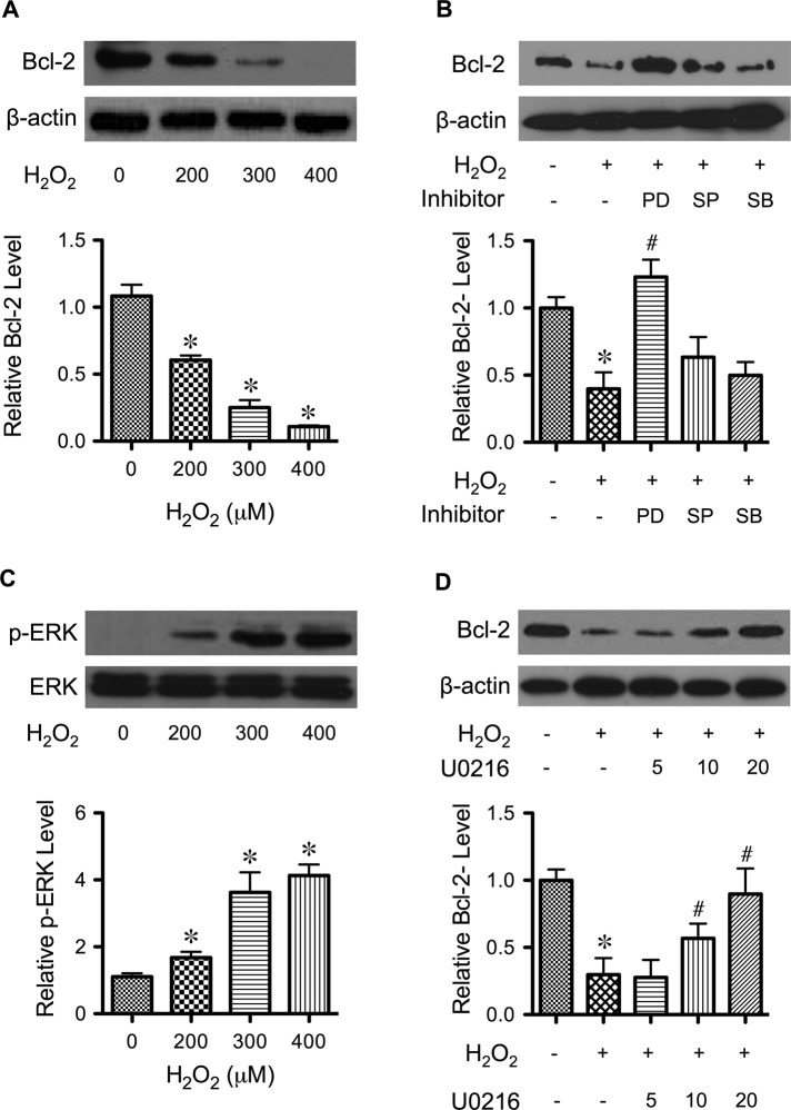
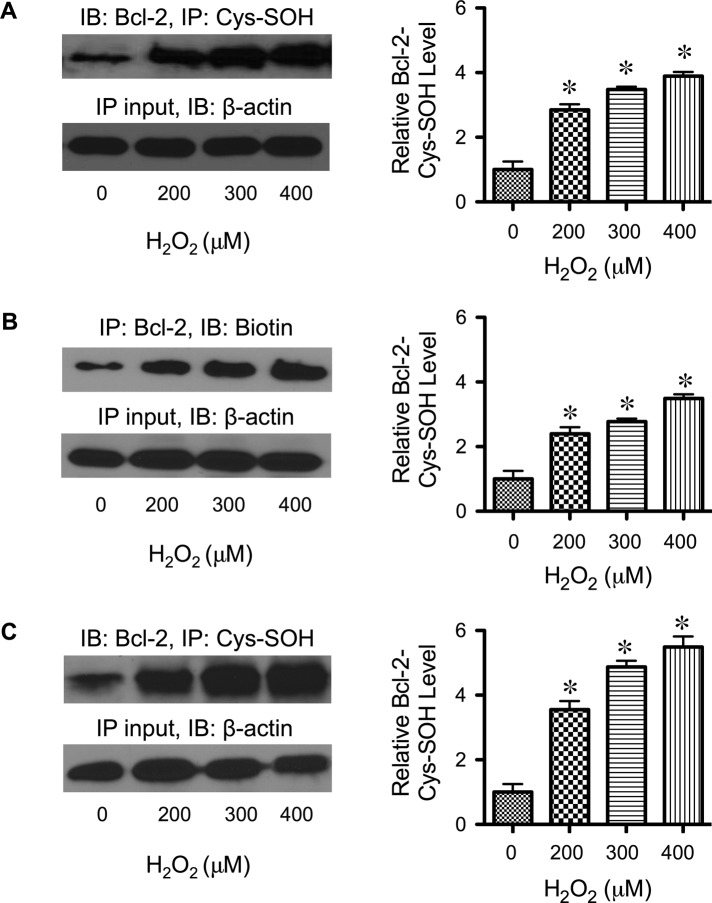
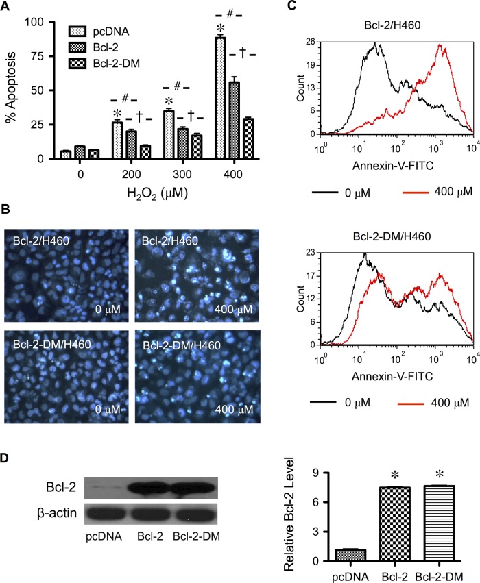
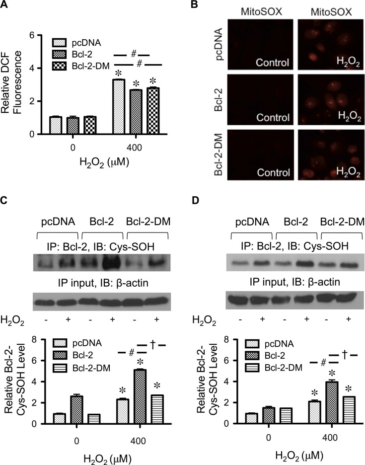
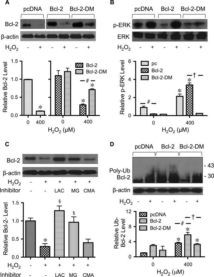
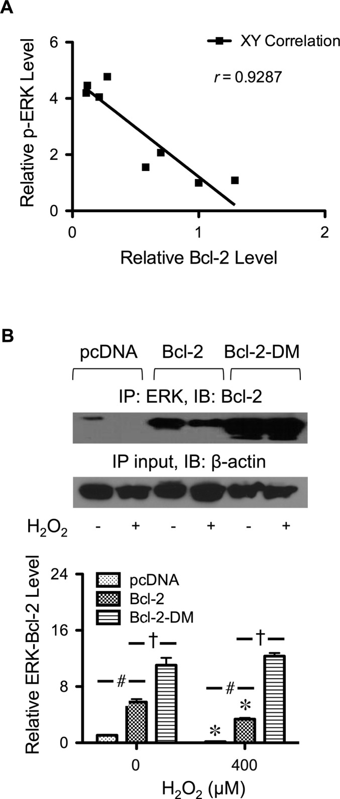
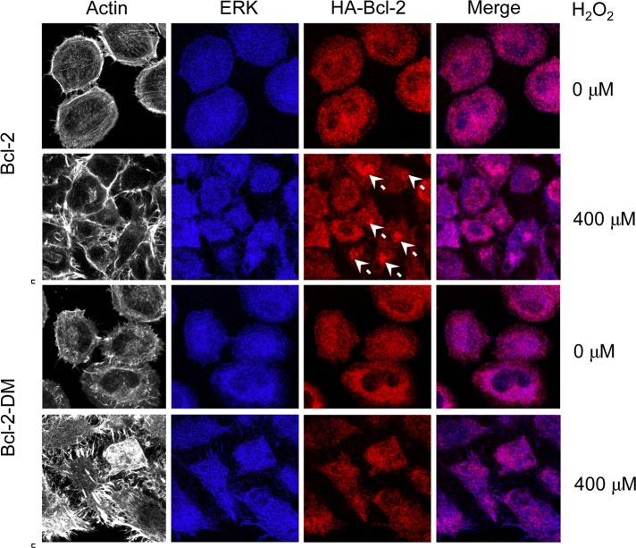
Hydrogen peroxide is a key mediator of oxidative stress known to be important in various cellular processes, including apoptosis. B-cell lymphoma-2 (Bcl-2) is an oxidative stress-responsive protein and a key regulator of apoptosis; however, the underlying mechanisms of oxidative regulation of Bcl-2 are not well understood. The present study investigates the direct effect of H2O2 on Bcl-2 cysteine oxidation as a potential mechanism of apoptosis regulation. Exposure of human lung epithelial cells to H2O2 induces apoptosis concomitant with cysteine oxidation and down-regulation of Bcl-2. Inhibition of Bcl-2 oxidation by antioxidants or by site-directed mutagenesis of Bcl-2 at Cys-158 and Cys-229 abrogates the effects of H2O2 on Bcl-2 and apoptosis. Immunoprecipitation and confocal microscopic studies show that Bcl-2 interacts with mitogen-activated protein kinase (extracellular signal-regulated kinase 1/2 [ERK1/2]) to suppress apoptosis and that this interaction is modulated by cysteine oxidation of Bcl-2. The H2O2-induced Bcl-2 cysteine oxidation interferes with Bcl-2 and ERK1/2 interaction. Mutation of the cysteine residues inhibits the disruption of Bcl-2-ERK complex, as well as the induction of apoptosis by H2O2. Taken together, these results demonstrate the critical role of Bcl-2 cysteine oxidation in the regulation of apoptosis through ERK signaling. This new finding reveals crucial redox regulatory mechanisms that control the antiapoptotic function of Bcl-2.


Figures

FIGURE 1:
FIGURE 1:
Hydrogen peroxide induces apoptosis of human lung epithelial H460 cells and its inhibition by antioxidants. (A) Subconfluent monolayers (80%) of H460 cells were treated with various pathophysiologically relevant concentrations of H2O2 (0–400 μM) for 24 h and analyzed for apoptosis by Hoechst 33342 assay. (B) Cells were similarly treated with H2O2 (300 μM) in the presence or absence of various antioxidants, including NAC (5 mM), GSH (5 mM), and catalase. (5000 U/ml), before analysis for apoptosis by Hoechst assay. (C) Cells were treated with various concentrations of H2O2 in the presence or absence of NAC (2.5 mM) for 2 h, and cellular ROS levels were determined fluorometrically using H2DCF-DA as a fluorescent probe. (D) Cells were treated with H2O2 (300 μM) in the presence or absence of NAC for 24 h and analyzed for apoptosis and necrosis by flow cytometry using annexin V and PI as probes. Histograms show raw data before correcting for background fluorescence, which accounts for ∼21.8% of the signal in the lower right quadrant. The results indicate apoptosis as the primary mode of H2O2-induced cell death. (E) Representative flow cytometric histograms of annexin V comparing cells treated with H2O2 alone and cells treated with H2O2 and NAC (400 μM vs. 400 μM + NAC). A shift to the left indicates the inhibitory effect of NAC on H2O2-induced apoptosis. Plots are mean ± SD (n = 3). *p < 0.05 vs. nontreated control. #p < 0.05 vs. H2O2-treated control (300 μM).
FIGURE 2:
FIGURE 2:
Effects of H2O2 on Bcl-2 expression and ERK activation. (A) H460 cells were treated with various concentrations of H2O2 (0–400 μM) for 24 h. Cell lysates were prepared and analyzed for Bcl-2 expression by Western blotting. Blots were reprobed with β-actin antibody to confirm equal loading of the samples. Immunoblot signals were quantified by densitometry, and mean data from three independent experiments (one of which is shown here) were normalized to the result obtained in cells without treatment (control). (B) Cells were treated with H2O2 (300 μM) for 12 h in the presence of PD98059 (ERK1/2 inhibitor, 25 μM), SP600125 (JNK inhibitor, 10 μM), and SB203580 (p38 inhibitor, 10 μM), and Bcl-2 expression was determined. (C) Cells were treated with H2O2 (0–400 μM) for 12 h and analyzed for ERK1/2 activation (phosphorylation) by Western blotting. Blots were reprobed for total ERK1/2 to confirm equal loading of the samples. (D) Cells were treated with H2O2 (300 μM) for 24 h in the presence of increasing concentrations of U0216 (0–20 μM), and Bcl-2 expression levels were measured as described. β-Actin was used as a loading control. Plots are mean ± SD (n = 3). *p < 0.05 vs. nontreated control. #p < 0.05 vs. H2O2-treated control (300 μM).
FIGURE 3:
FIGURE 3:
Effects of H2O2 on Bcl-2 Cys-SOH formation. (A) H460 cells were treated with H2O2 (0–400 μM) for 3 h, and cell lysates were prepared in the presence of dimedone (0.1 mM) and immunoprecipitated with anti–Bcl-2 antibody. The immune complexes were analyzed for cysteine sulfenic acid formation by Western blotting using anti–Cys-SOH antibody. (B) H460 cells were treated with H2O2 (0–400 μM) for 3 h, and cells were labeled with DCP-Bio1 (1 mM) and immunoprecipitated with anti–Bcl-2 antibody. The immune complexes were analyzed for cysteine sulfenic acid formation by Western blotting using anti-biotin antibody. (C) Analysis of Bcl-2 Cys-SOH formation was repeated in lung epithelial BEAS-2B cells using Cys-SOH antibody–based assay. Cells were treated with H2O2 (0–400 μM) for 3 h, and cell lysates were prepared in the presence of dimedone (0.1 mM) and immunoprecipitated with anti–Bcl-2 antibody. Cysteine sulfenic acid formation was analyzed by Western blotting using anti–Cys-SOH antibody (as described in A). Plots are mean ± SD (n = 3). *p < 0.05 vs. nontreated control.
FIGURE 4:
FIGURE 4:
Mutation of Bcl-2 cysteine residues suppresses H2O2-induced apoptosis. (A) Cells were transfected with mutant Bcl-2-DM, wild-type Bcl-2, or pcDNA3 control plasmid, as described in Materials and Methods. Transfected cells were treated with H2O2 (400 μM) for 24 h, and apoptosis was determined by Hoechst assay. (B) Fluorescence micrographs of treated cells stained with the Hoechst dye. Apoptotic cells exhibited condensed and/or fragmented nuclei. (C) Representative flow cytometric histograms of annexin V, comparing control and H2O2-treated cells (0 vs. 400 μM) in wild-type Bcl-2– and mutant Bcl-2-DM–expressing cells (top and bottom, respectively). (D) Untreated transfected cells were analyzed for Bcl-2 expression by Western blotting to confirm the overexpression. β-Actin was used as a loading control. Plots are mean ± SD (n = 3). *p < 0.05 vs. nontreated control. #p < 0.05 vs. H2O2-treated, vector-transfected control. p < 0.05 vs. H2O2-treated, Bcl-2-transfected control.
FIGURE 5:
FIGURE 5:
Effect of H2O2 treatment cellular ROS, mitochondrial superoxide, and Cys-SOH formation. (A) Cells were transfected with mutant Bcl-2-DM, wild-type Bcl-2, or control plasmid as described in Materials and Methods. Transfected cells were treated with H2O2 (400 μM) for 2 h, and cellular ROS levels were determined fluorometrically using H2DCF-DA as a fluorescent probe. (B) Transfected cells were treated with H2O2 (400 μM) for 1 h and analyzed for mitochondrial superoxide generation by fluorescence microscopy using MitoSOX Red as a specific probe. (C) Transfected cells were treated with H2O2 (400 μM) for 3 h, and cell lysates were prepared and immunoprecipitated with anti–Bcl-2 antibody. The immune complexes were analyzed for Cys-SOH by Western blotting. (D) Transfected cells were treated with H2O2 (400 μM) for 3 h, and cells were labeled with DCP-Bio1 (1 mM) and immunoprecipitated with anti–Bcl-2 antibody. The immune complexes were analyzed for Cys-SOH by Western blotting using anti-biotin antibody. Plots are mean ± SD (n = 3). *p < 0.05 vs. nontreated control. #p < 0.05 vs. vector-transfected control. p < 0.05 vs. Bcl-2–transfected control.
FIGURE 6:
FIGURE 6:
Effect of Bcl-2 cysteine residues on Bcl-2 and ERK expression. (A) Cells were transfected with mutant Bcl-2-DM, wild-type Bcl-2, or control plasmid as described in Materials and Methods. Transfected cells were treated with H2O2 (400 μM) for 24 h, and cell lysates were prepared and analyzed for Bcl-2 expression by Western blotting. β-Actin was used as a loading control. (B) Transfected cells were treated with H2O2 (400 μM) for 12 h and analyzed for ERK1/2 activation (phospho-ERK1/2) by Western blotting. Total ERK1/2 was used as a loading control. (C) Cells were treated with the proteasome inhibitors lactacystin (LAC; 10 μM) and MG132 (25 μM) or with the lysosomal inhibitor concanamycin A (CMA; 1 μM) for 1 h and then treated with H2O2 (400 μM) for 24 h. Bcl-2 expression was determined by Western blotting. (D) Cells were transfected with mutant Bcl-2-DM, wild-type Bcl-2, or control plasmid and then treated with H2O2 (400 μM) in the presence of lactacystin (10 μM; to prevent proteasomal degradation of Bcl-2). Cell lysates were immunoprecipitated with Bcl-2 antibody, and the immune complexes were analyzed for ubiquitin by Western blotting. Analysis of ubiquitin was performed at 6 h after treatment, in which ubiquitin was found to be maximal. Equal amounts of protein were loaded in each lane. Plots are mean ± SD (n = 3). *p < 0.05 vs. nontreated control. #p < 0.05 vs. vector-transfected control. p < 0.05 vs. Bcl-2-transfected control. §p < 0.05 vs. treated control.
FIGURE 7:
FIGURE 7:
Interaction of Bcl-2 with ERK. (A) Correlation analysis of the expression of Bcl-2 and phospho-ERK in response to H2O2 treatment. (B) Cells were transfected with mutant Bcl-2-DM, wild-type Bcl-2, or control plasmid as described in Materials and Methods. Transfected cells were treated with H2O2 (400 μM) for 6 h, and cell lysates were prepared and immunoprecipitated with anti-ERK1/2 antibody. The immunoblots were probed with anti–Bcl-2 antibody. Equal amounts of protein (60 μg) were loaded in each lane. Plots are mean ± SD (n = 3). *p < 0.05 vs. nontreated control. #p < 0.05 vs. vector-transfected control (400 μM). p < 0.05 vs. Bcl-2-transfected control.
FIGURE 8:
FIGURE 8:
Cellular localization of Bcl-2 and ERK. Cells were transfected with mutant Bcl-2-DM, wild-type Bcl-2, or control plasmid as described and seeded onto type I collagen–coated slides. The cells were treated with H2O2 (400 μM) for 6 h and were immunostained and analyzed for protein localization by confocal fluorescence microscopy. Actin was visualized by using phalloidin. Colocalization of ERK and Bcl-2 is shown in the merge display. White arrows indicate aggregations of Bcl-2.

Similar articles

Cited by

References

    1. Azad N, Iyer AK, Manosroi A, Wang L, Rojanasakul Y. Superoxide-mediated proteasomal degradation of Bcl-2 determined cell susceptibility to chromium (VI)-induced apoptosis. Carcinogenesis. 2008;29:1538–1545. - PMC - PubMed
    1. Ben-Ezra JM, Kornstein MJ, Grimes MM, Krystal G. Small cell carcinomas of the lung express the Bcl-2 protein. Am J Pathol. 1994;145:1036–1040. - PMC - PubMed
    1. Chanvorachote P, Nimmanit U, Stehlik C, Wang L, Jiang B, Ongpipatanakul B, Rojanasakul Y. Nitric oxide regulates cell sensitivity to cisplatin-induced apoptosis through S-nitrosylation and inhibition of Bcl-2 ubiquitination. Cancer Res. 2006;66:6353–6360. - PubMed
    1. Darnell GA, Antalis TM, Johnstone RW, Stringer BW, Ogbourne SM, Harrich D, Suhrbier Inhibition of retinoblastoma protein degradation by interaction with the serpin plasminogen activator inhibitor 2 via a novel consensus motif. Mol Cell Biol. 2003;23:6520–6532. - PMC - PubMed
    1. Dimmeler S, Breitschopf K, Haendeler J, Zeiher AM. Dephosphorylation targets Bcl-2 for ubiquitin-dependent degradation: a link between the apoptosome and the proteasome pathway. J Exp Med. 1999;189:1815–1822. - PMC - PubMed

Publication types

MeSH terms

LinkOut - more resources