Recessive loss of function of the neuronal ubiquitin hydrolase UCHL1 leads to early-onset progressive neurodegeneration
- PMID: 23359680
- PMCID: PMC3587195
- DOI: 10.1073/pnas.1222732110
Recessive loss of function of the neuronal ubiquitin hydrolase UCHL1 leads to early-onset progressive neurodegeneration
Abstract
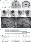
Ubiquitin C-terminal hydrolase-L1 (UCHL1), a neuron-specific de-ubiquitinating enzyme, is one of the most abundant proteins in the brain. We describe three siblings from a consanguineous union with a previously unreported early-onset progressive neurodegenerative syndrome featuring childhood onset blindness, cerebellar ataxia, nystagmus, dorsal column dysfuction, and spasticity with upper motor neuron dysfunction. Through homozygosity mapping of the affected individuals followed by whole-exome sequencing of the index case, we identified a previously undescribed homozygous missense mutation within the ubiquitin binding domain of UCHL1 (UCHL1(GLU7ALA)), shared by all affected subjects. As demonstrated by isothermal titration calorimetry, purified UCHL1(GLU7ALA), compared with WT, exhibited at least sevenfold reduced affinity for ubiquitin. In vitro, the mutation led to a near complete loss of UCHL1 hydrolase activity. The GLU7ALA variant is predicted to interfere with the substrate binding by restricting the proper positioning of the substrate for tunneling underneath the cross-over loop spanning the catalytic cleft of UCHL1. This interference with substrate binding, combined with near complete loss of hydrolase activity, resulted in a >100-fold reduction in the efficiency of UCHL1(GLU7ALA) relative to WT. These findings demonstrate a broad requirement of UCHL1 in the maintenance of the nervous system.
Conflict of interest statement
The authors declare no conflict of interest.
Figures


Similar articles
-
Novel UCHL1 mutations reveal new insights into ubiquitin processing.Hum Mol Genet. 2017 Mar 15;26(6):1031-1040. doi: 10.1093/hmg/ddw391. Hum Mol Genet. 2017. PMID: 28007905
-
Abolishing UCHL1's hydrolase activity exacerbates TBI-induced axonal injury and neuronal death in mice.Exp Neurol. 2021 Feb;336:113524. doi: 10.1016/j.expneurol.2020.113524. Epub 2020 Nov 4. Exp Neurol. 2021. PMID: 33159930 Free PMC article.
-
Phenotypic variability related to dominant UCHL1 mutations: about three families with optic atrophy and ataxia.J Neurol. 2024 Sep;271(9):6038-6044. doi: 10.1007/s00415-024-12574-z. Epub 2024 Jul 20. J Neurol. 2024. PMID: 39030458
-
Ubiquitin carboxy-terminal hydrolase L1 - physiology and pathology.Cell Biochem Funct. 2020 Jul;38(5):533-540. doi: 10.1002/cbf.3527. Epub 2020 Mar 24. Cell Biochem Funct. 2020. PMID: 32207552 Review.
-
Role of UCHL1 in the pathogenesis of neurodegenerative diseases and brain injury.Ageing Res Rev. 2023 Apr;86:101856. doi: 10.1016/j.arr.2023.101856. Epub 2023 Jan 19. Ageing Res Rev. 2023. PMID: 36681249 Free PMC article. Review.
Cited by
-
Challenges and Controversies in the Genetic Diagnosis of Hereditary Spastic Paraplegia.Curr Neurol Neurosci Rep. 2021 Feb 28;21(4):15. doi: 10.1007/s11910-021-01099-x. Curr Neurol Neurosci Rep. 2021. PMID: 33646413 Free PMC article. Review.
-
Expression analysis of the long non-coding RNA antisense to Uchl1 (AS Uchl1) during dopaminergic cells' differentiation in vitro and in neurochemical models of Parkinson's disease.Front Cell Neurosci. 2015 Apr 1;9:114. doi: 10.3389/fncel.2015.00114. eCollection 2015. Front Cell Neurosci. 2015. PMID: 25883552 Free PMC article.
-
The evolving spectrum of complex inherited neuropathies.Curr Opin Neurol. 2024 Oct 1;37(5):427-444. doi: 10.1097/WCO.0000000000001307. Epub 2024 Jul 31. Curr Opin Neurol. 2024. PMID: 39083076 Free PMC article. Review.
-
UCHL5 is a putative prognostic marker in renal cell carcinoma: a study of UCHL family.Mol Biomed. 2024 Jul 22;5(1):28. doi: 10.1186/s43556-024-00192-0. Mol Biomed. 2024. PMID: 39034372 Free PMC article.
-
Insights into Clinical, Genetic, and Pathological Aspects of Hereditary Spastic Paraplegias: A Comprehensive Overview.Front Mol Biosci. 2021 Nov 26;8:690899. doi: 10.3389/fmolb.2021.690899. eCollection 2021. Front Mol Biosci. 2021. PMID: 34901147 Free PMC article. Review.
References
Publication types
MeSH terms
Substances
Grants and funding
LinkOut - more resources
Full Text Sources
Other Literature Sources
Molecular Biology Databases
Research Materials
Miscellaneous

