

doi: 10.1038/cr.2013.17.
Epub 2013 Jan 29.
Structural insight into substrate recognition by histone demethylase LSD2/KDM1b
- PMID: 23357850
- PMCID: PMC3567815
- DOI: 10.1038/cr.2013.17
Item in Clipboard
Structural insight into substrate recognition by histone demethylase LSD2/KDM1b
Cell Res.
2013 Feb.
No abstract available
Figures
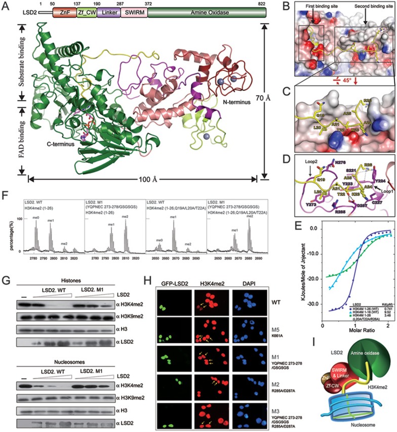
Structural insight into the substrate recognition of LSD2. (A) Overall structure of LSD2-H3K4M(1-26) is shown as a ribbon representation in two different views. The H3K4M peptide is colored in yellow. FAD is shown in stick representation (purple) and three zinc atoms are shown as grey balls. Schematic representation of the domain structure of human LSD2 with boundaries for each domain is indicated above the structure. The same color scheme is used in all structure figures of LSD2. (B) LSD2 is shown in electrostatic potential surface representation and the H3K4M peptide is shown in ribbon representation, with two substrate-binding sites highlighted by rectangles. (C) A zoom-in view of the interaction between the H3K4M peptide and the second binding site. (D) A close-up view as shown in C. Residues of H3K4M and LSD2 are labeled and colored in yellow and purple, respectively. Hydrogen bonds are shown as dashed lines. Two critical loops for the second binding site formation are indicated. (E) Superimposed ITC enthalpy plots for the binding of histone H3K4M peptides (syringe) to purified wild-type LSD2 protein (cell) with the estimated binding affinity (Kd) listed. (F)
In vitro histone demethylation assay using H3K4me2 peptides (residues 1-26, WT and Q19A/L20A/T22A) as substrates. LSD2 was used at a high protein concentration. MALDI-TOF mass spectrometry analyses of demethylation by LSD2 wild-type or mutant proteins are shown. (G)
In vitro histone demethylation assay using histones and nucleosomes as substrates, respectively. LSD2 wild-type or mutant proteins were used in three different concentrations and the reactions were monitored by immunoblotting with indicated antibodies. (H) Immunofluorescence analysis of H3K4me2 demethylase activities in 293T cells transfected with GFP fusion proteins as indicated. Green, GFP fusion protein; red, H3K4me2; blue, DAPI staining of nuclei. Transfected cells are indicated by arrows. The reduced levels of H3K4me2 were observed in cells expressing the wild-type LSD2, but not its mutants. (I) The catalytic cavity in the AO domain is the first substrate-binding site, which binds to the N-terminus of histone H3K4me2 for demethylation. The linker region forms the second binding site away from the catalytic cavity. This additional interaction is important for histone H3 recognition and essential for demethylation activity of LSD2.
Similar articles
-
Structure-function analysis reveals a novel mechanism for regulation of histone demethylase LSD2/AOF1/KDM1b.Cell Res. 2013 Feb;23(2):225-41. doi: 10.1038/cr.2012.177. Epub 2012 Dec 25. Cell Res. 2013. PMID: 23266887 Free PMC article.
-
Human LSD2/KDM1b/AOF1 regulates gene transcription by modulating intragenic H3K4me2 methylation.Mol Cell. 2010 Jul 30;39(2):222-33. doi: 10.1016/j.molcel.2010.07.008. Mol Cell. 2010. PMID: 20670891 Free PMC article.
-
LSD2 Is an Epigenetic Player in Multiple Types of Cancer and Beyond.Biomolecules. 2024 May 3;14(5):553. doi: 10.3390/biom14050553. Biomolecules. 2024. PMID: 38785960 Free PMC article. Review.
-
LSD2/KDM1B and its cofactor NPAC/GLYR1 endow a structural and molecular model for regulation of H3K4 demethylation.Mol Cell. 2013 Feb 7;49(3):558-70. doi: 10.1016/j.molcel.2012.11.019. Epub 2012 Dec 20. Mol Cell. 2013. PMID: 23260659 Free PMC article.
-
Reversal of histone methylation: biochemical and molecular mechanisms of histone demethylases.Annu Rev Biochem. 2010;79:155-79. doi: 10.1146/annurev.biochem.78.070907.103946. Annu Rev Biochem. 2010. PMID: 20373914 Review.
Cited by
-
Histone 3.3-related chromatinopathy: missense variants throughout H3-3A and H3-3B cause a range of functional consequences across species.Hum Genet. 2024 Apr;143(4):497-510. doi: 10.1007/s00439-023-02536-2. Epub 2023 Mar 3. Hum Genet. 2024. PMID: 36867246 Review.
-
Sample Volume Reduction Using the Schwarzschild Objective for a Circular Dichroism Spectrophotometer and an Application to the Structural Analysis of Lysine-36 Trimethylated Histone H3 Protein.Molecules. 2018 Nov 2;23(11):2865. doi: 10.3390/molecules23112865. Molecules. 2018. PMID: 30400257 Free PMC article.
-
Structural insight into inhibitors of flavin adenine dinucleotide-dependent lysine demethylases.Epigenetics. 2017 May 4;12(5):340-352. doi: 10.1080/15592294.2017.1290032. Epub 2017 Feb 10. Epigenetics. 2017. PMID: 28277979 Free PMC article. Review.
-
The emerging role of KDM5A in human cancer.J Hematol Oncol. 2021 Feb 17;14(1):30. doi: 10.1186/s13045-021-01041-1. J Hematol Oncol. 2021. PMID: 33596982 Free PMC article. Review.
-
The kinase NEK6 positively regulates LSD1 activity and accumulation in local chromatin sub-compartments.Commun Biol. 2024 Nov 10;7(1):1483. doi: 10.1038/s42003-024-07199-x. Commun Biol. 2024. PMID: 39523439 Free PMC article.
References
Publication types
MeSH terms
Substances
Grants and funding
LinkOut - more resources
Full Text Sources
Other Literature Sources
Molecular Biology Databases
