Diversity of ubiquitin and ISG15 specificity among nairoviruses' viral ovarian tumor domain proteases
- PMID: 23345508
- PMCID: PMC3624237
- DOI: 10.1128/JVI.03252-12
Diversity of ubiquitin and ISG15 specificity among nairoviruses' viral ovarian tumor domain proteases
Abstract
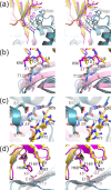
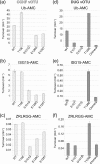
Nairoviruses are responsible for numerous diseases that affect both humans and animal. Recent work has implicated the viral ovarian tumor domain (vOTU) as a possible nairovirus virulence factor due to its ability to edit ubiquitin (Ub) bound to cellular proteins and, at least in the case of Crimean-Congo hemorrhagic fever virus (CCHFV), to cleave the Ub-like protein interferon-stimulated gene 15 (ISG15), a protein involved in the regulation of host immunity. The prospective roles of vOTUs in immune evasion have generated several questions concerning whether vOTUs act through a preserved specificity for Ub- and ISG15-conjugated proteins and where that specificity may originate. To gain insight into the substrate specificity of vOTUs, enzymological studies were conducted on vOTUs from Dugbe, CCHFV, and Erve nairoviruses. These studies revealed that vOTUs originating from different nairoviruses display a significant divergence in their preference toward Ub and ISG15. In addition, a recently identified vOTU from turnip yellow mosaic tymovirus was evaluated to elucidate any possible similarities between vOTUs originating from different viral families. Although possessing a similar preference for certain polymeric Ub moieties, its activity toward Ub in general was significantly less then those of nairoviruses. Lastly, the X-ray crystallographic structure of the vOTU from the Dugbe nairovirus was obtained in complex with Ub to reveal structural commonalities of vOTUs originating from nairoviruses. The structure suggests that divergences between nairovirus vOTUs specificity originate at the primary structural level. Comparison of this structure to that originating from CCHFV identified key residues that infer the substrate specificity of vOTUs.
Figures


Similar articles
-
Determining the molecular drivers of species-specific interferon-stimulated gene product 15 interactions with nairovirus ovarian tumor domain proteases.PLoS One. 2019 Dec 23;14(12):e0226415. doi: 10.1371/journal.pone.0226415. eCollection 2019. PLoS One. 2019. PMID: 31869347 Free PMC article.
-
Probing the impact of nairovirus genomic diversity on viral ovarian tumor domain protease (vOTU) structure and deubiquitinase activity.PLoS Pathog. 2019 Jan 10;15(1):e1007515. doi: 10.1371/journal.ppat.1007515. eCollection 2019 Jan. PLoS Pathog. 2019. PMID: 30629698 Free PMC article.
-
Biochemical and Structural Insights into the Preference of Nairoviral DeISGylases for Interferon-Stimulated Gene Product 15 Originating from Certain Species.J Virol. 2016 Aug 26;90(18):8314-27. doi: 10.1128/JVI.00975-16. Print 2016 Sep 15. J Virol. 2016. PMID: 27412597 Free PMC article.
-
Ub surprised: viral ovarian tumor domain proteases remove ubiquitin and ISG15 conjugates.Cell Host Microbe. 2007 Dec 13;2(6):367-9. doi: 10.1016/j.chom.2007.11.005. Cell Host Microbe. 2007. PMID: 18078688 Review.
-
Coronaviral PLpro proteases and the immunomodulatory roles of conjugated versus free Interferon Stimulated Gene product-15 (ISG15).Semin Cell Dev Biol. 2022 Dec;132:16-26. doi: 10.1016/j.semcdb.2022.06.005. Epub 2022 Jun 25. Semin Cell Dev Biol. 2022. PMID: 35764457 Free PMC article. Review.
Cited by
-
Role of Virally-Encoded Deubiquitinating Enzymes in Regulation of the Virus Life Cycle.Int J Mol Sci. 2021 Apr 23;22(9):4438. doi: 10.3390/ijms22094438. Int J Mol Sci. 2021. PMID: 33922750 Free PMC article. Review.
-
Fluorometric CCHFV OTU protease assay with potent inhibitors.Virus Genes. 2015 Oct;51(2):190-7. doi: 10.1007/s11262-015-1226-5. Epub 2015 Jul 9. Virus Genes. 2015. PMID: 26156848
-
Determining the molecular drivers of species-specific interferon-stimulated gene product 15 interactions with nairovirus ovarian tumor domain proteases.PLoS One. 2019 Dec 23;14(12):e0226415. doi: 10.1371/journal.pone.0226415. eCollection 2019. PLoS One. 2019. PMID: 31869347 Free PMC article.
-
Probing the impact of nairovirus genomic diversity on viral ovarian tumor domain protease (vOTU) structure and deubiquitinase activity.PLoS Pathog. 2019 Jan 10;15(1):e1007515. doi: 10.1371/journal.ppat.1007515. eCollection 2019 Jan. PLoS Pathog. 2019. PMID: 30629698 Free PMC article.
-
Turnip yellow mosaic virus protease binds ubiquitin suboptimally to fine-tune its deubiquitinase activity.J Biol Chem. 2020 Oct 2;295(40):13769-13783. doi: 10.1074/jbc.RA120.014628. Epub 2020 Jul 30. J Biol Chem. 2020. PMID: 32732284 Free PMC article.
References
-
- Mishra AC, Mehta M, Mourya DT, Gandhi S. 2011. Crimean-Congo haemorrhagic fever in India. Lancet 378:372. - PubMed
-
- Ozkaya E, Dincer E, Carhan A, Uyar Y, Ertek M, Whitehouse CA, Ozkul A. 2010. Molecular epidemiology of Crimean-Congo hemorrhagic fever virus in Turkey: occurrence of local topotype. Virus Res. 149:64–70 - PubMed
-
- World Health Organization 2001. Crimean-Congo haemorrhagic fever: fact sheet. WHO Media Centre, World Health Organization, Geneva, Switzerland
-
- World Health Organization 2003. Crimean-Congo haemorrhagic fever (CCHF) in Mauritania: update. World Health Organization, Geneva, Switzerland
Publication types
MeSH terms
Substances
Grants and funding
LinkOut - more resources
Full Text Sources
Other Literature Sources
Research Materials
Miscellaneous

