Overexpression of snail induces epithelial-mesenchymal transition and a cancer stem cell-like phenotype in human colorectal cancer cells
- PMID: 23342249
- PMCID: PMC3544430
- DOI: 10.1002/cam4.4
Overexpression of snail induces epithelial-mesenchymal transition and a cancer stem cell-like phenotype in human colorectal cancer cells
Abstract
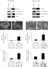
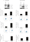
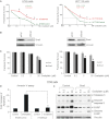
Epithelial-mesenchymal transition (EMT) is a critical process providing tumor cells with the ability to migrate and escape from the primary tumor and metastasize to distant sites. Recently, EMT was shown to be associated with the cancer stem cell (CSC) phenotype in breast cancer. Snail is a transcription factor that mediates EMT in a number of tumor types, including colorectal cancer (CRC). Our study was done to determine the role of Snail in mediating EMT and CSC function in CRC. Human CRC specimens were stained for Snail expression, and human CRC cell lines were transduced with a retroviral Snail construct or vector control. Cell proliferation and chemosensitivity to oxaliplatin of the infected cells were determined by the MTT (colorimetric 3-(4,5-dimethylthiazol-2-yl)-2,5-diphenyltetrazolium bromide) assay. Migration and invasion were determined in vitro using modified Boyden chamber assays. EMT and putative CSC markers were analyzed using Western blotting. Intravenous injection of tumor cells was done to evaluate their metastatic potential in mice. Snail was overexpressed in human CRC surgical specimens. This overexpression induced EMT and a CSC-like phenotype in human CRC cells and enhanced cell migration and invasion (P < 0.002 vs. control). Snail overexpression also led to an increase in metastasis formation in vivo (P < 0.002 vs. control). Furthermore, the Snail-overexpressing CRC cells were more chemoresistant to oxaliplatin than control cells. Increased Snail expression induces EMT and the CSC-like phenotype in CRC cells, which enhance cancer cell invasion and chemoresistance. Thus, Snail is a potential therapeutic target in metastatic CRC.
Keywords: Cancer stem cells; EMT; Snail; colorectal cancer; migration.
Figures


Similar articles
-
Twist mediates an aggressive phenotype in human colorectal cancer cells.Int J Oncol. 2016 Mar;48(3):1117-24. doi: 10.3892/ijo.2016.3342. Epub 2016 Jan 15. Int J Oncol. 2016. PMID: 26782761
-
Resveratrol suppresses epithelial-to-mesenchymal transition in colorectal cancer through TGF-β1/Smads signaling pathway mediated Snail/E-cadherin expression.BMC Cancer. 2015 Mar 5;15:97. doi: 10.1186/s12885-015-1119-y. BMC Cancer. 2015. PMID: 25884904 Free PMC article.
-
SNAIL regulates interleukin-8 expression, stem cell-like activity, and tumorigenicity of human colorectal carcinoma cells.Gastroenterology. 2011 Jul;141(1):279-91, 291.e1-5. doi: 10.1053/j.gastro.2011.04.008. Epub 2011 Apr 16. Gastroenterology. 2011. PMID: 21640118
-
Role of Epithelial to Mesenchymal Transition in Colorectal Cancer.Int J Mol Sci. 2023 Oct 1;24(19):14815. doi: 10.3390/ijms241914815. Int J Mol Sci. 2023. PMID: 37834263 Free PMC article. Review.
-
Research Progress of Epithelial-mesenchymal Transition Treatment and Drug Resistance in Colorectal Cancer.Technol Cancer Res Treat. 2022 Jan-Dec;21:15330338221081219. doi: 10.1177/15330338221081219. Technol Cancer Res Treat. 2022. PMID: 35435774 Free PMC article. Review.
Cited by
-
Molecular features and gene expression signature of metastatic colorectal cancer (Review).Oncol Rep. 2021 Apr;45(4):10. doi: 10.3892/or.2021.7961. Epub 2021 Mar 2. Oncol Rep. 2021. PMID: 33649827 Free PMC article. Review.
-
Establishment and characterization of models of chemotherapy resistance in colorectal cancer: Towards a predictive signature of chemoresistance.Mol Oncol. 2015 Jun;9(6):1169-85. doi: 10.1016/j.molonc.2015.02.008. Epub 2015 Feb 24. Mol Oncol. 2015. PMID: 25759163 Free PMC article.
-
Long Non-Coding RNAs as Potential Regulators of EMT-Related Transcription Factors in Colorectal Cancer-A Systematic Review and Bioinformatics Analysis.Cancers (Basel). 2022 May 3;14(9):2280. doi: 10.3390/cancers14092280. Cancers (Basel). 2022. PMID: 35565409 Free PMC article. Review.
-
Emerging Biological Principles of Metastasis.Cell. 2017 Feb 9;168(4):670-691. doi: 10.1016/j.cell.2016.11.037. Cell. 2017. PMID: 28187288 Free PMC article. Review.
-
Epithelial-to-mesenchymal transition in tumor progression.Med Oncol. 2017 Jul;34(7):122. doi: 10.1007/s12032-017-0980-8. Epub 2017 May 30. Med Oncol. 2017. PMID: 28560682 Review.
References
-
- Thiery JP, Sleeman JP. Complex networks orchestrate epithelial-mesenchymal transitions. Nat. Rev. Mol. Cell Biol. 2006;7:131–142. - PubMed
-
- Nieto MA. The snail superfamily of zinc-finger transcription factors. Nat. Rev. Mol. Cell Biol. 2002;3:155–166. - PubMed
-
- Yang J, Mani SA, Donaher JL, Ramaswamy S, Itzykson RA, Come C, et al. Twist, a master regulator of morphogenesis, plays an essential role in tumor metastasis. Cell. 2004;117:927–939. - PubMed
-
- Peinado H, Olmeda D, Cano A. Snail, Zeb and bHLH factors in tumour progression: an alliance against the epithelial phenotype? Nat. Rev. Cancer. 2007;7:415–428. - PubMed
Publication types
MeSH terms
Substances
Grants and funding
LinkOut - more resources
Full Text Sources
Medical
Research Materials

