. 2013 Jan 15;2(1):68-81.
doi: 10.1242/bio.20123012. Epub 2012 Oct 31.

Characterization of Fragile X Mental Retardation Protein granules formation and dynamics in Drosophila

Affiliations

Characterization of Fragile X Mental Retardation Protein granules formation and dynamics in Drosophila

Cristina Gareau et al. Biol Open. .

Abstract

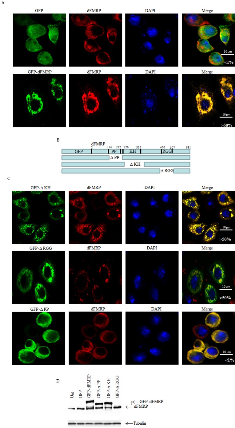
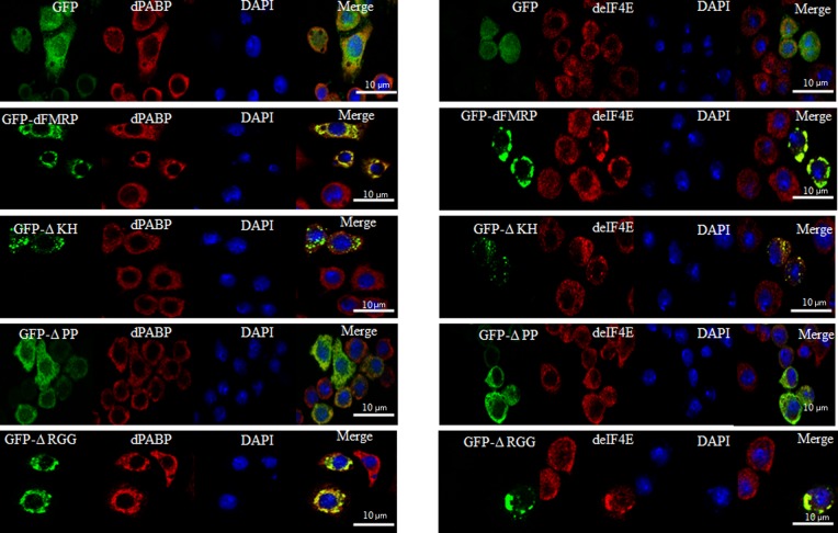
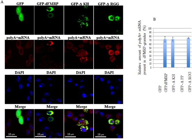
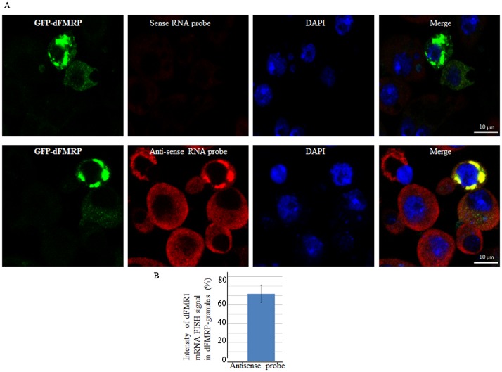
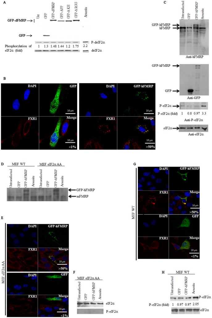
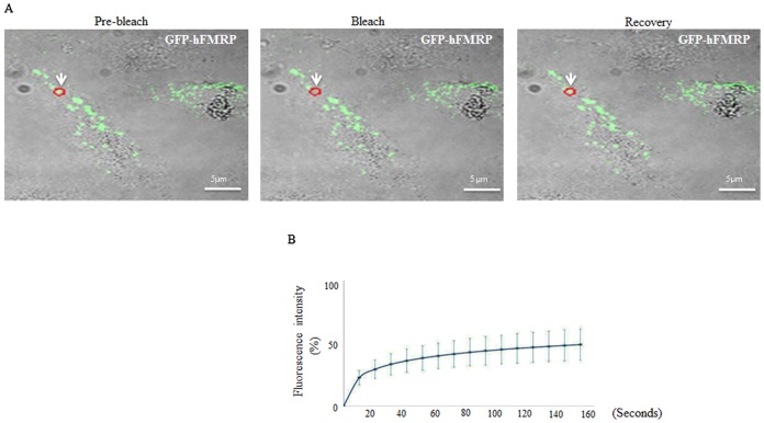
FMRP is an evolutionarily conserved protein that is highly expressed in neurons and its deficiency causes fragile X mental retardation syndrome. FMRP controls the translation of target mRNAs in part by promoting their dynamic transport in neuronal RNA granules. We have previously shown that high expression of mammalian FMRP induces formation of granules termed FMRP granules. These RNA granules are reminiscent of neuronal granules, of stress granules, as well as of the recently described in vitro-assembled granules. In contrast with mammalian FMRP, which has two paralog proteins, Drosophila FMRP (dFMRP) is encoded by a single gene that has no paralog. Using this genetically simple organism, we investigated formation and dynamics of FMRP granules. We found that increased expression of dFMRP in Drosophila cells induces the formation of dynamic dFMRP RNA granules. Mutagenesis studies identified the N-terminal protein-protein domain of dFMRP as a key determinant for FMRP granules formation. The RGG RNA binding motif of dFMRP is dispensable for dFMRP granules formation since its deletion does not prevent formation of those granules. Deletion of the RGG motif reduced, however, dFMRP trafficking between FMRP granules and the cytosol. Similarly, deletion of a large part of the KH RNA binding motif of dFMRP had no effect on formation of dFMRP-granules, but diminished the shuttling activity of dFMRP. Our results thus suggest that the mechanisms controlling formation of RNA granules and those promoting their dynamics are uncoupled. This study opens new avenues to further elucidate the molecular mechanisms controlling FMRP trafficking with its associated mRNAs in and out of RNA granules.

Keywords: FMRP; Protein translation; RNA.


Figures

Fig. 1.
Fig. 1.. Expression of dFMRP induces dFMRP granules: role of the PP domain.
(A) Schneider cells were transfected with either GFP or GFP-dFMRP constructs for 48 h. Cells were then processed for immunofluorescence to detect GFP or GFP-dFMRP (green). The intracellular localization of endogenous dFMRP (red) is revealed using specific anti-dFMRP antibodies. Scale bar: 10 µm. (B) Schematic representation of GFP-dFMRP (top) and its deletion versions. (C) Cells were transfected with GFP-dFMRP mutants and then processed for immunofluorescence as in Fig. 1A. Note that while the ΔPP mutant fails to induce dFMRP granules, both ΔKH and ΔRGG mutants induce granules that are positive for the corresponding GFP-dFMRP mutants and for endogenous dFMRP as well. All pictures were taken using a 63× objective at 1.5 zoom. The percentage of cells harboring FMRP granules from 5 different fields and 5 different experiments containing a total of 1,000 cells is indicated at the bottom of merged images. Scale bar: 10 µm. (D) Cells expressing GFP-dFMRP were collected and protein extracts were next analyzed by immunoblotting for GFP-dFMRP expression using dFMRP-specific antibodies. Tubulin was used as a loading control.
Fig. 2.
Fig. 2.. Co-localization of GFP-dFMRP mutants with the RNA granules markers deIF4E, dPABP and with poly(A)+ mRNA within dFMRP granules in Schneider cells.
Following transfection with the indicated GFP polypeptides, cells were fixed, permeabilized, and then processed for immunofluorescence to detect GFP or GFP-dFMRP (green). The intracellular localization of dPABP and deIF4E (red) is revealed using the appropriate specific antibodies. Scale bar: 10 µm.
Fig. 3.
Fig. 3.. Co-localization of GFP-dFMRP mutants with poly(A)+ mRNA within dFMRP granules in Schneider.
Following transfection with the indicated GFP polypeptides, cells were fixed, permeabilized, and then incubated with 0.2 µM of an Alexa Fluor 594-labeled oligo(dT) probe to detect poly(A)+ mRNA (red), as described in Materials and Methods. (A) dFMRP granules were visualized by confocal microscopy using anti-dFMRP antibodies (green). Scale bar: 10 µm. (B) Densitometry of FISH poly(A)+ mRNA signal with Adobe Photoshop software. The number of pixels and mean intensity were recorded for the selected regions (SG, cytoplasm and background). The mean intensity was multiplied by the number of pixels for the region selected in order to obtain the absolute intensity. The absolute intensity of the background region was subtracted from each region of interest. In order to compare the intensity of two given regions of interest, relative intensities were next calculated. The relative intensity corresponds to the absolute intensity normalized to the absolute intensity of the region of reference.
Fig. 4.
Fig. 4.. Detection of dFMR1 mRNA in dFMRP granules by FISH.
(A) Schneider cells were transfected with GFP-dFMRP. Cells were then fixed, permeabilized, and incubated with 3 nM of Alexa Fluor 488-labeled antisense RNA probe to detect dFMR1 mRNA (panels 6 and 8) or with Alexa Fluor 488-labeled sense probe (panels 2 and 4) as control. dFMRP granules were visualized as green fluorescence. Scale bar: 10 µm. (B) Densitometry quantification of FISH poly(A)+ mRNA signal was done with Photoshop software as above.
Fig. 5.
Fig. 5.. Expression of either dFMRP or human FMRP does not induce eIF2α phosphorylation.
(A) Analysis of eIF2α phosphorylation upon dFMRP expression. Following transfection with GFP-dFMRP or its mutants, Schneider cells were lysed and protein content was analyzed by immunoblotting for GFP-dFMRP expression using anti-GFP antibodies. Phosphorylation of eIF2α was analyzed by western blot (P-eIF2α; center panel) using specific antibodies. An extract isolated from arsenite-treated Schneider cells was used as a positive control for eIF2α phosphorylation. Total eIF2α was analyzed using the pan-eIF2α antibodies and used as a loading control (bottom panel). The amount of phosphorylated eIF2α was determined by quantitation of the film signals by densitometry using the Adobe Photoshop and expressed as a percentage of total eIF2α. The results are representative of 5 different experiments. (B) Expression of GFP-hFMRP in HeLa cells induces FMRP granules. Cells expressing GFP-hFMRP or GFP alone were processed for fluorescence to detect GFP or GFP-hFMRP (green). The intracellular localization of the FMRP partner FXR1 in FMRP granules (red) was revealed by immunofluorescence using specific antibodies. The indicated percentage of FMRP granules that are induced by expressing GFP-hFMRP in HeLa cells is representative of 3 different experiments. (C) Analysis of eIF2α phosphorylation upon dFMRP expression. HeLa cells expressing GFP-hFMRP or GFP alone were collected and their protein extracts prepared for western blot analysis of eIF2α phosphorylation and of eIF2α (as loading control) using specific antibodies (bottom panels). GFP-hFMRP and endogenous FMRP were detected using anti-FMRP antibodies (top panel). GFP was detected using anti-GFP antibodies, which also detect the GFP-hFMRP fusion (center panel). The indicated amount of phosphorylated eIF2α was quantified as described in Fig. 2A. (D–H) GFP-hFMRP induces FMRP granules independently of eIF2α phosphorylation. (D) MEF-eIF2α A/A and its WT counterpart cell line were transfected with either GFP or GFP-hFMRP. Cells were then collected and their protein extracts were analyzed by western blotting for the expression of GFP-hFMRP and endogenous mFMRP using anti-FMRP antibodies. (E,G) Expression of GFP-hFMRP induces FMRP granules in both MEF eIF2α A/A (E) and its WT MEF counterparts (G). Cells expressing GFP or GFP-hFMRP were processed for confocal microscopy for GFP and GFP-hFMRP detection (green). The intracellular localization of the FMRP partner FXR1 in FMRP granules (red) is revealed by immunofluorescence using specific antibodies. The indicated percentage of FMRP granules that are induced by expressing GFP-hFMRP in MEF-eIF2a A/A and their WT MEF counterparts is representative of 3 different experiments. (F,H) The indicated cells expressing GFP or GFP-hFMRP were collected and their protein extracts prepared for western blot analysis of eIF2α phosphorylation and of eIF2α as a loading control using specific antibodies. Arsenite treatment was used as a positive control for eIF2α phosphorylation. The indicated percentage of phosphorylated eIF2α was calculated as described in Fig. 5A. Scale bars in B,E,G: 10 µm.
Fig. 6.
Fig. 6.. Formation of FMRP granules involves FMRP dimerization.
(A–C) Co-immunoprecipitation of GFP-dFMRP mutant polypeptides with endogenous dFMRP. (A) Schneider cells were transfected with GFP-dFMRP or GFP-ΔPP. 48 h post transfection, cells were lysed and their extracts used to immunoprecipitate dFMRP using anti-GFP antibodies. IgG were used as a control immunoprecipitation. IP: immunoprecipitate; total represents 5% of the input used for immunoprecipitation. Immunoprecipitated proteins were analyzed by western blot for dFMRP using anti-dFMRP antibodies. The positions of GFP-dFMRP, GFP-ΔPP, and endogenous dFMRP are indicated by arrows. (B,C) Validation of GFP-dFMRP interaction with endogenous dFMRP. Schneider cells were first treated with siRNAs specific to the 3′UTR of dFMR1 mRNA and were then transfected with GFP-dFMRP 48 h later. Twenty-four h later, cells were lysed and their extracts used to immunoprecipitate dFMRP using anti-GFP antibodies. (B) Total: 5% of the input used for immunoprecipitation. Proteins were analyzed by western blot for dFMRP and GFP-dFMRP expression using anti-dFMRP antibodies Tubulin serves as a loading control. (C) IP: immunoprecipitate. Immunoprecipitated proteins were analyzed by western blot for dFMRP using anti-dFMRP antibodies. The positions of GFP-dFMRP and endogenous dFMRP are indicated by arrows. (D–G) GFP-I304N is a weak inducer of FMRP granules in STEK cells. MEF and STEK cells were transfected with either GFP-hFMRP or GFP-I304N (D,F) for 48 h. Cells were then processed for immunofluorescence to detect GFP fusion proteins (green). The intracellular localization of the FMRP partner FXR1 in FMRP granules (red) was revealed using specific antibodies. The indicated percentage of FMRP granules induced by expressing either GFP-hFMRP (D,E) or GFP-I304N (F,G) in MEFs and STEK is representative of 3 different experiments. Scale bars in D,F: 10 µm.
Fig. 7.
Fig. 7.. Analysis of dFMRP kinetics in dFMRP granules by FRAP.
Schneider cells were transfected with GFP-dFMRP and its mutant versions. (A–C) 48 h post transfection, a single dFMRP granule (red circle; indicated by arrow) was photobleached and fluorescence recovery was recorded over 140 s using a confocal microscope. The FRAP methodology is described in Materials and Methods. Scale bar: 10 µm. The images shown in (A) were selected for illustration and correspond to merged differential interference contrast (DIC) microscopy and fluorescence pictures. The recovery of dFMRP fluorescence in the photobleached area was quantified as described in Materials and Methods and plotted as a function of time as indicated in (B). Curves are representative of 3 independent experiments with a total of 100 photobleached granules for each indicated GFP fusion protein. (C) Bar graphs for the MF of each GFP fusion protein are shown with error bars corresponding to the SD of 3 independent experiments. The indicated P-values are calculated with unpaired Student's t-test (n = 3).
Fig. 8.
Fig. 8.. Dynamics of GFP-hFMRP within FMRP granules in HeLa cells by FRAP.
(A,B) Cells were transfected with GFP-hFMRP. Forty-eight h post transfection, a single FMRP-granule (red circle; indicated by arrow) was photobleached (A) and the fluorescence recovery (B) was recorded over 140 s using a confocal microscope as described in Fig. 7. Scale bar in A: 5 µm.

Similar articles

Cited by

References

    1. Anderson P., Kedersha N. (2009). RNA granules: post-transcriptional and epigenetic modulators of gene expression. Nat. Rev. Mol. Cell Biol. 10, 430–436 10.1038/nrm2694 - DOI - PubMed
    1. Ashley C. T., Sutcliffe J. S., Kunst C. B., Leiner H. A., Eichler E. E., Nelson D. L., Warren S. T. (1993). Human and murine FMR-1: alternative splicing and translational initiation downstream of the CGG-repeat. Nat. Genet. 4, 244–251 10.1038/ng0793-244 - DOI - PubMed
    1. Baguet A., Degot S., Cougot N., Bertrand E., Chenard M. P., Wendling C., Kessler P., Le Hir H., Rio M. C., Tomasetto C. (2007). The exon-junction-complex-component metastatic lymph node 51 functions in stress-granule assembly. J. Cell Sci. 120, 2774–2784 10.1242/jcs.009225 - DOI - PubMed
    1. Balagopal V., Parker R. (2009). Polysomes, P bodies and stress granules: states and fates of eukaryotic mRNAs. Curr. Opin. Cell Biol. 21, 403–408 10.1016/j.ceb.2009.03.005 - DOI - PMC - PubMed
    1. Barbee S. A., Estes P. S., Cziko A. M., Hillebrand J., Luedeman R. A., Coller J. M., Johnson N., Howlett I. C., Geng C., Ueda R.et al. (2006). Staufen- and FMRP-containing neuronal RNPs are structurally and functionally related to somatic P bodies. Neuron 52, 997–1009 10.1016/j.neuron.2006.10.028 - DOI - PMC - PubMed

LinkOut - more resources