Altering SARS coronavirus frameshift efficiency affects genomic and subgenomic RNA production
- PMID: 23334702
- PMCID: PMC3564121
- DOI: 10.3390/v5010279
Altering SARS coronavirus frameshift efficiency affects genomic and subgenomic RNA production
Abstract
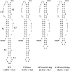
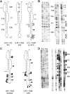
In previous studies, differences in the amount of genomic and subgenomic RNA produced by coronaviruses with mutations in the programmed ribosomal frameshift signal of ORF1a/b were observed. It was not clear if these differences were due to changes in genomic sequence, the protein sequence or the frequency of frameshifting. Here, viruses with synonymous codon changes are shown to produce different ratios of genomic and subgenomic RNA. These findings demonstrate that the protein sequence is not the primary cause of altered genomic and subgenomic RNA production. The synonymous codon changes affect both the structure of the frameshift signal and frameshifting efficiency. Small differences in frameshifting efficiency result in dramatic differences in genomic RNA production and TCID50 suggesting that the frameshifting frequency must stay above a certain threshold for optimal virus production. The data suggest that either the RNA sequence or the ratio of viral proteins resulting from different levels of frameshifting affects viral replication.
Figures


Similar articles
-
Achieving a golden mean: mechanisms by which coronaviruses ensure synthesis of the correct stoichiometric ratios of viral proteins.J Virol. 2010 May;84(9):4330-40. doi: 10.1128/JVI.02480-09. Epub 2010 Feb 17. J Virol. 2010. PMID: 20164235 Free PMC article.
-
The role of programmed-1 ribosomal frameshifting in coronavirus propagation.Front Biosci. 2008 May 1;13:4873-81. doi: 10.2741/3046. Front Biosci. 2008. PMID: 18508552 Free PMC article. Review.
-
A three-stemmed mRNA pseudoknot in the SARS coronavirus frameshift signal.PLoS Biol. 2005 Jun;3(6):e172. doi: 10.1371/journal.pbio.0030172. Epub 2005 May 17. PLoS Biol. 2005. PMID: 15884978 Free PMC article.
-
Programmed ribosomal frameshifting in HIV-1 and the SARS-CoV.Virus Res. 2006 Jul;119(1):29-42. doi: 10.1016/j.virusres.2005.10.008. Epub 2005 Nov 28. Virus Res. 2006. PMID: 16310880 Free PMC article. Review.
-
Interference of ribosomal frameshifting by antisense peptide nucleic acids suppresses SARS coronavirus replication.Antiviral Res. 2011 Jul;91(1):1-10. doi: 10.1016/j.antiviral.2011.04.009. Epub 2011 Apr 23. Antiviral Res. 2011. PMID: 21549154 Free PMC article.
Cited by
-
The molecular virology of coronaviruses.J Biol Chem. 2020 Sep 11;295(37):12910-12934. doi: 10.1074/jbc.REV120.013930. Epub 2020 Jul 13. J Biol Chem. 2020. PMID: 32661197 Free PMC article. Review.
-
Functional and structural characterization of the chikungunya virus translational recoding signals.J Biol Chem. 2018 Nov 9;293(45):17536-17545. doi: 10.1074/jbc.RA118.005606. Epub 2018 Sep 21. J Biol Chem. 2018. PMID: 30242123 Free PMC article.
-
Ribosomal frameshifting and transcriptional slippage: From genetic steganography and cryptography to adventitious use.Nucleic Acids Res. 2016 Sep 6;44(15):7007-78. doi: 10.1093/nar/gkw530. Epub 2016 Jul 19. Nucleic Acids Res. 2016. PMID: 27436286 Free PMC article. Review.
-
Anti-Frameshifting Ligand Active against SARS Coronavirus-2 Is Resistant to Natural Mutations of the Frameshift-Stimulatory Pseudoknot.J Mol Biol. 2020 Oct 2;432(21):5843-5847. doi: 10.1016/j.jmb.2020.09.006. Epub 2020 Sep 11. J Mol Biol. 2020. PMID: 32920049 Free PMC article.
-
Molecular Virology of SARS-CoV-2 and Related Coronaviruses.Microbiol Mol Biol Rev. 2022 Jun 15;86(2):e0002621. doi: 10.1128/mmbr.00026-21. Epub 2022 Mar 28. Microbiol Mol Biol Rev. 2022. PMID: 35343760 Free PMC article. Review.
References
-
- Nga P.T., Parquet M.C., Lauber C., Parida M., Nabeshima T., Yu F., Thuy N.T., Inoue S., Ito T., Okamoto K., et al. Discovery of the first insect nidovirus, a missing evolutionary link in the emergence of the largest RNA virus genomes. PLoS Pathog. 2011;7:e1002215. doi: 10.1371/journal.ppat.1002215. - DOI - PMC - PubMed
Publication types
MeSH terms
Substances
Grants and funding
LinkOut - more resources
Full Text Sources
Other Literature Sources
Miscellaneous

