. 2013 Feb 6;32(3):409-23.
doi: 10.1038/emboj.2012.356. Epub 2013 Jan 18.

A structural basis for kinetochore recruitment of the Ndc80 complex via two distinct centromere receptors

Affiliations

A structural basis for kinetochore recruitment of the Ndc80 complex via two distinct centromere receptors

Francesca Malvezzi et al. EMBO J. .

Abstract

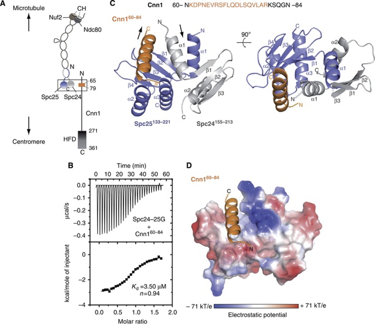
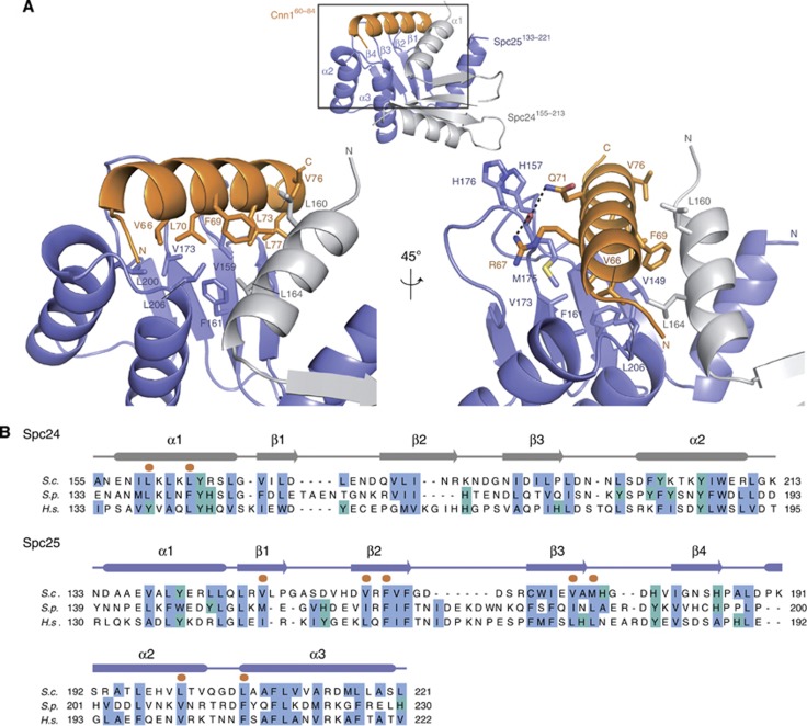
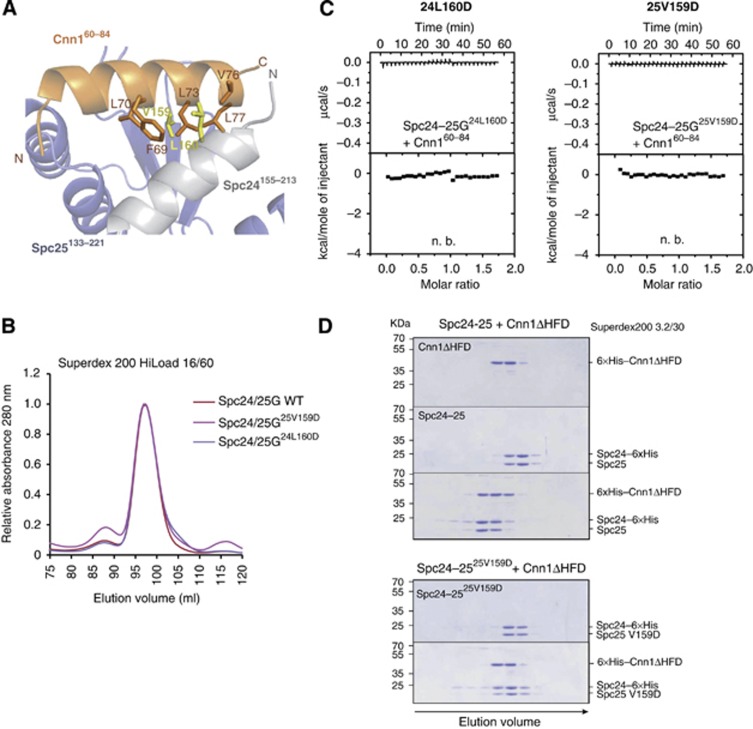
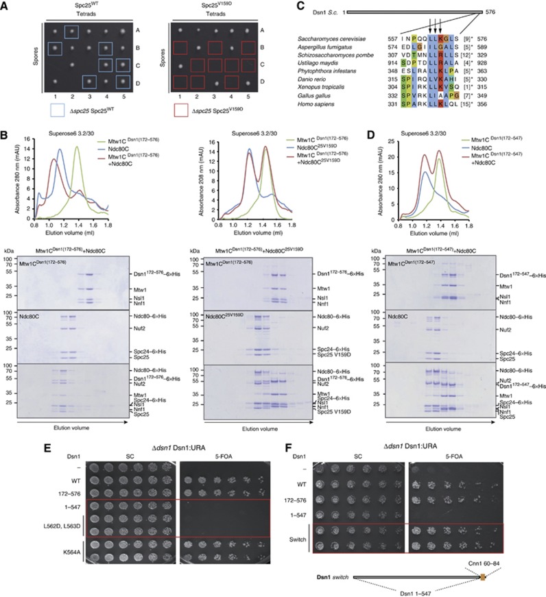
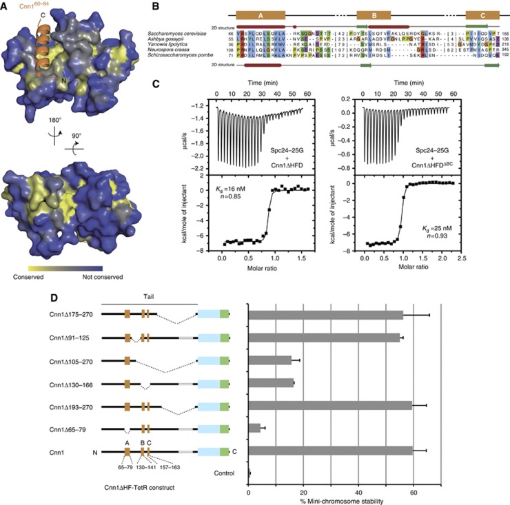
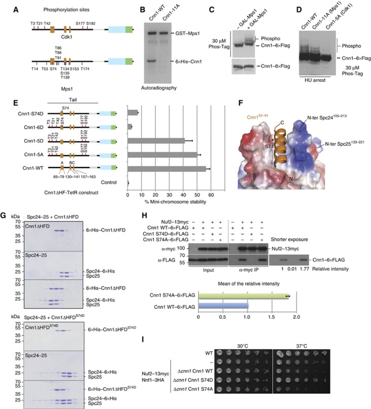
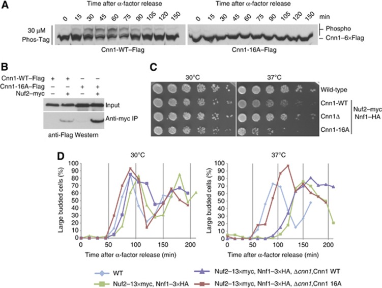
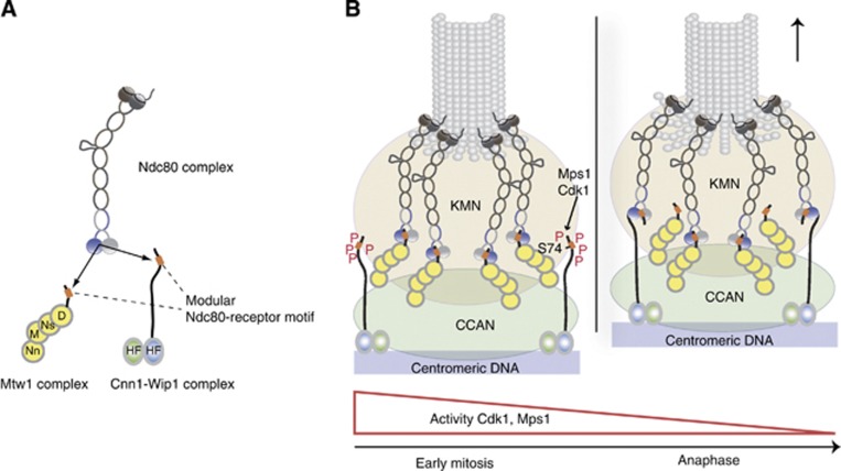
The Ndc80 complex is the key microtubule-binding element of the kinetochore. In contrast to the well-characterized interaction of Ndc80-Nuf2 heads with microtubules, little is known about how the Spc24-25 heterodimer connects to centromeric chromatin. Here, we present molecular details of Spc24-25 in complex with the histone-fold protein Cnn1/CENP-T illustrating how this connection ultimately links microtubules to chromosomes. The conserved Ndc80 receptor motif of Cnn1 is bound as an α helix in a hydrophobic cleft at the interface between Spc24 and Spc25. Point mutations that disrupt the Ndc80-Cnn1 interaction also abrogate binding to the Mtw1 complex and are lethal in yeast. We identify a Cnn1-related motif in the Dsn1 subunit of the Mtw1 complex, necessary for Ndc80 binding and essential for yeast growth. Replacing this region with the Cnn1 peptide restores viability demonstrating functionality of the Ndc80-binding module in different molecular contexts. Finally, phosphorylation of the Cnn1 N-terminus coordinates the binding of the two competing Ndc80 interaction partners. Together, our data provide structural insights into the modular binding mechanism of the Ndc80 complex to its centromere recruiters.


Conflict of interest statement

The authors declare that they have no conflict of interest.

Figures

Figure 1
Figure 1
Structure determination of the Spc24-25/Cnn1 interface. (A) Schematic representation illustrating the general centromere-microtubule orientation of the Cnn1–Ndc80 complex. Nuf2 and Ndc80 are shown in brown and dark grey, respectively, with the microtubule-binding calponin homology (CH) domains highlighted; Spc24 and Spc25 are shown in light grey and slate blue, respectively. The Cnn1 conserved N-terminal motif (65–79) is displayed in orange and its histone-fold domain (HFD) is highlighted. A rectangle indicates the interface visualized in the crystal structure. (B) Isothermal titration calorimetry performed by titrating the Spc24-25 globular domain (Spc24-25G) with a Cnn1 peptide including the Ndc80-binding motif (Cnn160–84). The Kd was estimated fitting a non-linear curve to the binding isotherms derived from the data. (C) Overall architecture of the Spc24155–213–Spc25133–221–Cnn160–84 crystal structure. Spc24, Spc25, and Cnn1 are represented according to their secondary structure and coloured in light grey, slate blue, and orange, respectively. Arrows indicate the opposite N-terminal/C-terminal orientation of Cnn1 relative to Spc24-25. A dotted line indicates the residue Lys184 of Spc24 not visible in the electron density (molecule B-A-F of the asymmetric unit). (D) Localization of Cnn160–84 in the hydrophobic pocket at the interface between Spc24155–213 and Spc25133–221, shown as electrostatic surface (transparency 20%). Cnn1 secondary structure is displayed in orange.
Figure 2
Figure 2
Details of the Spc24-25–Cnn1 interaction. (A) Close-up view of the Spc24155–213-25133–221–Cnn160–84 interaction network. The secondary structure of Spc24, Spc25, and Cnn1 is displayed in light grey, slate blue, and orange, respectively. The interacting residues are highlighted as sticks, with O in red, N in blue, and S in yellow. The established hydrogen bonds are represented as black dashed lines. (B) Sequence alignment of the Spc24 and Spc25 homologues from Saccharomyces cerevisiae (S.c.), Schizosaccharomyces pombe (S.p.), and Homo sapiens (H.s.). The conservation of non-polar residues is highlighted according to the Clustalx colouring scheme and Cnn160–84-interacting residues are marked with an orange dot. The secondary structure is derived from the X-ray coordinates of Spc24155–213–Spc25133–221–Cnn160–84 using the program STRIDE (Heinig and Frishman, 2004). See Supplementary Figure S5 for alignment of more sequences.
Figure 3
Figure 3
Validation of the Spc24-25/Cnn1 interface. (A) Role of L160 (Spc24) and V159 (Spc25) in Cnn160–84 binding. The secondary structure of Spc24, Spc25, and Cnn1 is displayed in light grey, slate blue, and orange, respectively (transparency 40%). The residues L160 (Spc24) and V159 (Spc25), mutated to aspartic acid for validating the Spc24155–213–Spc25133–221/Cnn160–84 interface, are shown as sticks and highlighted in yellow. The Cnn1 residues that establish van der Waals contacts with L160 or V159 are displayed as sticks. (B) Elution profiles of size-exclusion chromatography performed with Spc24-25 globular domain (Spc24-25G) wild-type (in red) or Cnn1-binding mutants, Spc24-25G25V159D (in violet) and Spc24-25G24L160D (in magenta). Identical elution profiles indicate a similar complex shape and integrity. (C) Isothermal titration calorimetry performed by titrating Spc24-25G25V159D or Spc24-25G24L160D with Cnn160–84 (n.b.=no binding). (D) Coomassie-stained gels of analytical size-exclusion chromatography using 12 μM Cnn1ΔHFD (1–270), 12 μM full-length Spc24-25 wild-type and their combination (upper part); 12 μM full-length Spc25-25V159D and its combination in equimolar amount with Cnn1ΔHFD (lower part). Complex formation is visualized by a shift to earlier elution volumes of both molecules.
Figure 4
Figure 4
Identification of an essential Ndc80–Mtw1 interface. (A) Asci dissection of Δspc25/Spc25 Spc25WT (left) and Δspc25/Spc25 Spc25V159D (right). The viable Δspc25 Spc25WT spores are highlighted with a light blue square. The Δspc25 Spc25V159D spores, marked with a red rectangle, failed to be recovered, indicating that V159D is a lethal Spc25 mutation. (B) Elution profiles and Coomassie-stained gels of analytical size-exclusion chromatography performed with 4 μM Mtw1Dsn1(172–576) complex (Mtw1CDsn1(172–576)), 4 μM wild-type Ndc80 complex (Ndc80C) and their combination (left); 4 μM Mtw1Dsn1(172–576) complex, 4 μM Ndc80 complex including the Spc25V159D mutant (Ndc80C25V159D) and their combination (right). Complex formation is visualized by a shift to earlier elution volumes of both molecules. (C) Multiple-sequence alignment of a conserved C-terminal motif in Dsn1 proteins. Background colouring of residues is based on the Clustalx colouring scheme. Amino acids omitted before the stop are given in brackets. Amino acids mutated and tested in the plasmid shuffle assays are marked with an arrow. (D) Elution profiles and Coomassie-stained gels of analytical size-exclusion chromatography performed with 4 μM Mtw1CDsn1(172–547), 4 μM wild-type Ndc80C and their combination. The lack of a shift to earlier elution volumes of both molecules indicates abolished complex formation. (E) Plasmid shuffle assays performed transforming Δdsn1 Dsn1:URA strain with Dsn1 wild type (WT), Dsn1172–576 (172–576), Dsn11–547 (1–547) and the mutants Dsn1L562D, L563D (L562D, L563D) and Dsn1K564A (K564A). Single clones were spotted in two-fold dilutions on minimal medium lacking (left panel, SC) or containing (right panel) 5-FOA. Red rectangles highlight the lethality of mutants Dsn11–547 and Dsn1L562D, L563D. (F) Plasmid shuffle assay executed as in (E) testing the functionality of a Dsn1 version where the last C-terminal 29 amino acids were substituted by the Cnn1 residues 60–84 (Dsn1 switch, in schematic representation underneath the panel). The red rectangle encloses the viable Dsn1 switch mutant.
Figure 5
Figure 5
Functional dissection of the Cnn1 N-tail. (A) Conservation of Spc24-25 plotted onto the structure of the Spc24-25 globular domain. Highly conserved surfaces are coloured in yellow and less conserved surfaces in blue. Note the presence of conserved residues in the Cnn1 binding pocket and on the opposite site of the globular domain. (B) Multiple sequence alignment of an N-terminal region in Cnn1 homologues. S. cerevisiae (above) and S. pombe (below) secondary structure predictions are derived from the Jpred 3 server (α helices in red and β strands in green) (Cole et al, 2008). Background colouring of the residues is based on the Clustalx colouring scheme. The number of omitted residues is indicated in brackets. See Supplementary Figure S5 for alignment of more sequences. (C) Isothermal titration calorimetry performed by titrating the Spc24-25 globular domain (Spc24-25G) with Cnn1 deleted of the histone-fold domain (Cnn1ΔHFD) on the left, or additionally deleted of residues 130–166 (Cnn1ΔHFDΔBC) on the right. Note the very similar Kd values, estimated by fitting a non-linear curve to the derived binding isotherms. (D) Stability of acentric URA3 mini-chromosomes segregated through artificial recruitment of Cnn1ΔHF-TetR. The respective Cnn1 construct is indicated in the left panel (dotted lines indicate the position of the deletion), the corresponding mini-chromosome stability on the right. Error bars denote s.e.m. (n=3).
Figure 6
Figure 6
Phospho-regulation of the Cnn1 N-tail by Cdk1 and Mps1 kinases. (A) Position of Cdk1 and Mps1 phosphorylation sites in Cnn1 as obtained from in vitro phosphorylation and mass spectrometry. See Supplementary Table 3 for a detailed description. (B) Mutation of the mapped Mps1 phosphorylation sites eliminates Mps1-dependent phosphorylation of Cnn1 in vitro. Autoradiography showing kinase reactions with recombinant wild-type and 11A-mutant Cnn1ΔHF phosphorylated by GST-Mps1. (C) Overexpression of Mps1 increases phospho-isoforms of Cnn1 in vivo. Phos-Tag western blot of Cnn1–6 × Flag in the absence or presence of Mps1 overexpression. (D) Alanine mutations of the mapped Cdk1 and Mps1 sites reduce or abolish Cnn1 phospho-isoforms as judged by Phos-Tag western blot. (E) Effects of phospho-eliminating or phospho-mimicking mutations on Cnn1-mediated mini-chromosome segregation. The Cnn1 construct is indicated in the left panel, the corresponding plasmid stability on the right. Error bars denote s.e.m. n=3. (F) Location of Ser74 in the Spc24-25–Cnn1 structure. Note that Ser74 is located in the Cnn1 binding pocket. Electrostatic potential coding: blue, −71 kT/e; red, +71 kT/e. (G) Coomassie-stained gels of analytical size-exclusion chromatography performed with 12 μM Spc24-25, 12 μM Cnn1ΔHFD, and their combination (above); 12 μM Spc24-25, 12 μM Cnn1ΔHFDS74D, and their combination (below). The abolished complex formation caused by S74D mutation is indicated by the lack of a shift to earlier elution volumes of both molecules. (H) Co-immunoprecipitation experiments from log-phase cell extracts visualizing the interaction between Cnn1–6 × Flag wild-type and S74D or S74A mutants with Nuf2–13 × myc (Ndc80 complex). The intensity of the bands relative to the wild-type Cnn1 is reported below the western blot and quantified for the S74A mutant compared to the wild type in the lower panel. Error bar denotes s.e.m. (n=3). (I) Spot assays performed by plating four-fold dilutions of the indicated strains at different temperatures on YPD plates.
Figure 7
Figure 7
Characterization of the phospho-inhibiting Cnn1-16A mutant. (A) Phos-Tag western blot showing wild-type or phosphorylation-deficient Cnn1 in the course of the cell cycle after an α-factor release. Note the absence of phospho-isoforms in the Cnn1-16A mutant. (B) Co-immnuoprecipitation experiment testing the association between Nuf2–myc (Ndc80 complex) and Cnn1 wild-type or phosphorylation-deficient Cnn1-16A mutant. (C) Serial dilution assay comparing growth of Cnn1 wild type, Cnn1 deletion, and non-phosphorylatable Cnn1 in a Nuf2−myc, Nnf1−HA, background. Note the temperature sensitivity of the Cnn1-16A mutant. (D) Percentage of large budded cells after α-factor release, counted for 100 cells at each time point at permissive or restrictive temperature (37°C).
Figure 8
Figure 8
Model for coordinated interaction of the Ndc80 complex with two centromere recruiters. (A) The Mtw1 complex and the histone-fold protein complex Cnn1-Wip1 are mutually exclusive binding partners for the Ndc80 complex, containing structurally related binding motifs (orange boxes) that interact with the same hydrophobic pocket in Spc24-25. (B) Coordination of Ndc80 binding requires phosphorylation of the Cnn1 N-terminus. During early mitosis, where the activity of Cdk1 and Mps1 is high, phosphorylation of the Cnn1 N-terminus decreases the affinity for Ndc80 binding and thus promotes formation of the essential Ndc80–Mtw1 linkage. With decreasing Cdk1 and Mps1 activity over the cell cycle, Cnn1 effectively competes with the Mtw1 complex for Ndc80 binding.

Similar articles

Cited by

References

    1. 4 CCPN. (1994) The CCP4 suite: programs for protein crystallography. Acta Crystallogr D Biol Crystallogr 50(Pt 5): 760–763 - PubMed
    1. Afonine PV, Grosse-Kunstleve RW, Echols N, Headd JJ, Moriarty NW, Mustyakimov M, Terwilliger TC, Urzhumtsev A, Zwart PH, Adams PD (2012) Towards automated crystallographic structure refinement with phenix.refine. Acta Crystallogr D Biol Crystallogr 68(Pt 4): 352–367 - PMC - PubMed
    1. Altschul SF, Madden TL, Schaffer AA, Zhang J, Zhang Z, Miller W, Lipman DJ (1997) Gapped BLAST and PSI-BLAST: a new generation of protein database search programs. Nucleic Acids Res 25: 3389–3402 - PMC - PubMed
    1. Alushin GM, Ramey VH, Pasqualato S, Ball DA, Grigorieff N, Musacchio A, Nogales E (2010) The Ndc80 kinetochore complex forms oligomeric arrays along microtubules. Nature 467: 805–810 - PMC - PubMed
    1. Bailey TL, Boden M, Buske FA, Frith M, Grant CE, Clementi L, Ren J, Li WW, Noble WS (2009) MEME SUITE: tools for motif discovery and searching. Nucleic Acids Res 37(Web Server issue): W202–W208 - PMC - PubMed

Publication types

MeSH terms

Substances

Associated data