

Premature death of TDP-43 (A315T) transgenic mice due to gastrointestinal complications prior to development of full neurological symptoms of amyotrophic lateral sclerosis
- PMID: 23317354
- PMCID: PMC3575874
- DOI: 10.1111/iep.12006
Premature death of TDP-43 (A315T) transgenic mice due to gastrointestinal complications prior to development of full neurological symptoms of amyotrophic lateral sclerosis
Abstract
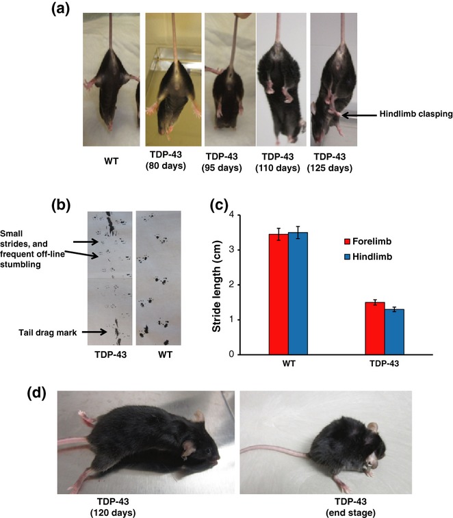
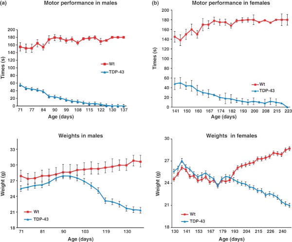
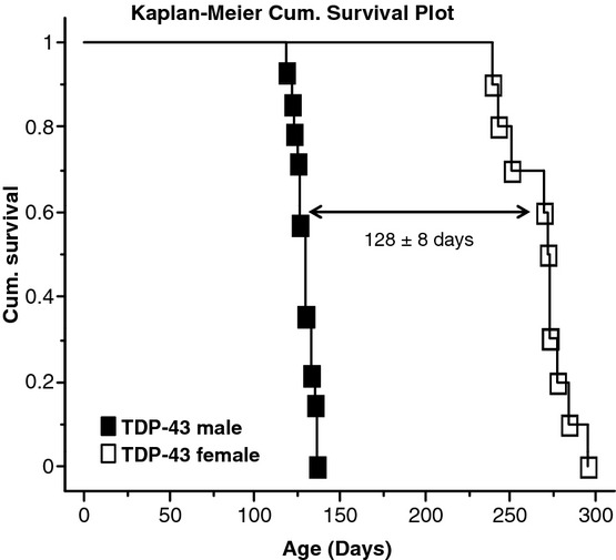
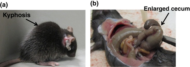
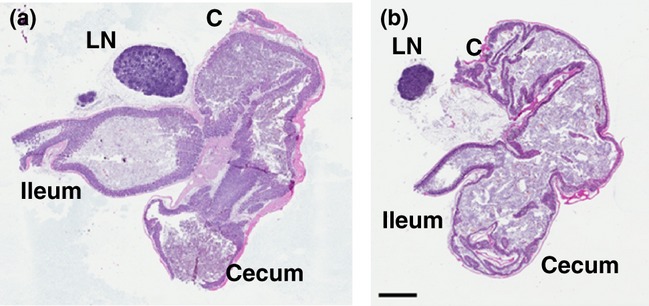
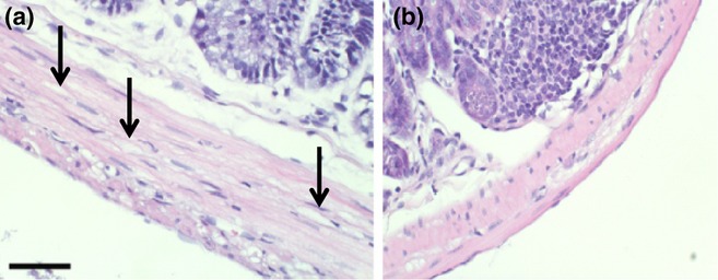
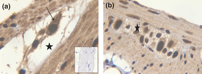
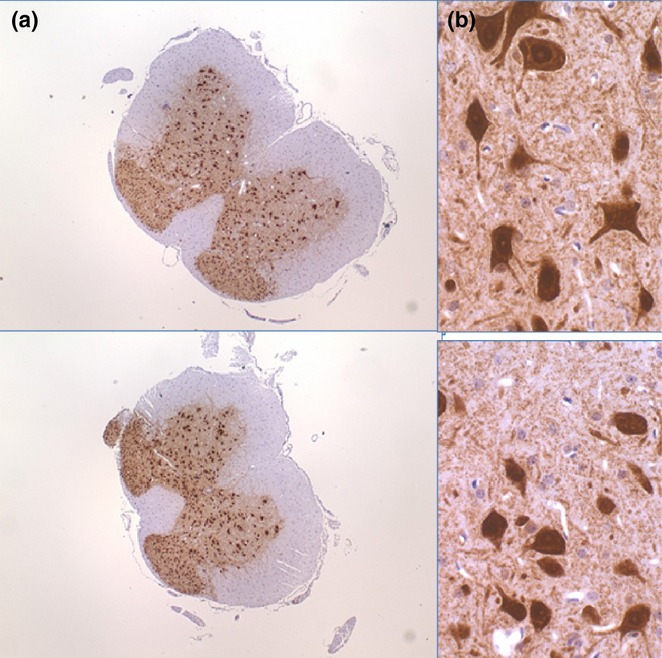
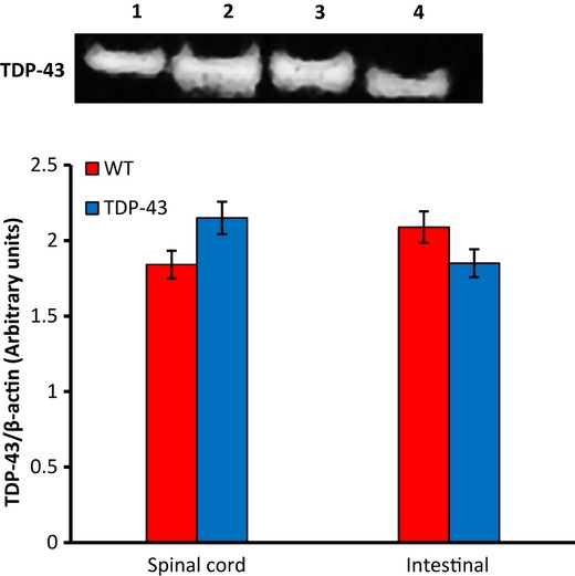
Abnormal distribution, modification and aggregation of transactivation response DNA-binding protein 43 (TDP-43) are the hallmarks of multiple neurodegenerative diseases, especially frontotemporal lobar degeneration with ubiquitin-positive inclusions (FTLD-U) and amyotrophic lateral sclerosis (ALS). Transgenic mouse lines overexpressing wild-type or mutant TDP-43 exhibit ALS-like symptom, motor abnormalities and early paralysis followed by death. Reports on lifespan and phenotypic behaviour in Prp-TDP-43 (A315T) vary, and these animals are not fully characterized. Although it has been proposed that the approximate 20% loss of motor neurons at end stage is responsible for the severe weakness and death in TDP-43 mice, this degree of neurologic damage appears insufficient to cause death. Hence we studied these mice to further characterize and determine the reason for the death. Our characterization of TDP-43 transgenic mice showed that these mice develop ALS-like symptoms that later become compounded by gastrointestinal (GI) complications that resulted in death. This is the first report of a set of pathological evidence in the GI track that is strong indicator for the cause of death of Prp-hTDP-43 (A315T) transgenic mice.
© 2012 The Authors. International Journal of Experimental Pathology © 2012 International Journal of Experimental Pathology.
Figures


Similar articles
-
A high-fat jelly diet restores bioenergetic balance and extends lifespan in the presence of motor dysfunction and lumbar spinal cord motor neuron loss in TDP-43A315T mutant C57BL6/J mice.Dis Model Mech. 2016 Sep 1;9(9):1029-37. doi: 10.1242/dmm.024786. Epub 2016 Aug 4. Dis Model Mech. 2016. PMID: 27491077 Free PMC article.
-
Withania somnifera Reverses Transactive Response DNA Binding Protein 43 Proteinopathy in a Mouse Model of Amyotrophic Lateral Sclerosis/Frontotemporal Lobar Degeneration.Neurotherapeutics. 2017 Apr;14(2):447-462. doi: 10.1007/s13311-016-0499-2. Neurotherapeutics. 2017. PMID: 27928708 Free PMC article.
-
Pathological hallmarks of amyotrophic lateral sclerosis/frontotemporal lobar degeneration in transgenic mice produced with TDP-43 genomic fragments.Brain. 2011 Sep;134(Pt 9):2610-26. doi: 10.1093/brain/awr159. Epub 2011 Jul 13. Brain. 2011. PMID: 21752789
-
Review: Prion-like mechanisms of transactive response DNA binding protein of 43 kDa (TDP-43) in amyotrophic lateral sclerosis (ALS).Neuropathol Appl Neurobiol. 2015 Aug;41(5):578-97. doi: 10.1111/nan.12206. Epub 2015 Apr 20. Neuropathol Appl Neurobiol. 2015. PMID: 25487060 Review.
-
Possible concurrence of TDP-43, tau and other proteins in amyotrophic lateral sclerosis/frontotemporal lobar degeneration.Neuropathology. 2018 Feb;38(1):72-81. doi: 10.1111/neup.12428. Epub 2017 Sep 27. Neuropathology. 2018. PMID: 28960544 Review.
Cited by
-
Mitochondrial dysregulation occurs early in ALS motor cortex with TDP-43 pathology and suggests maintaining NAD+ balance as a therapeutic strategy.Sci Rep. 2022 Mar 11;12(1):4287. doi: 10.1038/s41598-022-08068-5. Sci Rep. 2022. PMID: 35277554 Free PMC article.
-
Methodology Aspects of Colony Maintain for a Murine Model of Amyotrophic Lateral Sclerosis (ALS) TDP-43 Proteinopathy.Animals (Basel). 2020 Dec 7;10(12):2329. doi: 10.3390/ani10122329. Animals (Basel). 2020. PMID: 33297584 Free PMC article.
-
From Mouse Models to Human Disease: An Approach for Amyotrophic Lateral Sclerosis.In Vivo. 2018 Sep-Oct;32(5):983-998. doi: 10.21873/invivo.11339. In Vivo. 2018. PMID: 30150420 Free PMC article. Review.
-
The progress in C9orf72 research: ALS/FTD pathogenesis, functions and structure.Small GTPases. 2022 Jan;13(1):56-76. doi: 10.1080/21541248.2021.1892443. Epub 2021 Mar 5. Small GTPases. 2022. PMID: 33663328 Free PMC article. Review.
-
A Gut Feeling in Amyotrophic Lateral Sclerosis: Microbiome of Mice and Men.Front Cell Infect Microbiol. 2022 Mar 11;12:839526. doi: 10.3389/fcimb.2022.839526. eCollection 2022. Front Cell Infect Microbiol. 2022. PMID: 35360111 Free PMC article. Review.
References
-
- Agar J, Durham H. Relevance of oxidative injury in the pathogenesis of motor neuron diseases. Amyotroph. Lateral Scler. Other Motor Neuron Disord. 2003;4:232–242. - PubMed
-
- Almer G, Guegan C, Teismann P, et al. Increased expression of the pro-inflammatory enzyme cyclooxygenase-2 in amyotrophic lateral sclerosis. Ann. Neurol. 2001;49:176–185. - PubMed
-
- Arai T, Hasegawa M, Akiyama H, et al. TDP-43 is a component of ubiquitin-positive tau-negative inclusions in frontotemporal lobar degeneration and amyotrophic lateral sclerosis. Biochem. Biophys. Res. Commun. 2006;351:602–611. - PubMed
-
- Borchelt DR, Ratovitski T, van Lare J, et al. Accelerated amyloid deposition in the brains of transgenic mice coexpressing mutant presenilin 1 and amyloid precursor proteins. Neuron. 1997;19:939–945. - PubMed
-
- Bruijn LI, Houseweart MK, Kato S, et al. Aggregation and motor neuron toxicity of an ALS-linked SOD1 mutant independent from wild-type SOD1. Science. 1998;281:1851–1854. - PubMed
Publication types
MeSH terms
Substances
Grants and funding
LinkOut - more resources
Full Text Sources
Other Literature Sources
Medical
Research Materials
Miscellaneous
