. 2013 Mar 21;121(12):2352-62.
doi: 10.1182/blood-2012-05-424713. Epub 2013 Jan 11.

NRP1 acts cell autonomously in endothelium to promote tip cell function during sprouting angiogenesis

Affiliations

NRP1 acts cell autonomously in endothelium to promote tip cell function during sprouting angiogenesis

Alessandro Fantin et al. Blood. .

Abstract

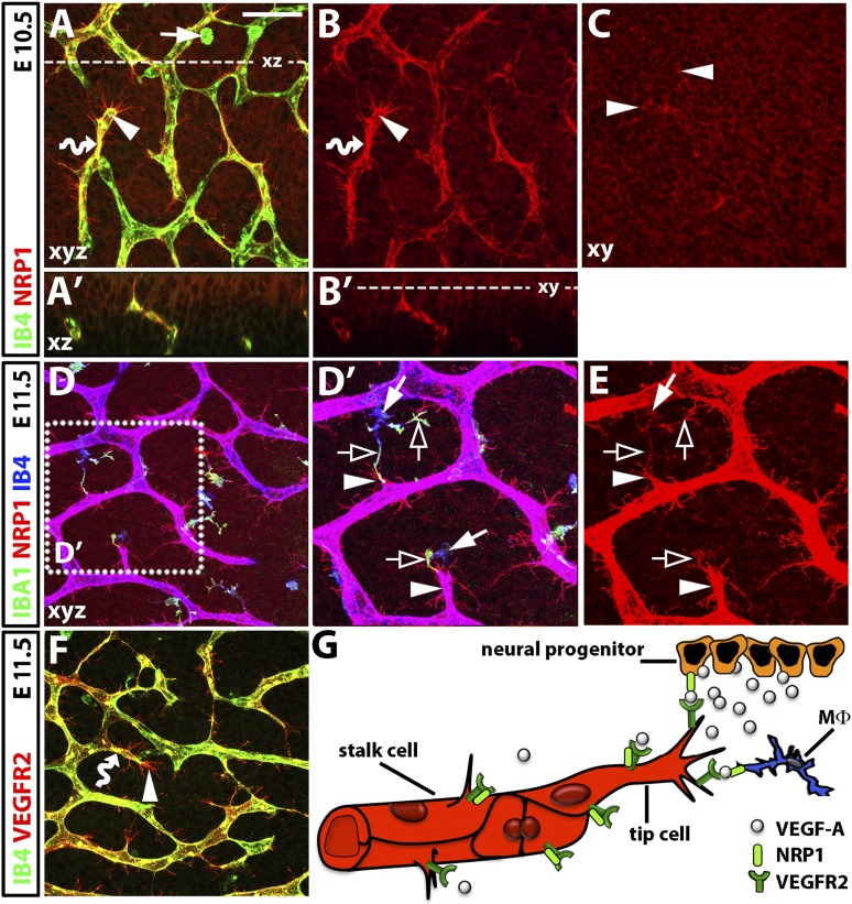
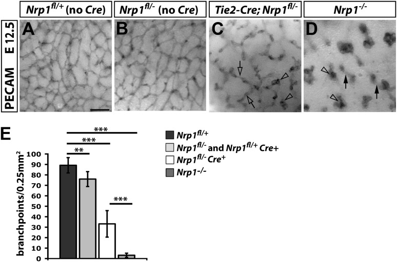
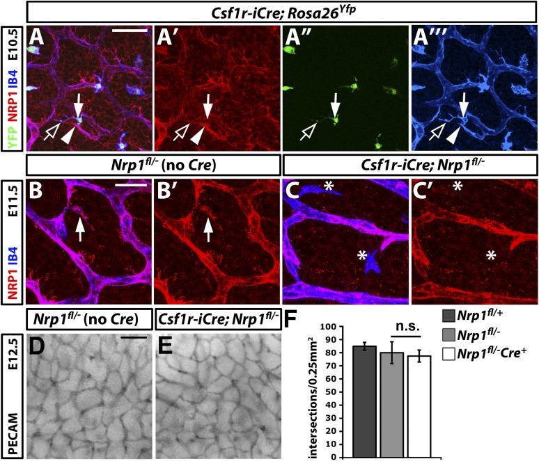
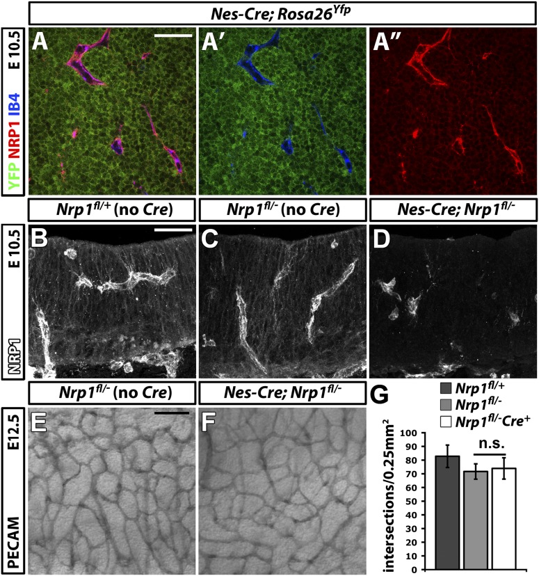
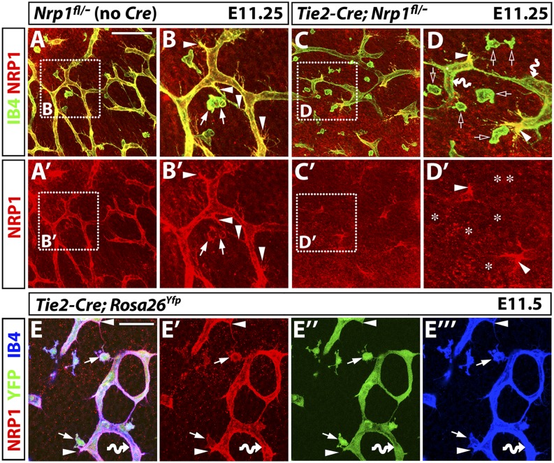
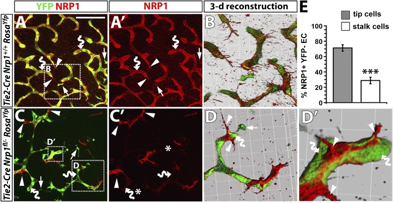
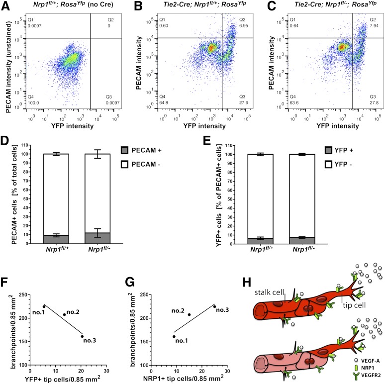
Neuropilin (NRP) 1 is a receptor for the vascular endothelial growth factor (VEGF)-A and is essential for normal angiogenesis. Previous in vitro experiments identified NRP1 interactions with VEGF-A's main signaling receptor VEGFR2 within endothelial cells, but also between nonendothelial NRP1 and endothelial VEGFR2. Consistent with an endothelial role for NRP1 in angiogenesis, we found that VEGFR2 and NRP1 were coexpressed in endothelial tip and stalk cells in the developing brain. In addition, NRP1 was expressed on two cell types that interact with growing brain vessels-the neural progenitors that secrete VEGF-A to stimulate tip cell activity and the pro-angiogenic macrophages that promote tip cell anastomosis. Selective targeting of Nrp1 in each of these cell types demonstrated that neural progenitor- and macrophage-derived NRP1 were dispensable, whereas endothelial NRP1 was essential for normal brain vessel growth. NRP1 therefore promotes brain angiogenesis cell autonomously in endothelium, independently of heterotypic interactions with nonendothelial cells. Genetic mosaic analyses demonstrated a key role for NRP1 in endothelial tip rather than stalk cells during vessel sprouting. Thus, NRP1-expressing endothelial cells attained the tip cell position when competing with NRP1-negative endothelial cells in chimeric vessel sprouts. Taken together, these findings demonstrate that NRP1 promotes endothelial tip cell function during angiogenesis.


Figures

Figure 1
Figure 1
NRP1 expression during hindbrain vascularization in the mouse. (A-F) Whole-mount immunofluorescence labeling of the mouse embryo hindbrain; scale bar (A-D and F) 50 μm. (A) Maximal projection (xy) of a confocal z stack through the E10.5 subventricular zone shows NRP1 on IB4-positive endothelial stalk and tip cells (wavy arrow and arrowhead, respectively; yellow indicates double labeling). NRP1 is also prominent on tip-cell filopodia (IB4 low) and neural progenitors (IB4 negative). (A′) Virtual transverse (xz) section through the z stack in (A) at the level indicated with a stippled line. (B,B′) Single NRP1 channel of (A,A′). Some IB4-positive tissue macrophages (arrow in A) are poorly ramified at E10.5 and do not obviously express NRP1 (compare A with B). (C) Single xy scan of the confocal z stack projection in (B) at the level indicated with a stippled line in (B'); clusters of tip cell filopodia protrude into the neural progenitor layer (arrowheads). (D-E) Confocal z stack through the E11.5 subventricular zone shows that IB4-positive endothelial cells and IBA1/IB4-positive ramified tissue macrophages express NRP1; purple indicates co-labeling with NRP1 and IB4. The boxed area in (D) is indicated at higher magnification in (D′) and as the single NRP1 channel in (E). NRP1 is high on filopodia-studded tip cells and on IBA1-enriched macrophage processes (arrowheads and clear arrows, respectively). NRP1 appears lower in neural progenitors at E11.5 (D) compared with E10.5 (A). (F) Z stack through the subventricular zone at E11.5 shows VEGFR2 expression on IB4-positive endothelial stalk and tip cells (wavy arrow and arrowhead, respectively; yellow indicates double labeling). VEGFR2 is high on filopodia but not obviously expressed by tissue macrophages or neural progenitors. (G) Schematic representation of NRP1 and VEGFR2 distribution and hypothetical interactions in hindbrain cell types during angiogenesis. NRP1 (light green) is co-expressed with VEGFR2 (dark green) on endothelial stalk and tip cells (red), which is a prerequisite for homotypic interactions. NRP1 is also expressed by neural progenitors (orange) and tissue macrophages (Mφ, blue), and therefore heterotypically and in trans relative to VEGFR2 in endothelial cells.
Figure 2
Figure 2
Vascular defects in Tie2-Cre conditional and full Nrp1-null mutants. (A-D) PECAM immunohistochemistry of E12.5 SVP vessels of the indicated genotypes. Clear arrowheads indicate examples of vascular tufts; clear arrows and solid arrows indicate examples of vascular interconnections in the SVP versus deeper brain layers, respectively. Scale bar represents 100 μm. (E) Quantitation of SVP branchpoints at E12.5; error bars represent SD; asterisks indicate P values; **P < .001; ***P < .0001.
Figure 3
Figure 3
NRP1 expression by tissue macrophages is not essential for brain vascularization. (A) A E10.5 hindbrain with a constitutively active Csf1r-iCre transgene and the Rosa26Yfp reporter was triple labeled for YFP (green), NRP1 (red), and IB4 (blue), shown together (A) and as single channels (A′-A′′′). The solid arrow indicates a tissue macrophage, the clear arrow a NRP1-positive macrophage process; the solid arrowhead indicates a tip cell. Scale bar represents 50 μm. (B,C) Double labeling of E11.5 control Nrp1fl/– and mutant Csf1r-iCre;Nrp1fl/– hindbrains for NRP1 (red) and IB4 (blue) shown together (B,C) and as single NRP1 channels (B′,C′). A NRP1-positive tissue macrophage is indicated with an arrow in (B,B′); NRP1-negative tissue macrophages are indicated with asterisks in (C,C′). Scale bar represents 25 μm. (D,E) PECAM immunohistochemistry of E12.5 littermate hindbrains of the indicated genotypes; scale bar represents 100 μm. (F) Quantitation of SVP branchpoints at E12.5; error bars represent SD; n.s., not significant.
Figure 4
Figure 4
NRP1 expression by neural progenitors is not essential for brain vascularization. (A-A″) Single xy scan through an E10.5 hindbrain carrying the constitutively active Nes-Cre transgene and the Rosa26Yfp reporter, labeled for YFP (green), NRP1 (red), and IB4 (blue), all shown together in (A) or as double YFP/IB4 (A′) and single NRP1 (A″) channels. Scale bar represents 50 μm. (B-D) Immunofluorescent staining for NRP1 of 20-μm thin, frozen sections from E10.5 control Nrp1fl/+, Nrp1fl/–, and mutant Nes-Cre;Nrp1fl/– hindbrains. Scale bar represents 50 μm. (E,F) PECAM immunohistochemistry of E12.5 littermate hindbrains of the indicated genotypes; scale bar represents 100 μm. (G) Quantitation of SVP branchpoints at E12.5; error bars represent SD; n.s., not significant.
Figure 5
Figure 5
Tie2-Cre;Nrp1fl/ mutants contain tip cells that retain NRP1 expression, even though Tie2-Cre effectively activates the Rosa26Yfp reporter in tissue macrophages as well as endothelial tip and stalk cells. (A-D) NRP1 (red) and IB4 (green) immunofluorescence staining of littermate E11.25 hindbrains lacking Cre or expressing a constitutively active Tie2-Cre transgene on an Nrp1fl/– background; single NRP1 channels are shown below each panel (A′-D′). (B and D) Higher magnifications of the boxed areas in (A and C). Arrowheads indicate examples of tip cells, arrows show examples of tissue macrophages expressing NRP1. Clear arrows in (D) and asterisks in (D′) indicate the position of macrophages lacking NRP1, and curved arrows indicate endothelial stalk cells lacking NRP1. Scale bar represents 100 μm for (A,A′,C,C′). (E) An E11.5 hindbrain carrying a constitutively active Tie2-Cre transgene and the Rosa26Yfp reporter was triple labeled for NRP1 (red), YFP (green), and IB4 (blue); single channels are shown in (E′-E′′′). Scale bar represents 50 μm.
Figure 6
Figure 6
NRP1 expression confers a selective advantage to endothelial cells competing for the tip cell position. (A-D) Immunofluorescence staining of littermate hindbrains of the indicated genotypes; YFP and NRP1 labeling are shown in (A,C), NRP1 only in (A′,C′). Scale bar represents 100 μm. Three-dimensional reconstructions of the boxed areas in (A,C) shown in (B,D,D′). Examples of tissue macrophages, endothelial tip cells, and endothelial stalk cells are indicated with arrows, arrowheads, and curved arrows, respectively; note that some vessels leave the plane of section, and the vessel therefore appears blunt-ended, terminating in a stalk cell. Asterisks in (C′) indicate the position of macrophages lacking NRP1. (E) Percentage of NRP1+ YFP– endothelial cells in the tip versus stalk cell position in Tie2-Cre;Nrp1fl/–;RosaYfp mutant hindbrains.
Figure 7
Figure 7
The number of NRP1-positive tip cells in blood vessels with mosaic expression of NRP1 is a good predictor of branching frequency. (A-C) Live cells from the indicated genotypes were FACS-sorted with a PECAM-APC antibody (y axis) and for YFP fluorescence (x axis); (A) Cells from a control embryo lacking YFP and not stained for PECAM; this FACS profile was used to determine gating parameters to identify YFP-positive, PECAM-stained cells in (B,C). (D) Percentage of total PECAM-positive (Q1 + Q2 quadrants) and PECAM-negative (Q3 + Q4 quadrants) cells in (B and C). (E) Percentage of YFP-positive, PECAM-positive cells (Q2 quadrant) and YFP-negative, PECAM-positive cells (Q1 quadrant) in (B,C). (F,G) The number of vessel branchpoints in three littermate mutants lacking NRP1 in the Tie2-Cre lineage correlates inversely with the number of YFP-positive (F) and positively with the number of NRP1-positive tip cells (G). (H) Schematic representation of NRP1 localization in wild-type (top) and chimeric (bottom) vessel sprouts; NRP1 is present on both tip and stalk cells in wild-types (bright red), whereas NRP1 is high on mutant tip cells (bright red) and low on mutant stalk cells (faded red) of chimeric vessels.

Similar articles

Cited by

References

    1. Ruhrberg C. Growing and shaping the vascular tree: multiple roles for VEGF. BioEssays. 2003;25(11):1052–1060. - PubMed
    1. Carmeliet P. Angiogenesis in life, disease and medicine. Nature. 2005;438(7070):932–936. - PubMed
    1. Koch S, Tugues S, Li X, Gualandi L, Claesson-Welsh L. Signal transduction by vascular endothelial growth factor receptors. Biochem J. 2011;437(2):169–183. - PubMed
    1. Shalaby F, Rossant J, Yamaguchi TP, et al. Failure of blood-island formation and vasculogenesis in Flk-1-deficient mice. Nature. 1995;376(6535):62–66. - PubMed
    1. Ruhrberg C, Gerhardt H, Golding M, et al. Spatially restricted patterning cues provided by heparin-binding VEGF-A control blood vessel branching morphogenesis. Genes Dev. 2002;16(20):2684–2698. - PMC - PubMed

Publication types

MeSH terms