

The CSF-1 receptor fashions the intestinal stem cell niche
- PMID: 23314290
- PMCID: PMC4096353
- DOI: 10.1016/j.scr.2012.12.001
The CSF-1 receptor fashions the intestinal stem cell niche
Abstract
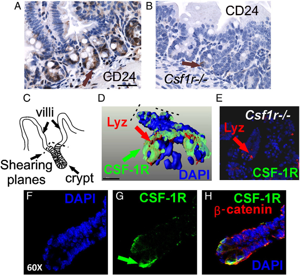
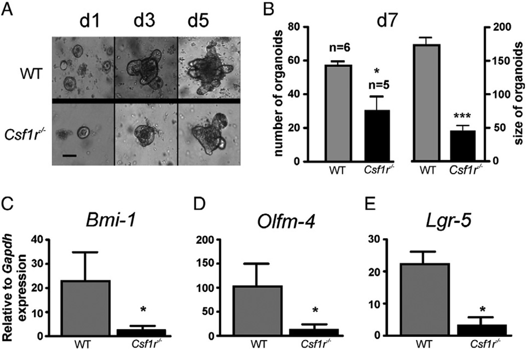
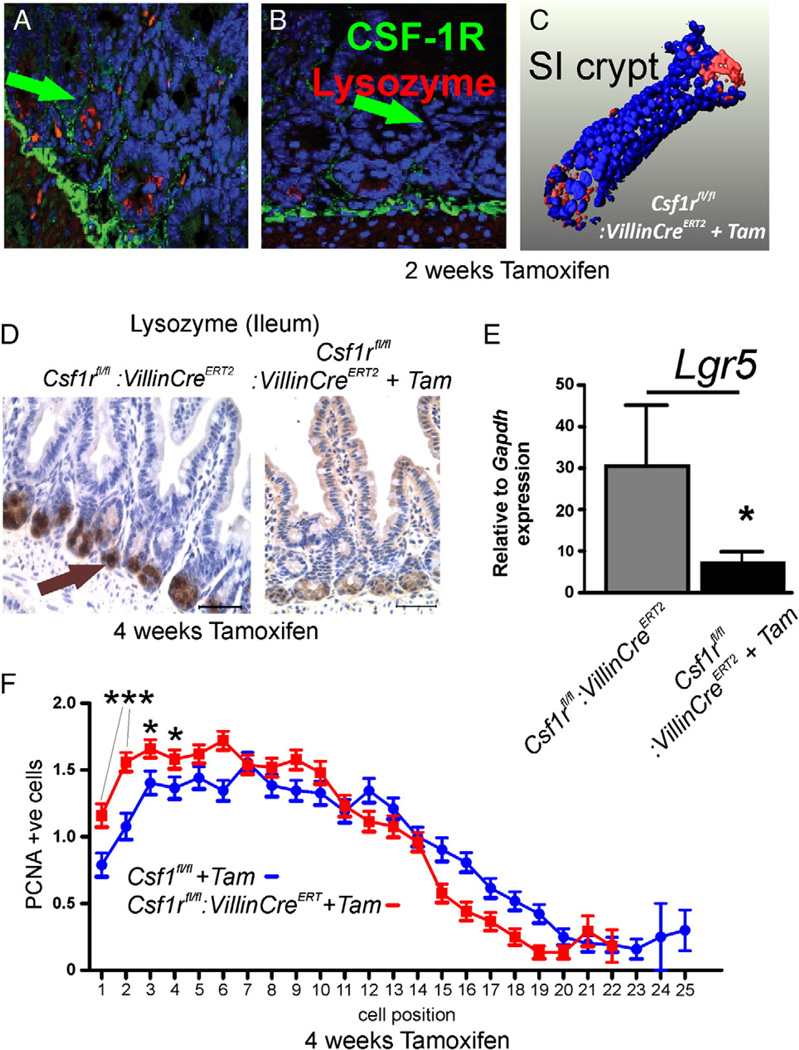
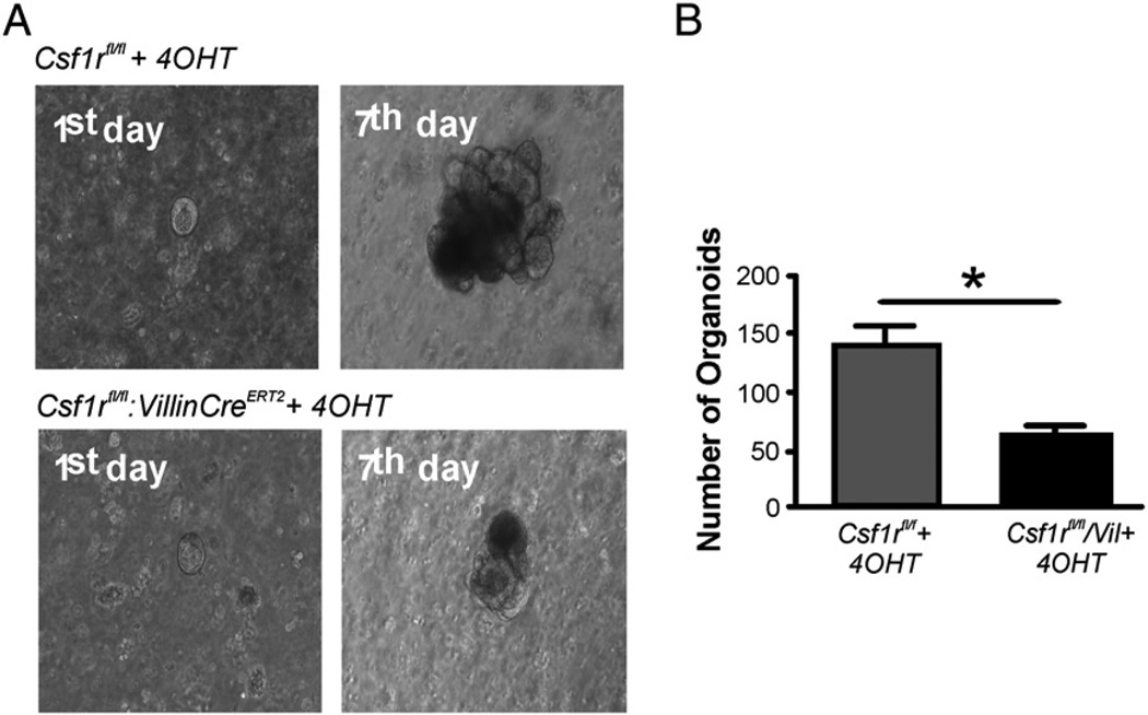
Gastrointestinal (GI) homeostasis requires the action of multiple pathways. There is some controversy regarding whether small intestine (SI) Paneth cells (PCs) play a central role in orchestrating crypt architecture and their relationship with Lgr5+ve stem cells. Nevertheless, we previously showed that germline CSF-1 receptor (Csf1r) knock out (KO) or Csf1 mutation is associated with an absence of mature PC, reduced crypt proliferation and lowered stem cell gene, Lgr5 expression. Here we show the additional loss of CD24, Bmi1 and Olfm4 expression in the KO crypts and a high resolution 3D localization of CSF-1R mainly to PC. The induction of GI-specific Csf1r deletion in young adult mice also led to PC loss over a period of weeks, in accord with the anticipated long life span of PC, changed distribution of proliferating cells and this was with a commensurate loss of Lgr5 and other stem cell marker gene expression. By culturing SI organoids, we further show that the Csf1r(-/-) defect in PC production is intrinsic to epithelial cells as well as definitively affecting stem cell activity. These results show that CSF-1R directly supports PC maturation and that in turn PCs fashion the intestinal stem cell niche.
Copyright © 2012 Elsevier B.V. All rights reserved.
Conflict of interest statement
The authors have no conflicts of interest.
Figures

Similar articles
-
Reg4+ deep crypt secretory cells function as epithelial niche for Lgr5+ stem cells in colon.Proc Natl Acad Sci U S A. 2016 Sep 13;113(37):E5399-407. doi: 10.1073/pnas.1607327113. Epub 2016 Aug 29. Proc Natl Acad Sci U S A. 2016. PMID: 27573849 Free PMC article.
-
Paneth cells constitute the niche for Lgr5 stem cells in intestinal crypts.Nature. 2011 Jan 20;469(7330):415-8. doi: 10.1038/nature09637. Epub 2010 Nov 28. Nature. 2011. PMID: 21113151 Free PMC article.
-
Colony stimulating factor-1 dependence of paneth cell development in the mouse small intestine.Gastroenterology. 2009 Jul;137(1):136-44, 144.e1-3. doi: 10.1053/j.gastro.2009.03.004. Epub 2009 Mar 18. Gastroenterology. 2009. PMID: 19303020 Free PMC article.
-
Regulation of intestinal stem cell fate specification.Sci China Life Sci. 2015 Jun;58(6):570-8. doi: 10.1007/s11427-015-4859-7. Epub 2015 May 8. Sci China Life Sci. 2015. PMID: 25951932 Review.
-
Modeling Intestinal Stem Cell Function with Organoids.Int J Mol Sci. 2021 Oct 9;22(20):10912. doi: 10.3390/ijms222010912. Int J Mol Sci. 2021. PMID: 34681571 Free PMC article. Review.
Cited by
-
Deletion of a Csf1r enhancer selectively impacts CSF1R expression and development of tissue macrophage populations.Nat Commun. 2019 Jul 19;10(1):3215. doi: 10.1038/s41467-019-11053-8. Nat Commun. 2019. PMID: 31324781 Free PMC article.
-
The Intestinal Stem Cell Niche: Homeostasis and Adaptations.Trends Cell Biol. 2018 Dec;28(12):1062-1078. doi: 10.1016/j.tcb.2018.08.001. Epub 2018 Sep 5. Trends Cell Biol. 2018. PMID: 30195922 Free PMC article. Review.
-
Pleiotropic effects of extended blockade of CSF1R signaling in adult mice.J Leukoc Biol. 2014 Aug;96(2):265-74. doi: 10.1189/jlb.2A0114-006R. Epub 2014 Mar 20. J Leukoc Biol. 2014. PMID: 24652541 Free PMC article.
-
Effects of Immune Cells on Intestinal Stem Cells: Prospects for Therapeutic Targets.Stem Cell Rev Rep. 2022 Oct;18(7):2296-2314. doi: 10.1007/s12015-022-10347-7. Epub 2022 Mar 12. Stem Cell Rev Rep. 2022. PMID: 35279803 Review.
-
Evidence for a direct effect of the autonomic nervous system on intestinal epithelial stem cell proliferation.Physiol Rep. 2018 Jun;6(12):e13745. doi: 10.14814/phy2.13745. Physiol Rep. 2018. PMID: 29932493 Free PMC article.
References
-
- Akazawa C, Ishibashi M, Shimizu C, Nakanishi S, Kageyama R. A mammalian helix–loop–helix factor structurally related to the product of Drosophila proneural gene atonal is a positive transcriptional regulator expressed in the developing nervous system. J. Biol. Chem. 1995;270:8730–8738. - PubMed
-
- Barker N, van Es JH, Kuipers J, Kujala P, van den Born M, Cozijnsen M, Haegebarth A, Korving J, Begthel H, Peters PJ, Clevers H. Identification of stem cells in small intestine and colon by marker gene Lgr5. Nature. 2007;449:1003–1007. - PubMed
-
- Bevins CL, Salzman NH. Paneth cells, antimicrobial peptides and maintenance of intestinal homeostasis. Nat. Rev. Microbiol. 2011;9:356–368. - PubMed
-
- Chitu V, Stanley ER. Colony-stimulating factor-1 in immunity and inflammation. Curr. Opin. Immunol. 2006;18:39–48. - PubMed
Publication types
MeSH terms
Substances
Grants and funding
LinkOut - more resources
Full Text Sources
Other Literature Sources
Medical
Molecular Biology Databases
Research Materials
Miscellaneous
