. 2013 Mar 7;121(10):1677-89.
doi: 10.1182/blood-2012-07-445189. Epub 2013 Jan 10.

Neutrophil differentiation into a unique hybrid population exhibiting dual phenotype and functionality of neutrophils and dendritic cells

Affiliations

Neutrophil differentiation into a unique hybrid population exhibiting dual phenotype and functionality of neutrophils and dendritic cells

Hironori Matsushima et al. Blood. .

Abstract

Neutrophils have been reported to acquire surface expression of MHC class II and co-stimulatory molecules as well as T-cell stimulatory activities when cultured with selected cytokines. However, cellular identity of those unusual neutrophils showing antigen presenting cell (APC)-like features still remains elusive. Here we show that both immature and mature neutrophils purified from mouse bone marrow differentiate into a previously unrecognized "hybrid" population showing dual properties of both neutrophils and dendritic cells (DCs) when cultured with granulocyte macrophage-colony-stimulating factor but not with other tested growth factors. The resulting hybrid cells express markers of both neutrophils (Ly6G, CXCR2, and 7/4) and DCs (CD11c, MHC II, CD80, and CD86). They also exhibit several properties typically reserved for DCs, including dendritic morphology, probing motion, podosome formation, production of interleukin-12 and other cytokines, and presentation of various forms of foreign protein antigens to naïve CD4 T cells. Importantly, they retain intrinsic abilities of neutrophils to capture exogenous material, extrude neutrophil extracellular traps, and kill bacteria via cathelicidin production. Not only do our results reinforce the notion that neutrophils can acquire APC-like properties, they also unveil a unique differentiation pathway of neutrophils into neutrophil-DC hybrids that can participate in both innate and adaptive immune responses.


Figures

Figure 1
Figure 1
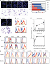
Purified band cells gain surface expression of CD11c and MHC II in GM-CSF–supplemented culture. (A) Gr-1high/CD48 band cells purified from BM of C57BL/6 mice (CD45.2) were examined for surface expression of the indicated markers. Blue lines indicate staining profiles with isotype-matched control IgG. (B-D) Band cells purified from C57BL/6 mice were cultured for 4 d (B) or 6 d (C) with BM feeder cells from B6 SJL mice (CD45.1) in the presence of GM-CSF, G-CSF, M-CSF, or Flt3L. The data show the expression profiles of CD11c (top panels) and Ly6G (bottom panels) within the CD45.2+ gated population and the recovery rates compared with the originally plated band cell number. (D) The data show the expression profiles of CD11c, MHC II, and Ly6G within the CD45.2+/CD45.1 gated population harvested on d 6 from GM-CSF–supplemented co-cultures. Data are representative of at least 3 independent experiments.
Figure 2
Figure 2
Differentiation of band cells into neutrophil-DC hybrids in culture. (A-B) Gr-1high/CD48 band cells purified from C57BL/6 mice were cultured with GM-CSF in the presence of crude BM feeder cells from B6 SJL mice. (A) CD45.2+/CD45.1 populations purified at the indicated time points were analyzed for nuclear shape (top panels) and morphology (bottom panels; bar represents 20 μm). (B) Changes in nuclear shape were examined by analyzing >2000 cells/time point after nuclear staining. Recovery rates of viable CD45.2+/CD45.1 cells relative to the originally plated cell numbers are shown on the right. (C-E) Crude BM cells from CD45.2 mice were cultured for 2 d with GM-CSF. Gr-1high/CD48 band cells were purified from these precultures and then co-cultured with BM feeder cells from CD45.1 mice in the presence of GM-CSF for an additional 6 d. (C) The starting population (left) and the CD45.2+/CD45.1 population purified from the co-culture (right) were analyzed for nuclear shape and morphology. The latter population was also examined for surface phenotype (D) and APC function to present OVA peptides to OT-II CD4 or OT-I CD8 T cells (means ± SD from triplicate cultures) (E). (F) CD15+/CD10/CD64/CD14 band cells purified from human BM samples were examined for the surface phenotype before (top) and after culturing for 7 d in the presence of GM-CSF, TNFα, and IL-4 (bottom). (G) The above-band cell population (top) and the CD14+ monocyte population purified from the same human BM samples (bottom) were cultured for 7 d in parallel and then examined for surface phenotype. Data are representative of 3 independent experiments.
Figure 3
Figure 3
Characterization of neutrophil-DC hybrids purified from crude BM culture. (A) BM cells from C57BL/6 mice were cultured with GM-CSF, Flt3L, or no added growth factor for the indicated periods, and resulting populations were examined for the expression of CD11c, Ly6G, and B220. The data show the numbers of total cells, CD11c+/Ly6G DCs, Ly6G+/CD11c+ neutrophil-DC hybrids, and B220+/CD11c+ pDCs (means ± SD from triplicate cultures). (B-E) Ly6G+/CD11c/MHC II neutrophils, Ly6G+/CD11c+/MHC II+ neutrophil-DC hybrids, and Ly6G/CD11c+/MHC II+ traditional DCs were purified on d 6 from GM-CSF–supplemented BM culture. These populations were examined under light microscopy after HEMA-3 staining (B), under differential interference contrast microscopy (C), or under confocal microscopy after staining with phalloidin and antivinculin mAb (D) or with anti-MPO mAb (E). All the images are representative of at least 3 independent experiments (bar represents 20 μm). Time-lapse images recorded under differential interference contrast microscope are shown in supplemental Video 1.
Figure 4
Figure 4
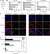
Neutrophil-DC hybrids retain functionality as professional phagocytes. (A) Cells harvested from GM-CSF–supplemented BM culture (d 6) were incubated for 30 min at 4°C or 37°C with FITC-DX, live E. coli expressing GFP, or fluorescent beads and then examined for the mean fluorescence intensity (MFI) or percent bead+ cells within the Ly6G+/CD11c neutrophil, Ly6G+/CD11c+ hybrid, and Ly6G/CD11c+ traditional DC populations. Some samples were tested after a 2-h pretreatment with PMA (100 nM). (B-D) Ly6G+/CD11c/MHC II neutrophils, Ly6G+/CD11c+/MHC II+ neutrophil-DC hybrids, and Ly6G/CD11c+/MHC II+ traditional DCs purified from GM-CSF–supplemented BM culture (d 6) were examined for NET formation. The purified samples were incubated for 2 h with phosphate-buffered saline alone (B) or PMA (C) on cover slips and then stained for DNA and histone 2A. Arrows indicate NET-forming cells (bar represents 20 μm). The same images are shown at a higher magnification in supplemental Figure 11. (D) The purified samples were incubated for 2 h with phosphate-buffered saline alone, LPS, or PMA and then examined for the magnitudes of NET formation by measuring DNA release (means ± SD from triplicate cultures). Data are representative of 2 independent experiments. **P < .01, ***P < .001 between the indicated samples.
Figure 5
Figure 5
Neutrophil-DC hybrids acquire functionality as APCs. (A) Ly6G+/CD11c/MHC II neutrophils, Ly6G+/CD11c+/MHC II+ neutrophil-DC hybrids, and Ly6G/CD11c+/MHC II+ traditional DCs purified from GM-CSF–supplemented BM culture (d 6) were examined for TLR mRNA expression profiles by quantitative polymerase chain reaction (means ± SD from triplicate samples). (B-C) The same 3 purified populations were cultured for 24 h with phosphate-buffered saline alone or each of the indicated TLR agonists. The samples were then examined for cytokine release measured by the cytometric bead array system (means ± SD from triplicate cultures) (B) and surface expression of CD40 (C). (D) The same 3 purified populations were pulsed for 60 min with the indicated form of OVA antigen and then co-cultured with OVA-specific CD4 T cells from OT-II TG mice (means ± SD from triplicate cultures). *P < .05, **P < .01, ***P < .001 compared with control group treated with PBS alone. Data are representative of at least 2 independent experiments.
Figure 6
Figure 6
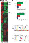
Identification of unique properties that distinguish neutrophil-DC hybrids from traditional DCs. (A) Ly6G+/CD11c+/MHC II+ neutrophil-DC hybrids and Ly6G/CD11c+/MHC II+ traditional DCs purified from the same GM-CSF–supplemented BM cultures (d 6) were compared for global gene expression profiles. The heat map was created from 3 independent pairs to show the genes that are predominantly expressed (>2-fold and P < .05). The whole-gene datasets have been deposited in Gene Expression Omnibus with accession number GSE28408. (B-D) Ly6G+/CD11c/MHC II neutrophils, Ly6G+/CD11c+/MHC II+ hybrids, and Ly6G/CD11c+/MHC II+ traditional DCs were examined for MMP9 mRNA expression by quantitative polymerase chain reaction and MMP9 protein elaboration by enzyme-linked immunosorbent assay (means ± SD from triplicate samples) (B) and for surface expression of CD62L (C) and CXCR2 (D). Data are representative of 3 independent experiments.
Figure 7
Figure 7
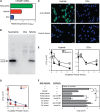
Neutrophil-DC hybrids kill bacteria rapidly by a CRAMP-mediated mechanism. (A-C) Ly6G+/CD11c/MHC II neutrophils, Ly6G+/CD11c+/MHC II+ hybrids, and/or Ly6G/CD11c+/MHC II+ traditional DCs purified from GM-CSF–supplemented BM culture (d 6) were compared for CRAMP mRNA expression by quantitative polymerase chain reaction (means ± SD from triplicate samples) (A) and for CRAMP protein production by western blotting (B) and immunofluorescence staining (bar represents 20 μm) (C). (D) The above hybrid and traditional DC populations were incubated for 60 min with live E. coli. After killing extracellular bacteria by exposure to kanamycin (time 0), the samples were incubated for an additional 0.5-3.5 h to test intracellular bacterial killing (means ± SD from triplicate samples). (E) Neutrophil-DC hybrids (left panel) and traditional DCs (right panel) purified from wild-type mice or CRAMP-deficient mice were examined for intracellular bacterial killing activities (means ± SD from triplicate samples). (F) E. coli were cultured for 4 h with a synthetic CRAMP peptide or whole protein extracts from neutrophil-DC hybrids or traditional DCs in the presence or absence of neutralizing anti-CRAMP antibodies or control antibodies. The starting bacterial number before culturing is shown in the top bar (means ± SD from triplicate bacterial cultures). *P < .05, **P < .01, ***P < .001 between the indicated samples (F) or compared with traditional DCs (E) or to the starting bacterial numbers at time 0 (E). Data are representative of at least 2 independent experiments.

Similar articles

Cited by

References

    1. Borregaard N. Neutrophils, from marrow to microbes. Immunity. 2010;33(5):657–670. - PubMed
    1. Mantovani A, Cassatella MA, Costantini C, et al. Neutrophils in the activation and regulation of innate and adaptive immunity. Nat Rev Immunol. 2011;11(8):519–531. - PubMed
    1. van Gisbergen KP, Sanchez-Hernandez M, Geijtenbeek TB, et al. Neutrophils mediate immune modulation of dendritic cells through glycosylation-dependent interactions between Mac-1 and DC-SIGN. J Exp Med. 2005;201(8):1281–1292. - PMC - PubMed
    1. Pelletier M, Maggi L, Micheletti A, et al. Evidence for a cross-talk between human neutrophils and Th17 cells. Blood. 2010;115(2):335–343. - PubMed
    1. Fanger NA, Liu C, Guyre PM, et al. Activation of human T cells by major histocompatability complex class II expressing neutrophils: proliferation in the presence of superantigen, but not tetanus toxoid. Blood. 1997;89(11):4128–4135. - PubMed

Publication types

MeSH terms