. 2013 Mar 14;121(11):2074-83.
doi: 10.1182/blood-2012-05-432088. Epub 2013 Jan 9.

Eosinophil extracellular DNA trap cell death mediates lytic release of free secretion-competent eosinophil granules in humans

Affiliations

Eosinophil extracellular DNA trap cell death mediates lytic release of free secretion-competent eosinophil granules in humans

Shigeharu Ueki et al. Blood. .

Abstract

Eosinophils release their granule proteins extracellularly through exocytosis, piecemeal degranulation, or cytolytic degranulation. Findings in diverse human eosinophilic diseases of intact extracellular eosinophil granules, either free or clustered, indicate that eosinophil cytolysis occurs in vivo, but the mechanisms and consequences of lytic eosinophil degranulation are poorly understood. We demonstrate that activated human eosinophils can undergo extracellular DNA trap cell death (ETosis) that cytolytically releases free eosinophil granules. Eosinophil ETosis (EETosis), in response to immobilized immunoglobulins (IgG, IgA), cytokines with platelet activating factor, calcium ionophore, or phorbol myristate acetate, develops within 120 minutes in a reduced NADP (NADPH) oxidase-dependent manner. Initially, nuclear lobular formation is lost and some granules are released by budding off from the cell as plasma membrane-enveloped clusters. Following nuclear chromatolysis, plasma membrane lysis liberates DNA that forms weblike extracellular DNA nets and releases free intact granules. EETosis-released eosinophil granules, still retaining eosinophil cationic granule proteins, can be activated to secrete when stimulated with CC chemokine ligand 11 (eotaxin-1). Our results indicate that an active NADPH oxidase-dependent mechanism of cytolytic, nonapoptotic eosinophil death initiates nuclear chromatolysis that eventuates in the release of intact secretion-competent granules and the formation of extracellular DNA nets.


Figures
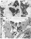
Figure 1
Figure 1
Transmission electron micrographs of human tissue eosinophils from (A) allergic and (B) hypereosinophilic patients. Biopsy samples from the left frontal sinus and the skin were obtained from a patient with allergic sinusitis and a patient with hypereosinophilic syndrome (negative for FIP1-like 1/platelet-derived growth factor-α mutation), respectively. In (Ai), a biopsy of the frontal sinus shows infiltrated eosinophils. Note the disrupting nuclei (Nu) and extracellular free secretory granules. (Aii) is the boxed area of (Ai) seen at higher magnification. In (B), both free granules (Gr) and disrupted nuclei (Nu) are also observed in eosinophils from a skin biopsy. Arrowheads in (Aii) and (B) indicate releasing decondensed chromatin. Samples were fixed in a mixture of glutaraldehyde and PFO and prepared for conventional TEM as in “Materials and methods.” Scale bars represent 1.2µm (Ai), 700 nm (Aii), 600 nm (B).
Figure 2
Figure 2
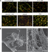
EETosis releases weblike chromatin structures from lytic cells. (A) Eosinophils were stimulated with the indicated stimuli in 0.1% BSA medium for 120 minutes or in A23187 for 60 minutes and fixed. EETosis was detected using extracellular histone staining without permeabilization, as described in “Materials and methods” and supplemental Figure 3. Green indicates histones, and red indicates DNA. Images were obtained using a BX62 microscope (20×, equipped with a Qimaging Rolera EM-C2 cooled digital camera (Surrey, BC, Canada) in conjunction with SlideBook 5.0 image analysis software (Intelligent Imaging Innovations, Denver, CO), or equipped with a Qimaging Retiga EXi cooled digital camera in conjunction with iVision image analysis software). Experiments were repeated with eosinophils from 3 independent donors with similar results. (B) Eosinophils were stimulated with 2 μM A23187 for 60 minutes and processed for SEM. Large DNA nets were released from lytic cells and often were associated with originating cells (outlined by arrows, left). Weblike DNA nets consisted of 25- to 35-nm diameter fibers aggregated into larger fibers (right). Scale bars represent 20 μm (A), 10 μm (B, left panel), and 500 nm (B, right panel).
Figure 3
Figure 3
EETosis developing within 120 minutes in an NADPH oxidase-dependent manner. Eosinophils (in 0.1% BSA and CYTOX containing RPMI medium) incubated with (A) 1 mg/mL immobilized IgG and (B) 2 μM A23187 with the indicated stimuli concentrations induced temporal EETosis cytolysis (extracellular CYTOX intensity) attributable to DPI-inhibitable NADPH oxidase activity. For (A), data are means ± SD, from 3 different donors, and for (B), data are means ± SD from 4 different donors.
Figure 4
Figure 4
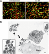
Intact granules and limited granule protein localization at extracellular DNA nets released through EETosis-mediated cell lysis. (A) Eosinophil EETosis was induced using 1 mg/mL immobilized IgG (120 minutes) or 2 μM A23187 (60 minutes) and fixed and permeabilized, then stained for MBP (green) and DNA (PI, red). Most released DNA nets did not contain free granule MBP protein and instead showed punctate or cell-associated staining. Images were obtained with a BX62 Olympus upright microscope, 20× UPlanApo objective with a numerical aperture of 0.70, coupled to a Hamamatsu Orca-AG fire-wire cooled digital camera (Hamamatsu Photonics, Hamamatsu, Japan; images were acquired using iVision software). Data are representative of >3 experiments from independent donors with similar results. (B) Cells were processed for TEM directly on slide surfaces. A23187-induced (2 μM, 60 minutes) EETosis eosinophils exhibit disrupted plasma membranes with clusters of released secretory granules (arrows) and DNA nets (Nets). Extracellular free granules (B, insert) show their typical morphologies with full crystalloid cores and matrices and intact granule-delimiting membranes. Some granules were entrapped in DNA nets. Scale bars represent 20 μm (A), 1 μm (B), and 700 nm (B, insert).
Figure 5
Figure 5
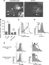
Characteristics of EETosis-induced intact eosinophil granule release. (A) Several cell-free granules were released by budding from PMs of eosinophils undergoing ETosis. Eosinophils were studied with time-lapse, phase-contrast imaging every 3 seconds following 1 mg/mL immobilized IgG or 2 μM A23187 stimulation. Images were captured from supplemental Video 1 and supplemental Video 2 (Nikon Eclipse TE300, 100× PlanApo objective with a numerical aperture of 1.40, equipped with a Qimaging Retiga EXi cooled digital camera in conjunction with iVision image analysis software). Eosinophils that initially showed a typical bilobed nucleus then demonstrated that peripheral small PM protrusions developed. Arrowheads show releasing/released granules. After nuclear membrane disintegration to release DNA nets within the cytoplasm, eosinophils gradually turned into a rounded shape, and granule movement slowed with deflected intracellular distribution of the granules. Several extracellular granules remained attached to the culture plate. Data representative of >3 experiments of independent donors with similar results are shown. (B) EETosis-elicited production of subcellular structures (⩽4 μm) was inhibited by DPI. Eosinophils were stimulated with A23187 (2 μM) in the presence of DPI in 0.1% BSA containing RPMI medium for 60 minutes. Subcellular structures in culture medium were counted as described in “Materials and methods” and supplemental Figure 2. Data (triplicate, mean ± SD) are representative of 3 experiments from independent donors with similar results. *P < .05. (C) Released cell-free structures retained the lysosomal dye AO. AO-loaded eosinophils were stimulated with A23187 for 1 hour. The structures (⩽4 μm) in culture medium from different preparations were subjected for flow cytometry. The graph shows the structures from AO-loaded cells (black line); fixed (2% PFO) and permeabilized (0.1% saponin) structures from AO-loaded cells (gray line); and structures from cells without AO staining (filled histogram). Data are representative of 3 experiments from independent donors with similar results. (D) A23187-induced subcellular structures were not apoptotic bodies. Subcellular structures (⩽4 μm) from A23187 and anti-Fas Ab–stimulated cells were stained with annexin V, followed by flow cytometric analysis. Fas-stimulated apoptotic cells produce annexin V–positive subcellular structures (ie, apoptotic bodies, filled histogram), but A23187-stimulated ETosis cells did not (open histogram). Apoptosis was induced, as described in supplemental Figure 4. Data are representative of 3 experiments from independent donors with similar results. (E) Isolated granule-rich subcellular structures retained the granule protein MBP; some were variably associated with PM-derived MHC class I proteins, regardless of size. Culture medium of A23187-stimulated cells was collected, and residual cells were removed by centrifugation (200 × g for 10 minutes). DNA was removed by DNase treatment, and buoyant vesicles and membranes were removed by further centrifugation (2500 × g for 10 minutes). Isolated granule-rich structures were stained for MBP and MHC class I (open histograms). Total structures (⩽4 μm) and small structures (⩽1 μm) were gated. Filled histograms show isotype-matched controls. Data are representative of 3 experiments from independent donors with similar results. Scale bars represent 5 μm (A). NS, not significant.
Figure 6
Figure 6
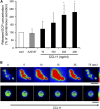
Released granules were secretion competent in response to CCL11. (A) Culture medium of A23187-stimulated cells (2 μM, 90 minutes) was collected, and granule-rich subcellular structures were isolated. The cell-free structures were stimulated with 2 μM A23187 or the indicated concentrations of CCL11 for 60 minutes. Secreted ECP levels, assessed by enzyme-linked immunosorbent assay, were normalized with spontaneous ECP release (100%), and data are expressed as means ± SD, from 3 different donors. *P < .05 vs nonstimulated controls. The spontaneous secretion levels were 10.0 ± 4.4% of the total ECP in the structures. (B) AO-loaded eosinophils were stimulated with 2 μM A23187 to induce EETosis, followed by the isolation of subcellular granule structures. Among the 80 single granules or groups of granules, following CCL11 stimulation, significant responses with intense transient fluorescent flashes indicative of the release of monomeric AO were observed from 14 nonenveloped granules (17.5%). The secretory response was not observed by likely PM-bound clusters of granules (lower panels). Images were obtained with a Hamamatsu Orca-AG fire-wire cooled digital camera coupled to a BX62 Olympus microscope using a 60× PlanApo objective with a numerical aperture of 1.42. Fluorescence intensity was analyzed by iVision software and pseudocolored with red to represent the greatest intensity as indicated by the scale color. Experiments were repeated with eosinophils from 8 independent donors. The scale bar represents 3 μm (B).
Figure 7
Figure 7
EETosis induces the release of intact cell-free granules through budding and cell lysis. The graphic shows the temporal course of the morphologic changes exhibited by eosinophils undergoing EETosis. Stimuli-elicited NADPH oxidase activation leads to the common process of eosinophil cytolysis, followed by the loss of the typically bilobed nuclei into a single round nuclei. During this time, some eosinophil granules were released extracellularly as PM-enveloped structures. Thereafter, nuclei disintegrated to form intracellular DNA nets. Subsequently, the eosinophils’ PMs rupture, releasing both a weblike chromatin structure and free eosinophil granules. EETosis-mediated cytolysis leads to the extracellular liberation of intact granules, some of which retained their capacity for secretory responses.

Similar articles

Cited by

References

    1. Klion AD, Nutman TB. The role of eosinophils in host defense against helminth parasites. J Allergy Clin Immunol. 2004;113(1):30–37. - PubMed
    1. Kay AB. The role of eosinophils in the pathogenesis of asthma. Trends Mol Med. 2005;11(4):148–152. - PubMed
    1. Lee JJ, Lee NA. Eosinophil degranulation: an evolutionary vestige or a universally destructive effector function? Clin Exp Allergy. 2005;35(8):986–994. - PubMed
    1. Neves JS, Weller PF. Functional extracellular eosinophil granules: novel implications in eosinophil immunobiology. Curr Opin Immunol. 2009;21(6):694–699. - PMC - PubMed
    1. Moqbel R, Coughlin JJ. Differential secretion of cytokines. Sci STKE. 2006;2006(338):pe26. - PubMed

Publication types

MeSH terms