. 2013 Mar;54(3):744-753.
doi: 10.1194/jlr.M033548. Epub 2013 Jan 7.

Quantitative fluorescence imaging reveals point of release for lipoproteins during LDLR-dependent uptake

Affiliations

Quantitative fluorescence imaging reveals point of release for lipoproteins during LDLR-dependent uptake

Shanica Pompey et al. J Lipid Res. 2013 Mar.

Abstract

The LDL receptor (LDLR) supports efficient uptake of both LDL and VLDL remnants by binding lipoprotein at the cell surface, internalizing lipoprotein through coated pits, and releasing lipoprotein in endocytic compartments before returning to the surface for further rounds of uptake. While many aspects of lipoprotein binding and receptor entry are well understood, it is less clear where, when, and how the LDLR releases lipoprotein. To address these questions, the current study employed quantitative fluorescence imaging to visualize the uptake and endosomal processing of LDL and the VLDL remnant β-VLDL. We find that lipoprotein release is rapid, with most release occurring prior to entry of lipoprotein into early endosomes. Published biochemical studies have identified two mechanisms of lipoprotein release: one that involves the β-propeller module of the LDLR and a second that is independent of this module. Quantitative imaging comparing uptake supported by the normal LDLR or by an LDLR variant incapable of β-propeller-dependent release shows that the β-propeller-independent process is sufficient for release for both lipoproteins but that the β-propeller process accelerates both LDL and β-VLDL release. Together these findings define where, when, and how lipoprotein release occurs and provide a generalizable methodology for visualizing endocytic handling in situ.


Figures

Fig. 1.
Fig. 1.
Quantitative imaging of clathrin, LDLR, and lipoprotein prior to uptake. Cells were treated or not with saturating concentrations of Alexa546-labeled LDL or β-VLDL at 4°C, washed, fixed, and counterstained for LDLR and clathrin by indirect immunofluorescence. A: MC and PC for LDLR-lipoprotein colocalization. Fifteen fields from three separate experiments were processed for MC of LDL with LDLR and β-VLDL with LDLR (MC LwR); MC of LDLR with LDL and LDLR with β-VLDL (MC RwL); and PC of LDLR with LDL and LDLR with β-VLDL (PC). Data are means ± SEM, n = 15. *P < 0.05 for β-VLDL compared with LDL. B: MC and PC for LDLR-clathrin colocalization. Fifteen fields from three experiments were processed for MC of LDLR with clathrin (MC RwC); MC of clathrin with LDLR (MC CwR); and PC of LDLR with clathrin in the presence or absence of LDL or β-VLDL. Data are means ± SEM, n = 15. *P < 0.05 compared with no lipoprotein.
Fig. 2.
Fig. 2.
LDL dissociates from the LDLR and enters EEA1-positive endosomes faster than β-VLDL. Surface LDLRs were saturated with Alexa546-LDL or β-VLDL at 4°C, washed, and shifted to 37°C for the indicated times. At each time point, cells were rapidly chilled to 4°C, fixed, and immunostained for LDLR and EEA1. Thirty fields from three separate experiments were processed for MC and PC of LDLR with LDL or β-VLDL (A and B) or for MC and PC of EEA1 with LDL or β-VLDL (C and D). Data are means ± SEM, n = 30.
Fig. 3.
Fig. 3.
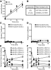
Lipoprotein release occurs prior to lipoprotein entry into early endosomes. A: LDL and β-VLDL internalize with similar kinetics. Surface LDLRs were saturated with 125I-LDL or 125I-β-VLDL at 4°C and then shifted to 37°C in the presence of 125I-lipoprotein for the indicated times. Surface-bound lipoprotein was then released by protease K, and surface and cell-associated (internal) lipoprotein was separated by centrifugation. Data are means ± SD, n = 4. B: Rates derived from linear regression of the data in (A). C and D: Lipoprotein internalization does not chase LDLRs into early endosomes. Data from Fig. 2 were processed for MC and PC of LDLR with EEA1 in the presence of LDL or β-VLDL. Data are means ± SEM, n = 30. E and F: Lipoprotein releases from the LDLR prior to entry of lipoprotein into early endosomes. Lipoprotein isosurfaces from three pulse-chase experiments were generated as described in Materials and Methods and in supplementary Fig. II. Isosurfaces were classified into four bins: surfaces that lack both LDLR and EEA1; surfaces that contain LDLR but not EEA1; surfaces that contain both LDLR and EEA1; and surfaces that contain EEA1 but not LDLR. Percentages of total lipoprotein surfaces for each bin were calculated for each experiment and are reported as means ± SEM, n = 3.
Fig. 4.
Fig. 4.
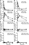
Deletion of the BC region slows both release and entry of lipoprotein into early endosomes. WT LDLR cells and LDLR-ΔBC cells were incubated with saturating concentration of Alexa546 LDL or β-VLDL at 4°C, washed, and shifted to 37°C for the indicated times. At each time point, cells were rapidly chilled to 4°C, washed, fixed and immunostained for LDLR and EEA1. Thirty fields from three experiments were used to generate MC and PC for colocalization. A and B: PC for the indicated receptors with LDL (A) or β-VLDL (B). C and D: PC for the EEA1 with LDL (C) or β-VLDL (D) in the indicated cells. E and F: PC for EEA1 with the indicated receptor in the presence of either LDL (E) or β-VLDL (F). G and H: MC for EEA1 with WT-LDLR or LDLR-ΔBC in the presence of either LDL (G) or β-VLDL (H). Data are means ± SEM, n = 30.
Fig. 5.
Fig. 5.
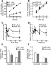
Deletion of the BC region increases LDL retro-endocytosis. The indicated cells were assayed for LDL and β-VLDL accumulation (A and B), total fluorescence intensity (C and D), or degradation and excretion of lipoprotein (E and F). A and B: Cells were incubated with saturating concentrations of Alexa546-LDL or β-VLDL for the indicated times at 37°C, washed, fixed and processed by flow cytometry. Data was normalized to cellular fluorescence of WT cells following 4 h of uptake. Data are means ± SD from three independent experiments. C and D: Total integrated fluorescence from each experiment described in Fig. 4 was quantified. Data are means ± SD, n = 3. E and F: Cells were incubated with saturating concentrations of 125I-LDL or 125I-β-VLDL at 4°C, washed, and either assayed for surface-bound lipoprotein or incubated at 37°C for 4 h. Media from cells incubated at 37°C were assayed for degradation products of LDL (TCA soluble counts) or excreted LDL (TCA insoluble counts). Data are shown as a fraction of initially surface-bound lipoprotein and are means ± SEM, n = 9. *P < 0.05 for LDLR-ΔBC compared with WT LDLR.
Fig. 6.
Fig. 6.
Co-internalization of LDL and β-VLDL reveals that LDL and β-VLDL use common endocytic compartments. WT LDLR cells were incubated with 10 μg/ml Alexa488-LDL, 5 μg/ml Alexa546-β-VLDL, or a mixture of 10 μg/ml Alexa488-LDL and 1.25 μg/ml Alexa546-β-VLDL at 4°C, and then washed and incubated at 37°C for the indicated times. The 10:1.25 ratio resulted in a ∼50:50 ratio of LDL and β-VLDL binding. At each time point, cells were rapidly chilled to 4°C, washed, fixed and immunostained for either EEA1 (A and C) or LDLR (B). A: MC and PC for colocalization of LDL with β-VLDL for cells incubated with a mixture of the two lipoproteins. B: PC for LDLR with LDL alone, with LDL in the presence of β-VLDL, with β-VLDL alone, or with β-VLDL in the presence of LDL. C: PC for EEA1 with LDL alone, with LDL in the presence of β-VLDL, with β-VLDL alone, or with β-VLDL in the presence of LDL. D: Degradation assays in which 125I-LDL or β-VLDL were incubated with WT LDLR cells at 4°C in the absence or presence of sufficient unlabeled β-VLDL or LDL, respectively, to decrease 125I-lipoprotein binding in half. +β-VLDL and +LDL indicate assays in which unlabeled β-VLDL or LDL were present. Concentrations of unlabeled lipoprotein were titrated to reduce radiolabeled lipoprotein binding by 50%. Cells were then washed and incubated at 37°C for the indicated times and assayed for 125I-degradation products. Data was normalized to initially bound 125I-lipoprotein and are means ± SD, n = 4.

Similar articles

Cited by

References

    1. Simonsen A., Lippe R., Christoforidis S., Gaullier J. M., Brech A., Callaghan J., Toh B. H., Murphy C., Zerial M., Stenmark H. 1998. EEA1 links PI(3)K function to Rab5 regulation of endosome fusion. Nature. 394: 494–498 - PubMed
    1. Mills I. G., Jones A. T., Clague M. J. 1999. Regulation of endosome fusion. Mol. Membr. Biol. 16: 73–79 - PubMed
    1. Tycko B., Maxfield F. R. 1982. Rapid acidification of endocytic vesicles containing alpha 2-macroglobulin. Cell. 28: 643–651 - PubMed
    1. Galloway C. J., Dean G. E., Marsh M., Rudnick G., Mellman I. 1983. Acidification of macrophage and fibroblast endocytic vesicles in vitro. Proc. Natl. Acad. Sci. USA. 80: 3334–3338 - PMC - PubMed
    1. Gerasimenko J. V., Tepikin A. V., Petersen O. H., Gerasimenko O. V. 1998. Calcium uptake via endocytosis with rapid release from acidifying endosomes. Curr. Biol. 8: 1335–1338 - PubMed

Publication types

LinkOut - more resources