. 2012;7(12):e52790.
doi: 10.1371/journal.pone.0052790. Epub 2012 Dec 28.

Piperidinols that show anti-tubercular activity as inhibitors of arylamine N-acetyltransferase: an essential enzyme for mycobacterial survival inside macrophages

Affiliations

Piperidinols that show anti-tubercular activity as inhibitors of arylamine N-acetyltransferase: an essential enzyme for mycobacterial survival inside macrophages

Areej Abuhammad et al. PLoS One. 2012.

Abstract

Latent M. tuberculosis infection presents one of the major obstacles in the global eradication of tuberculosis (TB). Cholesterol plays a critical role in the persistence of M. tuberculosis within the macrophage during latent infection. Catabolism of cholesterol contributes to the pool of propionyl-CoA, a precursor that is incorporated into cell-wall lipids. Arylamine N-acetyltransferase (NAT) is encoded within a gene cluster that is involved in the cholesterol sterol-ring degradation and is essential for intracellular survival. The ability of the NAT from M. tuberculosis (TBNAT) to utilise propionyl-CoA links it to the cholesterol-catabolism pathway. Deleting the nat gene or inhibiting the NAT enzyme prevents intracellular survival and results in depletion of cell-wall lipids. TBNAT has been investigated as a potential target for TB therapies. From a previous high-throughput screen, 3-benzoyl-4-phenyl-1-methylpiperidinol was identified as a selective inhibitor of prokaryotic NAT that exhibited antimycobacterial activity. The compound resulted in time-dependent irreversible inhibition of the NAT activity when tested against NAT from M. marinum (MMNAT). To further evaluate the antimycobacterial activity and the NAT inhibition of this compound, four piperidinol analogues were tested. All five compounds exert potent antimycobacterial activity against M. tuberculosis with MIC values of 2.3-16.9 µM. Treatment of the MMNAT enzyme with this set of inhibitors resulted in an irreversible time-dependent inhibition of NAT activity. Here we investigate the mechanism of NAT inhibition by studying protein-ligand interactions using mass spectrometry in combination with enzyme analysis and structure determination. We propose a covalent mechanism of NAT inhibition that involves the formation of a reactive intermediate and selective cysteine residue modification. These piperidinols present a unique class of antimycobacterial compounds that have a novel mode of action different from known anti-tubercular drugs.


Conflict of interest statement

Competing Interests: The authors have the following interests. E.S. and S.B. are members of the MRC UK TB Drug Discovery consortium, TBD-UK (http://www.tbd-uk.org.uk/). This does not alter the authors' adherence to all the PLOS ONE policies on sharing data and materials, as detailed online in the guide for authors.

Figures

Figure 1
Figure 1. The gene cluster that encodes for the nat gene in M. tuberculosis and M. bovis BCG and its relation to cholesterol catabolism.
The accession numbers, detailed at http://genolist.pasteur.fr/TubercuList/, for these genes in M. tuberculosis H37Rv are as follows: Rv3570c (hsaA), Rv3569c (hsaD), Rv3568c (hsaC), Rv3567c (hsaB), Rv3566A (possible pseudogene) and Rv3566c (nat). The gene cluster is virtually identical in M. tuberculosis and M. bovis BCG.
Figure 2
Figure 2. Specificity of compound 1 for prokaryotic NAT enzymes.
Compound 1 was tested at 30 µM against pure recombinant NAT enzymes from M. smegmatis (MSNAT), P. aeruginosa (PANAT), S. typhimurium (STNAT), MMNAT and TBNAT, and also against two eukaryotic enzymes, hamster NAT2 (shNAT2) and human NAT1. The results are shown as the mean ± S.D. of triplicate determinations of the percentage inhibition of hydrolysis of Ac-CoA in the presence of 5-aminosalicylic acid (5ASA) and against TBNAT using hydralazine as a substrate. The inhibition is represented as a percentage compared to an uninhibited control from triplicate measurements. The structure of compound 1 is shown and the piperidinol nucleus is highlighted by the shaded area.
Figure 3
Figure 3. Reversibility of the inhibition of TBNAT and MMNAT by compound 1.
Each enzyme (MMNAT, TBNAT, 0.07 mM, 50 µL) was preincubated either alone or with 15-fold molar excess 1 at 24°C for 1 h. Each sample was then dialysed against 1 L fresh assay buffer (20 mM Tris-HCl pH 8) at 4°C for 16 h. The enzyme activities of the samples were measured before dialysis and then measured after dialysis by measuring the rate of Ac-CoA hydrolysis in the presence of HLZ as described in Methods. The mean ± S.D. of three measurements of the activity is shown. Loss of enzyme activity upon dialysis is likely to be due to the oxidation of the active site sulfhydryl group, especially since dialysis was performed in the absence of dithiothreitol.
Figure 4
Figure 4. The time-dependent inhibition of the MMNAT and TBNAT by the piperidinols.
Semi-logarithmic plots showing the time-dependent inactivation of (A) MMNAT by various concentrations of 1 and (B) TBNAT by compound 3 at 23.8 µM. The enzyme activity was measured using the protocol described in Figure S1. The results are presented as the mean ± S.D. of triplicate measurements. The residual activity is shown as a percentage of a control prepared as described in Figure S1. The data were fitted against the incubation-time using the Semilog line (X is linear, Y is Log) module of GraphPad Prism 5.0. The slope of each line is equivalent to (−kobs/2.303) at each inhibitor concentration. The error bars are within the symbols.
Figure 5
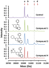
Figure 5. The ESI mass spectrum of MMNAT in the presence of 1, 3 and 4.
MMNAT was mixed with an equimolar sample (1∶1 ratio) of each inhibitor (50 µM) in 20 mM Tris-HCl, pH 8, and 5% (v/v) DMSO, and the ESI-MS was performed after 30 min of incubation. The masses correspond to each peak according to MMNAT with compound 3 chromatogram are: a = 30915 (Δm = 0 Da), b = 30955.5 (Δm = 40 Da), c = 31046.5 (Δm = 131.5 Da), and d = 31087.5 Da (Δm = 172.2 Da). The mass corresponding to the addition of a 132 Da-fragment is marked with a dashed line. Δm of +40 Da is likely to correspond to a potassium ion (38 Da). A mass spectrum of the protein in the absence of any inhibitor is shown as control in the top panel.
Figure 6
Figure 6. The chemical transformation of 1 to the corresponding phenyl vinyl ketone (PVK) and the subsequent modification of a thiol containing residue by the PVK.
(A) A proposed pathway of the formation of bis-Mannich bases from the rigid cyclic piperidinol. The bis-Mannich base can undergo a β-elimination of the amino group forming a reactive phenyl vinyl ketone (PVK). (B) The PVK reaction with thiols resulted in the addition of a 3-phenyl-3-oxopropyl moiety (POP) (when R1 is H) or a 3-(4-chlorophenyl)-3-oxopropyl moiety (when R1 is Cl). The expected Δm values of the added fragments are +132.07 Da and +166 Da, respectively. The shaded areas highlight the Michael acceptor moiety. Since the PVK binding species is transient, the second order rate constant cannot be determined without major assumptions being made.
Figure 7
Figure 7. A comparison of the 3D-shape of compounds 1 and 6 and their inhibition activity.
(A) The 3D-shape of compounds 1 and 6 are shown in a mesh view of the Van der Waals surface. Overlapping 3D-shapes of 1 (in white) and 6 are also shown. Energy minimisation of compounds 1 and 6 was performed using Grade (http://grade.globalphasing.org). The structure of 6 is shown. (B) The activity of MMNAT in the presence of 50 µM compound 1 or 6. The activity of MMNAT was measured after incubation with 50 µM of each inhibitor for 20 min before and after a 200-fold dilution. The NAT activity was measured by the NAT-inhibition assay using 150 µM of HLZ and 120 µM Ac-CoA. The percentage of enzyme activity was measured in the presence of 50 µM inhibitor and compared to the un-inhibited control. The results are presented as the mean ± S.D. from triplicate measurements at 24°C.
Figure 8
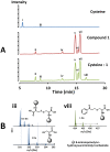
Figure 8. LC/MS analysis of the reaction of compound 1 with free cysteine.
(A) The total ion current chromatogram (from liquid chromatography LC) of 100 µM cysteine, 100 µM compound 1 and 100 µM cysteine: 1 (1∶1 mixture) in 20 mM MOPS buffer, pH 8 after 16 h incubation at 24°C. All samples were treated with 6-aminoquinolyl-n-hydroxysuccinimidyl carbamate before analysis. (B) The ESI-MS spectra of fractions collected from the peaks in the chromatogram (in A) corresponding to i: cysteine (m/z = 291.9 Da), iii: cystine (m/z = 290.9) and viii: the product of the reaction of cysteine with 1 (m/z = 423.9 Da). The chemical structures of the compounds corresponding to each peak are shown. The round symbol represents the aminoquinolyl carbamate moiety.
Figure 9
Figure 9. The active site electron density observed in the MMNAT-POP complex.
The crystal structure of MMNAT after reaction with compound 1 showed excess electron density connected to Cys70, into which a 3-phenyl-3-oxopropyl (POP) modification was modelled with full occupancy. All three cysteine residues in the MMNAT structure and the covalent modification (in pink) are shown with the electron density shown using blue 2Fo–Fc electron density contoured at 1 σ. This observation is compatible with the MS data, since the excess inhibitor was washed out prior to crystallisation and the native state of the protein was preserved throughout the structure determination process. The figures were prepared using PyMOL .
Figure 10
Figure 10. Reagents and conditions: (i) MeNH2.HCl, paraformaldehyde, MeCN, cat. HCl, Δ, 16 h.
Figure 11
Figure 11. The chemical dehydration of compound 1.

Similar articles

Cited by

References

    1. WHO (2011) World Health Organisation: Global tuberculosis control report 2011.
    1. Young DB, Gideon HP, Wilkinson RJ (2009) Eliminating latent tuberculosis. Trends in Microbiology 17: 183–188. - PubMed
    1. Gideon HP, Flynn JL (2011) Latent tuberculosis: what the host “sees”? Immunologic Research 50: 202–212. - PMC - PubMed
    1. Astarie-Dequeker C, Le Guyader L, Malaga W, Seaphanh FK, Chalut C, et al. (2009) Phthiocerol dimycocerosates of M. tuberculosis participate in macrophage invasion by inducing changes in the organization of plasma membrane lipids. PLoS Pathogens 5: e1000289. - PMC - PubMed
    1. Mishra AK, Driessen NN, Appelmelk BJ, Besra GS (2011) Lipoarabinomannan and related glycoconjugates: structure, biogenesis and role in Mycobacterium tuberculosis physiology and host–pathogen interaction. FEMS Microbiology Reviews 35: 1126–1157. - PMC - PubMed

Publication types

MeSH terms

Associated data