. 2013 Jan 7;200(1):45-60.
doi: 10.1083/jcb.201210106. Epub 2012 Dec 31.

The CCAN recruits CENP-A to the centromere and forms the structural core for kinetochore assembly

Affiliations

The CCAN recruits CENP-A to the centromere and forms the structural core for kinetochore assembly

Tetsuya Hori et al. J Cell Biol. .

Abstract

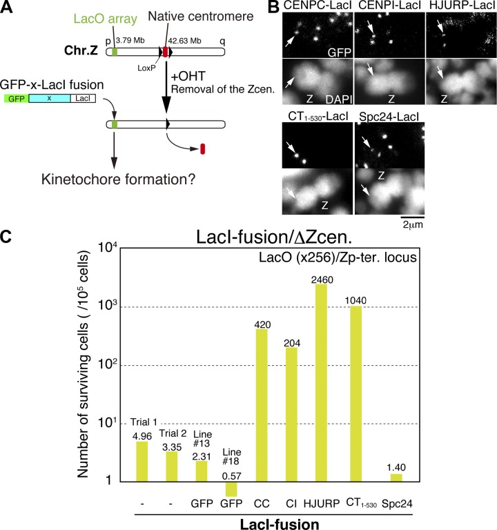
CENP-A acts as an important epigenetic marker for kinetochore specification. However, the mechanisms by which CENP-A is incorporated into centromeres and the structural basis for kinetochore formation downstream of CENP-A remain unclear. Here, we used a unique chromosome-engineering system in which kinetochore proteins are targeted to a noncentromeric site after the endogenous centromere is conditionally removed. Using this system, we created two distinct types of engineered kinetochores, both of which were stably maintained in chicken DT40 cells. Ectopic targeting of full-length HJURP, CENP-C, CENP-I, or the CENP-C C terminus generated engineered kinetochores containing major kinetochore components, including CENP-A. In contrast, ectopic targeting of the CENP-T or CENP-C N terminus generated functional kinetochores that recruit the microtubule-binding Ndc80 complex and chromosome passenger complex (CPC), but lack CENP-A and most constitutive centromere-associated network (CCAN) proteins. Based on the analysis of these different engineered kinetochores, we conclude that the CCAN has two distinct roles: recruiting CENP-A to establish the kinetochore and serving as a structural core to directly recruit kinetochore proteins.


Figures

Figure 1.
Figure 1.
Ectopic localization of CCAN proteins causes efficient kinetochore formation at a noncentromere locus. (A) An experimental design to examine kinetochore formation at a noncentromeric locus after removal of endogenous centromere of chromosome Z. The 256 copies of LacO repeats are integrated on the 3.79-Mb locus of chromosome Z in DT40 cells (LacO locus). GFP/LacI double fusion constructs with multiple different proteins (X) were expressed in cells with the LacO locus. The endogenous centromere of chromosome Z was removed by activation of Cre-recombinase (OHT addition). After removal of endogenous centromere, surviving clones were isolated. Surviving cells are expected to have an ectopic kinetochore at the LacO locus on chromosome Z. Experimental timeline is shown in Fig. S1. (B) Localization of GFP/LacI double fusion with multiple different proteins at the LacO locus before removal of endogenous centromere. Arrows show the LacO locus. All tested fusion proteins localize into the LacO locus. Z, Z chromosome. (C) The survival rate of cells after removal of endogenous centromere of chromosome Z in each assay. The survival rate was increased in cells expressing LacI fusion to CENP-C (CC), CENP-I (CI), HJURP, or CENP-T N terminus (CT1–530), compared with that of cells expressing GFP-LacI or lacking LacI fusion protein. The survival rate was not increased in cells expressing LacI-Spc24. The assay was completed once for each experiment except for CENP-C fusion. In the case of CENP-C, the assay was done twice.
Figure 2.
Figure 2.
Two distinct types of engineered kinetochores at the LacO locus were created. (A) Growth curve of the stable selected cell lines with the ectopic kinetochore at the LacO locus after removal of endogenous centromere of chromosome Z in the presence (red line) or absence of IPTG (black line). Each cell line was referred to as CC-LacI/ΔZcen (for CENP-C–LacI expression), CI-LacI/ΔZcen (for CENP-I–LacI expression), or HJ-LacI/ΔZcen (for HJURP-LacI expression). This measurement was completed once in each cell line. (B) Representative images of chromosome Z (Z) stained by the Z-specific probe (red) during anaphase of CC-LacI/ΔZcen, CI-LacI/ΔZcen, or HJ-LacI/ΔZcen cells after addition of IPTG. Arrows indicate FISH signals by a Z chromosome–specific satellite probe. (C) Localization of CENP-A, CENP-C, CENP-T, and Ndc80 at the LacO locus in CC-LacI/ΔZcen, CI-LacI/ΔZcen, or HJ-LacI/ΔZcen cells. These proteins were detected at the LacO locus as red signals. To define the chromosome Z, the Z-specific satellite probes are hybridized (green) on the q arm of chromosome Z (Zq). These experiments were performed in the absence of IPTG. Arrows indicate red signals stained with each antibody. (D) CENP-A–associated DNAs were isolated by ChIP with anti–CENP-A antibodies from CC-LacI/ΔZcen, CI-LacI/ΔZcen, or HJ-LacI/ΔZcen cells. Cells expressing GFP-LacI were also used as a control. The chromatin was isolated as a mononucleosome fraction digested with MNase. The CENP-A–associated DNAs were hybridized with a probe containing the LacO sequence or a centromere DNA from chromosome 5. LacO sequences were detected in CENP-A immunoprecipitates in CC-LacI/ΔZcen, CI-LacI/ΔZcen, or HJ-LacI/ΔZcen cells, but not in GFP-LacI cells. (E) Growth curve of CT1–530-LacI/ΔZcen cells in the presence or absence of IPTG. After addition of IPTG, these cells rapidly died (red solid line). This measurement was completed once in each condition. (F) Representative images of lagging chromosome Z stained by the Z-specific probe (red) during anaphase of CT1–530-LacI/ΔZcen cells after addition of IPTG. (G) Examination of loss or gain of chromosome Z after addition of IPTG to CT1–530-LacI/ΔZcen cells. As a control, numbers of chromosome 1 were also scored. Chromosome Z was lost in >80% of cells at 24 h and 48 h after the addition of IPTG (top). Loss or gain of chromosome 1 was not observed after addition of IPTG to CT1–530-LacI/ΔZcen cells (bottom). This experiment was completed once in each condition (CT1–530-LacI/+Zcen +IPTG, n = 223; CT1–530-LacI/ΔZcen 0 h, n = 140; 24 h, n = 201; 48 h, n = 94). (H) CENP-A–associated DNAs isolated by ChIP with anti–CENP-A antibodies from CT1–530-LacI/ΔZcen cells were hybridized with a probe containing the LacO sequence or a centromere DNA from chromosome 5. LacO sequences were not detected in CENP-A immunoprecipitates in CT1–530-LacI/ΔZcen cells.
Figure 3.
Figure 3.
Characterization of the CENP-C–derived kinetochores. (A) Diagram showing the chicken CENP-C sequence and the tested deletion constructs for the LacO–LacI–based kinetochore induction assay shown in Fig. 1. The yellow box shows a putative binding domain with the Mis12 complex. Mif2 II and Mif2 III show homology regions with yeast Mif2 (red). The survival rate of cells after removal of native centromere of chromosome Z in cells expressing each LacI fusion construct with multiple deletions is shown in the diagram (right). The survival rate was increased in cells expressing LacI fusion with CENP-C (1–643) or CENP-C (601–864). The assay was completed twice for CC full-length, once for CC1–643, once for CC74–643, once for CC1–165, twice for CC601–761, and three times for CC601–864. (B) Growth curve of CC601–864-LacI/ΔZcen cells in the presence (red line) or absence of IPTG (black line). This measurement was completed once in each condition. (C) Localization of CENP-A, CENP-C, CENP-T, and Ndc80 at the LacO locus in CC601–864-LacI/ΔZcen cells. These experiments were performed in the absence of IPTG. Arrows indicate red signals stained with each antibody. Zq, Z chromosome. (D) CENP-A–associated DNAs isolated by ChIP with anti–CENP-A antibodies from CC601–864-LacI/ΔZcen cells were hybridized with a probe containing the LacO sequence or a centromere DNA from chromosome 5. (E) Growth curve of CC1–643-LacI/ΔZcen cells in the presence or absence of IPTG. This measurement was completed once in each condition. (F) Representative images of lagging chromosome Z stained by the Z-specific probe (red) during anaphase of CC1–643-LacI/ΔZcen cells after addition of IPTG. (G) Examination of loss or gain of chromosome Z after addition of IPTG to CC1–643-LacI/ΔZcen cells (top). As a control, numbers of chromosome 1 were also scored (bottom). Chromosome Z was lost in >80% of cells at 48 h after the addition of IPTG. Loss or gain of chromosome 1 was not observed after addition of IPTG to CC1–643-LacI/ΔZcen cells. This experiment was completed once in each condition (CC1–643-LacI/+Zcen +IPTG, n = 229; CC1–643-LacI/ΔZcen 0 h, n = 281; 24 h, n = 235; 48 h, n = 204). (H) CENP-A–associated DNAs isolated by ChIP with anti-CENP-A antibodies from CC1–643-LacI/ΔZcen cells were hybridized with a probe containing the LacO sequence or a centromere DNA from chromosome 5. LacO sequences were not detected in CENP-A immunoprecipitates in CC1–643-LacI/ΔZcen cells. (I) Localization of Mis12 at the LacO locus in cells expressing LacI fusions with CENP-C (1–643), CENP-C (74–643), or CENP-C (1–165). Mis12 signals were detected in cells expressing LacI fusion with CENP-C (1–643), but not detected in cells expressing LacI fusions with CENP-C (74–643) or CENP-C (1–165). Arrows indicate the position of the LacO sequence. Z, Z chromosome.
Figure 4.
Figure 4.
The CENP-T and CENP-C N terminus–derived kinetochores are functional. (A) Mitotic progression of CT1–530-LacI/ΔZcen, CC1–643-LacI/ΔZcen, or wild-type DT40 cells observed by live-cell imaging under a microscope in the absence of IPTG. Selected images are shown. (B) Numbers of anaphase cells with normal or abnormal segregation of chromosome Z in CT1–530-LacI/ΔZcen or CC1–643-LacI/ΔZcen cells (in the absence of IPTG). It is rare to detect cells with abnormal segregation of chromosome Z in the absence of IPTG. GFP-LacI/Zp-ter LacO cells were used as a control. This measurement was completed once (GFP-LacI/Zp-ter LacO, n = 139; CT1–530-LacI/ΔZcen, n = 122; CC1–643-LacI/ΔZcen, n = 229). (C) Localization of CCAN (top), outer kinetochore (middle), and checkpoint proteins (bottom) at the LacO locus in CT1–530-LacI/ΔZcen cells. Each tested protein is shown in red and the LacI fusion with CENP-T (1–530) is shown in green. All tested CCAN proteins were not detected at the LacO locus, but outer kinetochore and checkpoint proteins were detected. Data were also summarized in F. (D) Localization of CCAN (top), outer kinetochore (middle), and checkpoint proteins (bottom) at the LacO locus in CC1–643-LacI/ΔZcen cells. Each tested protein is shown in red and the LacI fusion with CENP-C (1–643) is shown in green. Arrows indicate the position of the LacO sequence. Z, Z chromosome. (E) Relative signal intensities of Ndc80 or Mis12 at the LacO locus compared with those at endogenous kinetochores in CT1–530-LacI/ΔZcen or CC1–643-LacI/ΔZcen cells. Error bars indicate mean ± SD. (F) Summary of the localization results of various kinetochore proteins at the LacO locus in CT1–530-LacI/ΔZcen or CC1–643-LacI/ΔZcen cells. +, positive localization at the LacO locus; −, negative localization at the LacO locus.
Figure 5.
Figure 5.
The kinetochore-like structure is generated on the LacO-containing plasmid DNA in cells expressing LacI fusion with CENP-T N terminus. (A) The LacO-containing plasmids were transiently introduced into cells expressing LacI fusion with CENP-T N terminus (1–530). After introduction of the plasmids, cells were characterized at each time point. (B) Percentage of cells retaining the LacO plasmids at each time point after introduction of the plasmids into cells expressing LacI fusion with CENP-T N terminus (1–530). The plasmids were also introduced into cells expressing GFP-LacI fusion. The plasmids were retained in 51.1% of cells expressing LacI fusion with CENP-T N terminus (1–530) at 32 h after transfection, whereas all plasmids were lost in all cells expressing GFP-LacI fusion at 32 h after transfection. This experiment was completed once (cell with CT1–530, n > 340; cell with GFP-LacI, n > 160). (C) Behavior of the LacO-containing plasmids (green) during mitosis of cell expressing LacI fusion with CENP-T N terminus (1–530) (top) or GFP-LacI fusion (bottom). Cells were observed by live-cell imaging under a microscope. Selected images are shown. Many plasmids were segregated into daughter cells expressing LacI fusion with CENP-T N terminus (1–530) (see 9 min of top panel), whereas many plasmids were lost in cells expressing GFP-LacI fusion. (D) Localization of Ndc80 on the LacO-containing plasmids. Colocalization of CENP-T–LacI with Ndc80 was observed (left and middle), whereas colocalization of GFP-LacI with Ndc80 was not observed (right).
Figure 6.
Figure 6.
The CPC and KNL1 are recruited into the CENP-T or CENP-C N terminus–derived kinetochores. (A) Localization of Aurora B, Survivin, and phosphorylated T3 of histone H3 (H3T3ph) at the LacO locus in CT1–530-LacI/ΔZcen or CC1–643-LacI/ΔZcen cells. Each protein is shown in red and CENP-T–LacI or CENP-C–LacI signals are shown in green. Aurora B, Survivin, and H3T3ph are detected in the inner centromere between sister kinetochores induced by both CENP-T and CENP-C N termini at the LacO locus. An antibody against H3T3ph was provided by H. Kimura (Osaka University, Osaka, Japan). (B) Localization of Ndc80, Nsl1, KNL1, Bub1, Aurora B, and H3T3ph at the LacO locus in cells expressing LacI fusion with CENP-T (1–530) (top), CENP-T (1–120) (middle), or CENP-T (90–530) (bottom). Each tested protein is shown in red. CENP-T derivatives are shown in green at the LacO locus. Data were summarized in Fig. S5 E. Z, Z chromosome. (C) Localization of Ndc80, Mis12, KNL1, Bub1, Aurora B, and H3T3ph at the LacO locus in cells expressing LacI fusion with CENP-C (1–643) (top) or CENP-C (74–643) (bottom). Each tested protein is shown in red. CENP-C derivatives are shown in green at the LacO locus. Arrows indicate the position of the LacO sequence. Data are summarized in Fig. S5 F. (D) Localization of Bub1, Aurora B, or H3T3ph in KNL1-deficient DT40 cells (bottom, KNL1 OFF). Top panels show localization of Bub1, Aurora B, or H3T3ph in DT40 cells expressing KNL1 (KNL1 ON). Bub1 localization was abolished in KNL1 OFF cells (bottom). Whereas Aurora B and H3T3ph signals were concentrated on inner centromere in KNL1 ON cells, these signals were diffused along chromosome axis in KNL1 OFF cells. Arrows indicate the position of the centromere.
Figure 7.
Figure 7.
Model of kinetochore assembly in the engineered kinetochores. (A) Molecular architecture of the CENP-T or CENP-C N terminus–derived kinetochores. CENP-A and CCAN proteins are not detected in both the CENP-T or CENP-C N terminus–derived kinetochores. Function of these kinetochores is sensitive to IPTG addition. The CENP-T N terminus directly binds to the Ndc80 complex and the CENP-C N terminus binds to the Mis12 complex, which associates with the Ndc80 complex. Although both kinetochores do not contain CENP-A and CCAN, the CPC is recruited. If CENP-T or CENP-C N terminus were steadily supplied to the LacO locus, functional kinetochores would be formed. This indicates that CENP-T or CENP-C N terminus is sufficient for formation of functional kinetochores in the absence of CENP-A and most CCAN proteins. (B) Kinetochore formation at the LacO locus induced by LacI fusion with HJURP, CENP-I, full-length CENP-C, or CENP-C C terminus (601–864). These kinetochores are resistant to addition of IPTG, and full kinetochore components are recruited. HJURP is known as a CENP-A–specific chaperone. CENP-I and CENP-C C terminus function as a mark for CENP-A incorporation. Once a full kinetochore is formed at the LacO locus, Lac-I fusions with HJURP, CENP-I, or CENP-C proteins are dispensable. Characterization of this type of kinetochore suggests that a major function of CENP-C or CENP-I is recruitment of CENP-A.

Comment in

Similar articles

Cited by

References

    1. Alushin G.M., Ramey V.H., Pasqualato S., Ball D.A., Grigorieff N., Musacchio A., Nogales E. 2010. The Ndc80 kinetochore complex forms oligomeric arrays along microtubules. Nature. 467:805–810 10.1038/nature09423 - DOI - PMC - PubMed
    1. Barnhart M.C., Kuich P.H., Stellfox M.E., Ward J.A., Bassett E.A., Black B.E., Foltz D.R. 2011. HJURP is a CENP-A chromatin assembly factor sufficient to form a functional de novo kinetochore. J. Cell Biol. 194:229–243 10.1083/jcb.201012017 - DOI - PMC - PubMed
    1. Black B.E., Cleveland D.W. 2011. Epigenetic centromere propagation and the nature of CENP-a nucleosomes. Cell. 144:471–479 10.1016/j.cell.2011.02.002 - DOI - PMC - PubMed
    1. Brown M.T. 1995. Sequence similarities between the yeast chromosome segregation protein Mif2 and the mammalian centromere protein CENP-C. Gene. 160:111–116 10.1016/0378-1119(95)00163-Z - DOI - PubMed
    1. Burrack L.S., Berman J. 2012. Flexibility of centromere and kinetochore structures. Trends Genet. 28:204–212 10.1016/j.tig.2012.02.003 - DOI - PMC - PubMed

Publication types

MeSH terms