

doi: 10.1083/jcb.201207180.
Cab45 is required for Ca(2+)-dependent secretory cargo sorting at the trans-Golgi network
Affiliations
- PMID: 23266954
- PMCID: PMC3529532
- DOI: 10.1083/jcb.201207180
Item in Clipboard
Cab45 is required for Ca(2+)-dependent secretory cargo sorting at the trans-Golgi network
J Cell Biol.
.
Abstract
Ca(2+) import into the lumen of the trans-Golgi network (TGN) by the secretory pathway calcium ATPase1 (SPCA1) is required for the sorting of secretory cargo. How is Ca(2+) retained in the lumen of the Golgi, and what is its role in cargo sorting? We show here that a soluble, lumenal Golgi resident protein, Cab45, is required for SPCA1-dependent Ca(2+) import into the TGN; it binds secretory cargo in a Ca(2+)-dependent reaction and is required for its sorting at the TGN.
Figures
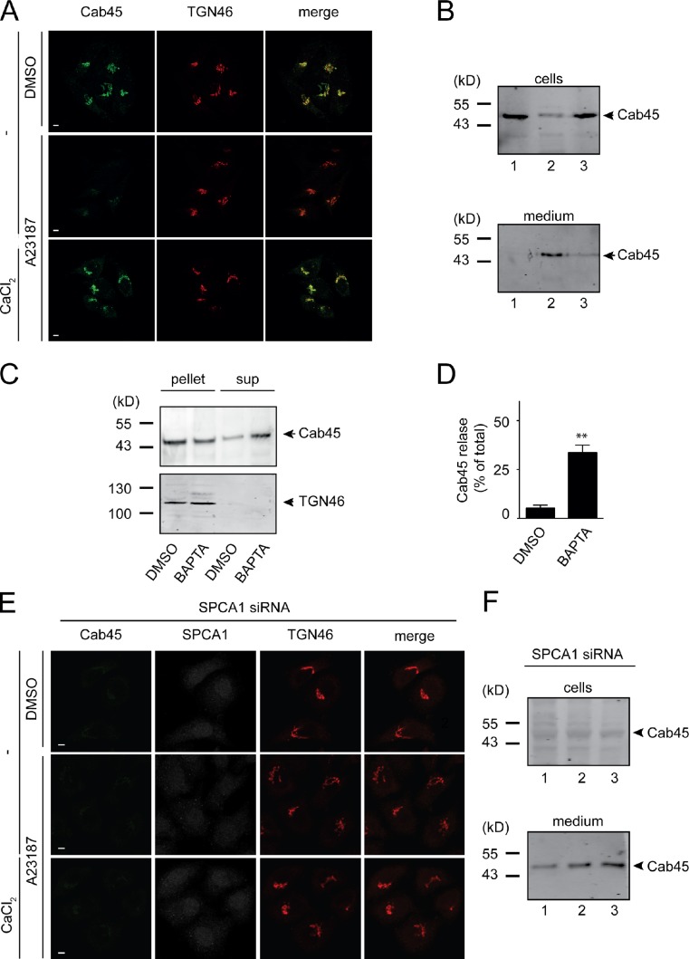
Ca2+ is required for retention of Cab45 in the Golgi apparatus. (A) HeLa cells were incubated in a Ca2+-free medium. After 1 h, cells were incubated for 5 min with A23187 with or without 10 mM CaCl2, after which the localization of Cab45 and TGN46 was visualized by immunofluorescence microscopy. Bars, 5 µm. (B) The experiment described in A was repeated; the cell lysates and the corresponding media were analyzed by Western blotting with an anti-Cab45 antibody (lane 1, DMSO; lane 2, 2.5 µM A23187; lane 3, 2.5 µM A23187 + 10 mM CaCl2). (C) Golgi membranes isolated from HeLa cells were incubated with DMSO or 25 µM BAPTA for 15 min. The membranes were subjected to five cycles of rapid freeze-thaw and finally centrifuged at 100,000 g. The pellets and the supernatants (sup) were analyzed by Western blotting with anti-Cab45 (top) and anti-TGN46 (bottom) antibodies, respectively. (D) Western blot bands from three independent experiments were quantified by densitometry using the ImageJ software. Bar graphs represent the densitometry values of external Cab45 (released from the Golgi membranes) normalized to the internal Cab45 values. Error bars show the mean ± SD densitometric values of three independent experiments. **, P < 0.01. (E) HeLa cells were transfected with SPCA1 siRNA. After 72 h at 37°C, the cells were incubated in Ca2+-free medium and treated with A23187 in the absence or presence of 10 mM CaCl2 and visualized by fluorescence microscopy as described in A. (F) The experiment was repeated as described in E; the cell lysates and the corresponding media were analyzed by Western blotting with an anti-Cab45 antibody (lane 1, DMSO; lane 2, 2.5 µM A23187; lane 3, 2.5 µM A23187 + 10 mM CaCl2).
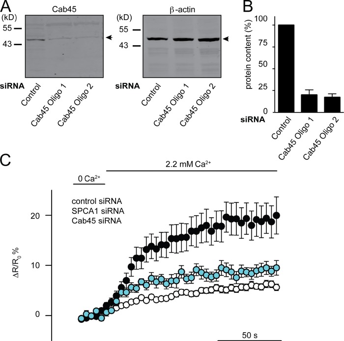
Ca2+ homeostasis of the TGN requires Cab45. (A) Lysates of HeLa cells transfected with control or Cab45-specific siRNA (Oligo1 and Oligo2) were Western blotted with anti-Cab45 (left) and β-actin antibodies (right). (B) The knockdown efficiency of Cab45 from three different experiments was quantitated by densitometry (histograms). Bar graphs represent the mean ± SD of triplicate experiments (error bars). Compared datasets were statistically significant (**) when P < 0.01. (C) HeLa cells were transfected with scrambled (control), Cab45 (Oligo1), and SPCA1-specific siRNA. After 48 h, cells were transfected with the TGN-specific Ca2+ FRET sensor GoD1cpv. After 12 h, cells were depleted of Ca2+ by incubation in a Ca2+-free solution and 1 µM Ionomycin for 1 h at 4°C. Subsequently, TGN Ca2+ influx was measured by detecting FRET signals of YFP (520 nm) and CFP (480) laser lines using an inverted confocal microscope (TCS SP5; Leica). Images were taken at 400 Hz at 20°C. TGN [Ca2+] fluorescent signals were presented as ΔR/R0 (R0 is the value measured before of 2.2 mM Ca2+ addition).
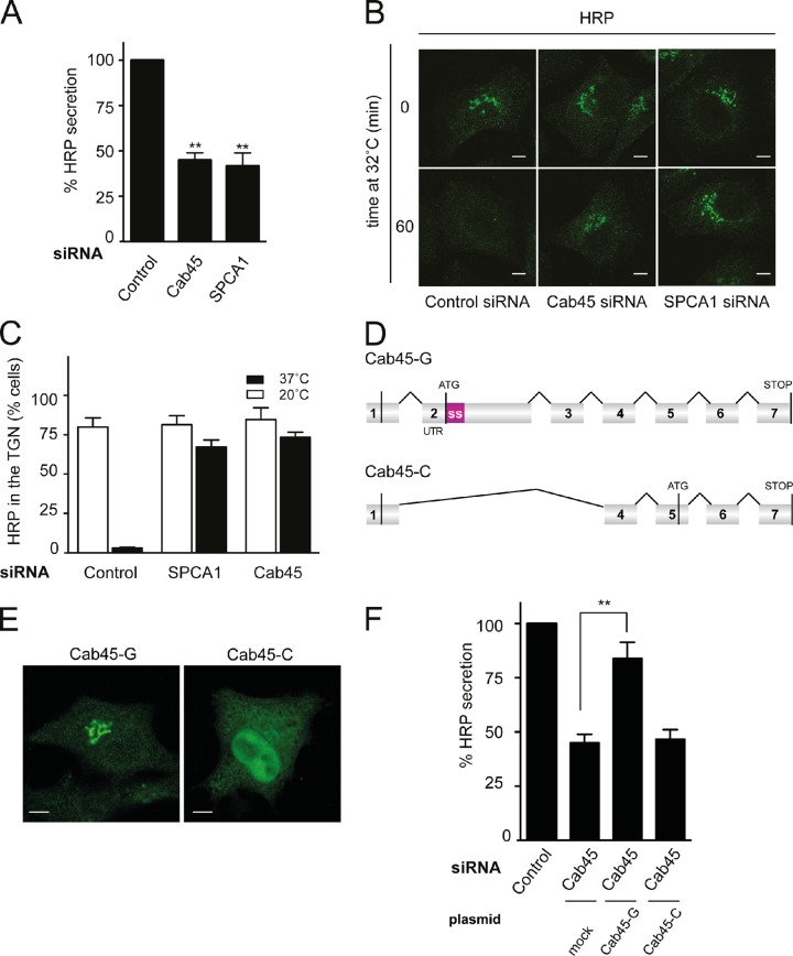
The siRNA knockdown of the Golgi isoform of Cab45 causes missorting of secretory cargo. (A) HeLa ss-HRP–expressing cells were transfected with scrambled (control), Cab45, or SPCA1 siRNA for 72 h. The cell culture supernatant from the respective cells was analyzed for HRP activity by chemiluminescence. Error bars show mean ± SD of external HRP activity normalized to internal HRP activity of three independent experiments. Datasets were statistically significant when P < 0.01 (**). (B) HeLa cells were transfected as described in A and incubated at 20°C for 2 h. Subsequently, the temperature was changed to 32°C, and the cellular distribution of HRP was monitored by fluorescence microscopy using an anti-HRP antibody. Bars, 5 µm. (C) HRP location was quantitated in 100 cells for each of the conditions described in B. Bar graphs represent data from at least three different experiments. (D) Schematic presentation of the mRNAs encoding full-length Cab45 (Cab45-G contained in the Golgi membrane) or Cab45 C (the cytoplasmic isoform), as reported previously (Lam et al., 2007). The exons are numbered 1–7; ATG, methionine start codon; STOP, stop codon; SS, cleavable amino-terminal signal sequence of Cab45G. The diagram is modified from Lam et al. (2007). (E) Cells expressing HA-Cab45-G or HA-Cab45-C were stained with an HA antibody and analyzed by confocal microscopy. Bars, 5 µm. (F) HeLa cells were transfected with control or Cab45 siRNA. After 48 h, cells were transfected with ss-HRP–Flag and siRNA-resistant HA-Cab45-G or HA-Cab45-C. Media from these cells was analyzed for HRP activity by chemiluminescence. Error bars indicate the mean ± SD of HRP activity in the medium normalized to HRP activity in cell lysates measured in at least three independent experiments. **, P < 0.01.
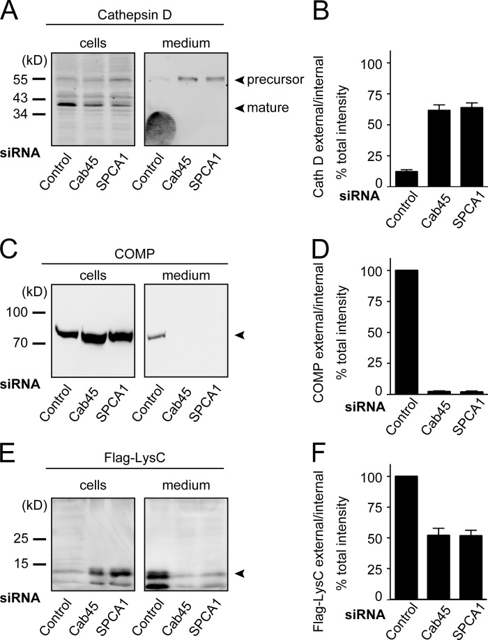
Cab45 knockdown by siRNA causes missorting of Cathepsin D, COMP, and LysC. (A and C) HeLa cells were stably transfected with scrambled (control), Cab45, or SPCA1 siRNA for 72 h as described in Fig. 3 A. Media and cell lysates from the respective cells were Western blotted with specific antibodies against Cathepsin D (A) or COMP (C). (B and D) Western blots from three independent experiments were quantified by densitometry using the ImageJ software. Bar graphs represent the densitometry values of external Cathepsin D and COMP normalized to internal Cathepsin D and COMP values, respectively. (E) The medium and the lysates of HeLa cells expressing Flag-LysC were analyzed by Western blotting with an anti-Flag antibody. (F) Western blots from three independent experiments were quantified by densitometry using the ImageJ software. Bar graphs represent the densitometric values of external Flag-LysC normalized to the internal Flag-LysC values. Error bars show mean ± SD of the densitometric values of three independent experiments.
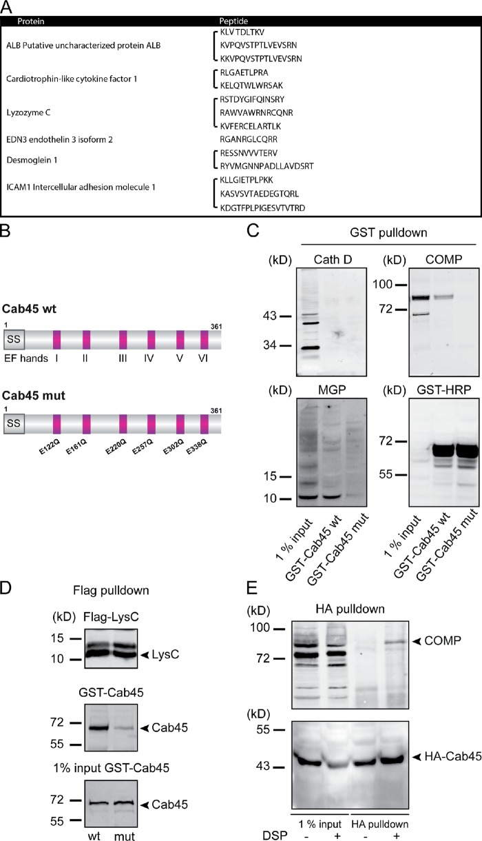
Cab45 binds secretory cargo. (A) GST and GST Cab45 expressed in E. coli were purified on GST beads. The respective preparations of beads were incubated with HeLa cell lysate for 4 h at 4°C, washed extensively, and finally incubated with SDS-PAGE sample buffer to release the bound proteins, which were then analyzed by SDS-PAGE. Gels were stained with Coomassie blue, and protein bands were excised and analyzed by mass spectrometry. The full list of identified proteins is shown in Table S1 . This pool contained several proteins that are secreted or transported to the plasma membranes and are shown here. (B) Schematic presentation of Cab45 wt and the amino acids mutated in the EF hand domains of Cab45 mutant. (C) Recombinant purified GST-Cab45 wt or GST-Cab45 mutant bound to glutathione Sepharose beads were incubated with HeLa cell lysates. Beads were washed extensively and then incubated with SDS-PAGE sample buffer to elute the bound proteins, which were Western blotted with antibodies to Cathepsin D, COMP, MGP, and GST. (D) Cells expressing Flag-LysC were lysed and incubated with anti-Flag beads for 1 h at 4°C. Beads were then incubated with recombinant GST-Cab45 wt or GST Cab45 mutant. Bound proteins were eluted with SDS-PAGE sample buffer and Western blotted with anti-GST and anti-Flag antibodies. (E) HeLa cells stably expressing HA-Cab45 wt were incubated with PBS-DMSO or 1 mM PBS-DSP for 30 min at room temperature. The reaction was terminated and the HA-tagged Cab45 was immunoprecipitated and analyzed by Western blotting with antibodies against HA and COMP.
Similar articles
-
Secretory cargo sorting by Ca2+-dependent Cab45 oligomerization at the trans-Golgi network.J Cell Biol. 2016 May 9;213(3):305-14. doi: 10.1083/jcb.201601089. Epub 2016 May 2. J Cell Biol. 2016. PMID: 27138253 Free PMC article.
-
Cab45-Unraveling key features of a novel secretory cargo sorter at the trans-Golgi network.Eur J Cell Biol. 2017 Aug;96(5):383-390. doi: 10.1016/j.ejcb.2017.03.001. Epub 2017 Mar 18. Eur J Cell Biol. 2017. PMID: 28372832 Review.
-
Exploring new routes for secretory protein export from the trans-Golgi network.Mol Biol Cell. 2018 Feb 1;29(3):235-240. doi: 10.1091/mbc.E17-02-0117. Mol Biol Cell. 2018. PMID: 29382805 Free PMC article.
-
Activity of the SPCA1 Calcium Pump Couples Sphingomyelin Synthesis to Sorting of Secretory Proteins in the Trans-Golgi Network.Dev Cell. 2018 Nov 19;47(4):464-478.e8. doi: 10.1016/j.devcel.2018.10.012. Epub 2018 Nov 1. Dev Cell. 2018. PMID: 30393074 Free PMC article.
-
Secretory cargo sorting at the trans-Golgi network.Trends Cell Biol. 2014 Oct;24(10):584-93. doi: 10.1016/j.tcb.2014.04.007. Epub 2014 May 16. Trends Cell Biol. 2014. PMID: 24841758 Review.
Cited by
-
Vti1a/b regulate synaptic vesicle and dense core vesicle secretion via protein sorting at the Golgi.Nat Commun. 2018 Aug 24;9(1):3421. doi: 10.1038/s41467-018-05699-z. Nat Commun. 2018. PMID: 30143604 Free PMC article.
-
Receptor-mediated transport of vacuolar proteins: a critical analysis and a new model.Protoplasma. 2014 Jan;251(1):247-64. doi: 10.1007/s00709-013-0542-7. Epub 2013 Sep 10. Protoplasma. 2014. PMID: 24019013
-
Molecular mechanisms of polarized transport to the apical plasma membrane.Front Cell Dev Biol. 2024 Sep 26;12:1477173. doi: 10.3389/fcell.2024.1477173. eCollection 2024. Front Cell Dev Biol. 2024. PMID: 39445332 Free PMC article. Review.
-
Lipid-dependent coupling of secretory cargo sorting and trafficking at the trans-Golgi network.FEBS Lett. 2019 Sep;593(17):2412-2427. doi: 10.1002/1873-3468.13552. Epub 2019 Jul 30. FEBS Lett. 2019. PMID: 31344259 Free PMC article. Review.
-
The Trans Golgi Region is a Labile Intracellular Ca2+ Store Sensitive to Emetine.Sci Rep. 2018 Nov 21;8(1):17143. doi: 10.1038/s41598-018-35280-z. Sci Rep. 2018. PMID: 30464185 Free PMC article.
References
Publication types
MeSH terms
Substances
LinkOut - more resources
Full Text Sources
Other Literature Sources
Miscellaneous
