. 2013 Mar;87(5):2549-62.
doi: 10.1128/JVI.03104-12. Epub 2012 Dec 19.

The highly conserved layer-3 component of the HIV-1 gp120 inner domain is critical for CD4-required conformational transitions

Affiliations

The highly conserved layer-3 component of the HIV-1 gp120 inner domain is critical for CD4-required conformational transitions

Anik Désormeaux et al. J Virol. 2013 Mar.

Abstract

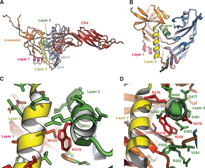
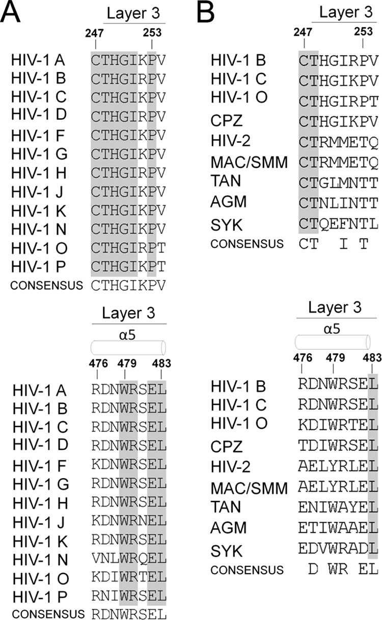
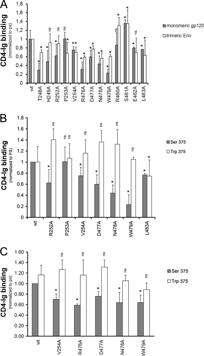
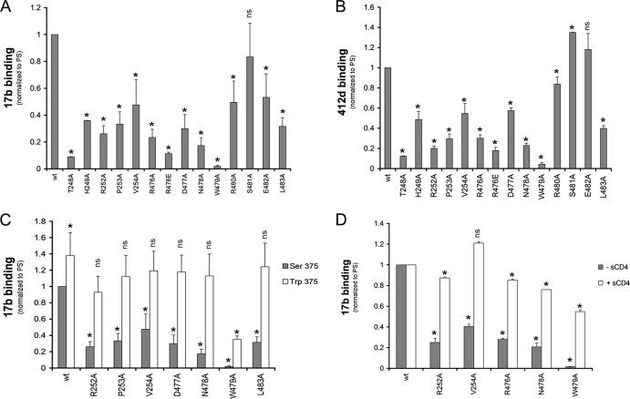
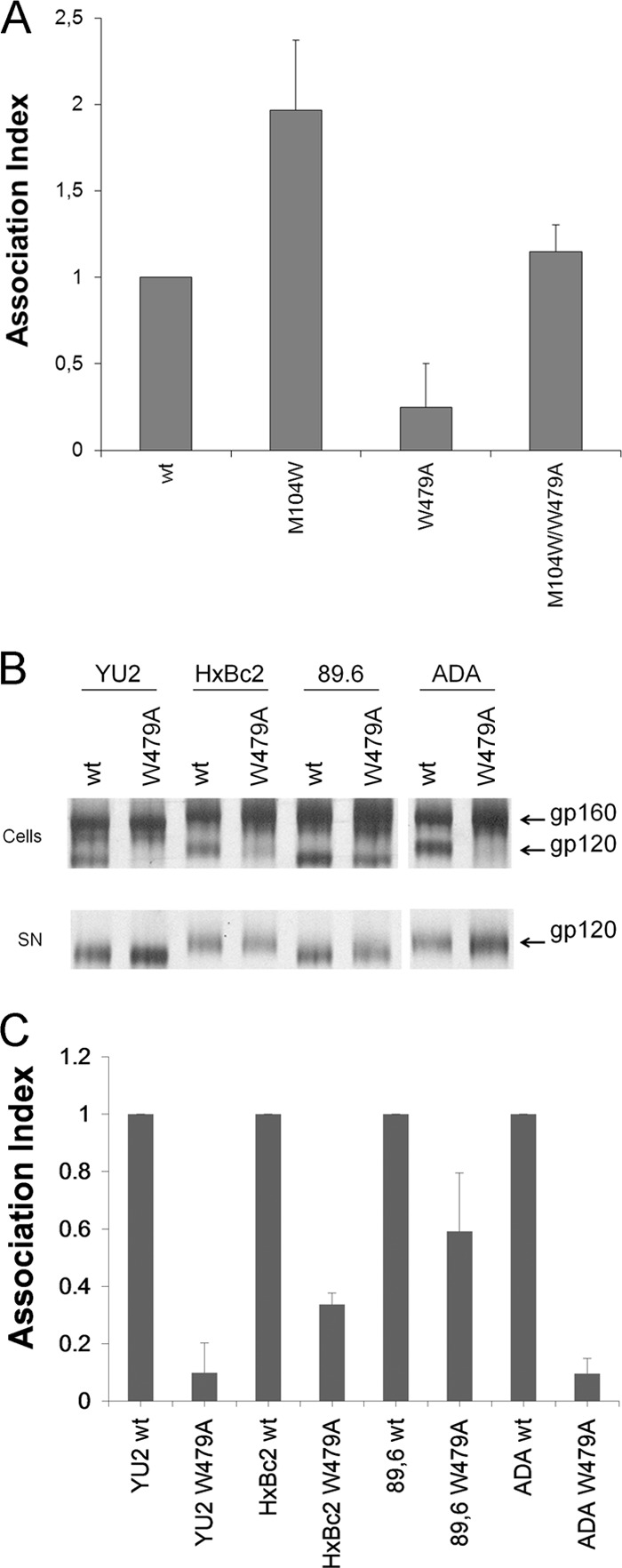
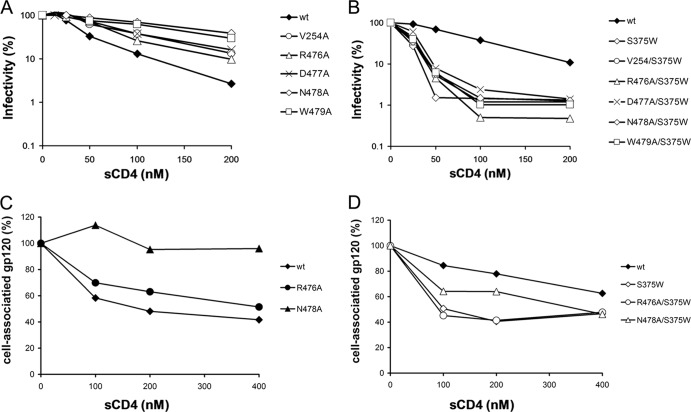
The trimeric envelope glycoprotein (Env) of human immunodeficiency virus type 1 (HIV-1) mediates virus entry into host cells. CD4 engagement with the gp120 exterior envelope glycoprotein subunit represents the first step during HIV-1 entry. CD4-induced conformational changes in the gp120 inner domain involve three potentially flexible topological layers (layers 1, 2, and 3). Structural rearrangements between layer 1 and layer 2 have been shown to facilitate the transition of the envelope glycoprotein trimer from the unliganded to the CD4-bound state and to stabilize gp120-CD4 interaction. However, our understanding of CD4-induced conformational changes in the gp120 inner domain remains incomplete. Here, we report that a highly conserved element of the gp120 inner domain, layer 3, plays a pivot-like role in these allosteric changes. In the unliganded state, layer 3 modulates the association of gp120 with the Env trimer, probably by influencing the relationship of the gp120 inner and outer domains. Importantly, layer 3 governs the efficiency of the initial gp120 interaction with CD4, a function that can also be fulfilled by filling the Phe43 cavity. This work defines the functional importance of layer 3 and completes a picture detailing the role of the gp120 inner domain in CD4-induced conformational transitions in the HIV-1 Env trimer.


Figures

Fig 1
Fig 1
Structure of the inner domain of HIV-1 gp120 in the CD4-bound conformation. (A) The structure (27) of HIV-1HXBc2 gp120 (ribbon) complexed with two-domain CD4 (red) is shown from the perspective of the Env trimer axis. The β20-β21 strands of gp120 project from the outer domain and compose two of the four strands of the bridging sheet. The other two strands of the bridging sheet are derived from the distal portion of layer 2. (B) A close-up view of the conformation adopted by the inner domain layers 1, 2, and 3. (C) A close-up view of the interactions between layers 2 and 3. The side chain residues that were altered in this and a previous study (3) are colored according to the gp120-trimer association index (red, association index of <0.5; green, association index of ≥0.7). (D) The side chain residues are colored according to CD4-Ig binding ability (red, relative CD4-Ig binding of ≤0.5; green, relative CD4-Ig binding of >0.5).
Fig 2
Fig 2
Sequences of HIV-1 and primate immunodeficiency virus Env. Primary sequence alignment of layer 3 gp120 residues from representative HIV-1 A (accession number ABB29387.1), HIV-1 B (accession number K03455), HIV-1 C (AAB36507.1), HIV-1 D (P04581.1), HIV-1 F (ACR27173.1), HIV-1 G (ACO91925.1), HIV-1 H (AAF18394.1), HIV-1 J (ABR20452.1), HIV-1 K (CAB59009.1), HIV-1 N (AAT08775.1), HIV-1 O (AAA99883.1), and HIV-1 P (ACY40659.1) viruses (A) and from primate immunodeficiency lineages SIVcpz (accession number ABD19490.1), HIV-2 (AAC95347.1), SIVmac/smm (AAA47637.1), SIVtan (AAC57057.1), SIVagm (AAA91919.1), and SIVsyk (AAA74712.1) (4, 5) (B). Secondary structure elements are shown above the sequences (4, 5). The shading highlights residues that are conserved.
Fig 3
Fig 3
Recognition of layer 3 gp120 variants by CD4. The effects of alterations in layer 3 on gp120 recognition by CD4-Ig were examined for monomeric soluble gp120 by immunoprecipitation or for cellular-expressed trimeric Env by cell-based ELISA (A). In both contexts, the gp120 variants were normalized by patient serum (PS) and to the signal obtained for their wt counterparts. The effect of filling the Phe43 cavity with a tryptophan (S375W) on layer 3 variants was analyzed by immunoprecipitation (B) or by cell-based-ELISA (C) with CD4-Ig. Data shown represent the means ± standard errors of the means of at least two independent experiments. *, P < 0.05 by a Mann-Whitney rank sum test. Differences with P values of ≥0.05 were not considered significant (ns).
Fig 4
Fig 4
Recognition of soluble gp120 variants by CD4i monoclonal antibodies. Comparable amounts of radiolabeled wt and mutant gp120 were incubated with 13 nm of 17b (A) or 412d (B) CD4i Abs for 1 h at 37°C. Alternatively, the effect of filling the Phe43 cavity with a tryptophan (S375W) (C) or of adding sCD4 (D) to layer 3 on 17b recognition was assessed by immunoprecipitation. gp120 variants were normalized by PS and to the signal obtained for their wt counterparts. Precipitates were analyzed by SDS-PAGE and densitometry. Incubation with sCD4 increased the binding of wt gp120 to 17b 1.5-fold. Data shown represent the means ± standard errors of the means of at least two independent experiments. *, P < 0.05 by a Mann-Whitney rank sum test. Differences with P values of ≥0.05 were not considered significant (ns).
Fig 5
Fig 5
Recognition of cellular-expressed trimeric HIV-1 Env variants by a CD4i monoclonal antibody. The effects of alterations in layer 3 on gp120 recognition by 17b, a CD4i antibody, were examined in the absence or presence of CD4 (13 nM CD4-Ig) by cell-based ELISA (A). To avoid recognition of the Ig portion of the CD4-Ig fusion protein by a secondary antibody, 17b was directly conjugated to HRP, as described in Materials and Methods. The effect of filling the Phe43 cavity with a tryptophan (S375W) on layer 3 variants was also analyzed by cell-based-ELISA (B). HIV-1 Env variants were normalized by PS and to the signal obtained for the wt. The S375W mutant, known to interact better with CD4i Abs (50), was introduced for comparison. Data shown represent the means ± standard errors of the means of at least four independent experiments. (C and D) Conformational fixation followed by ligand selection. The ratio of ligand binding to cross-linked versus untreated HIV-1 Env trimer is shown for wt and layer 3 variants without (C) or with (D) filling of the Phe43 cavity. Signals were normalized to those obtained for the wt. Data shown are representative of at least three independent experiments performed in duplicate. *, P < 0.05 by a Mann-Whitney rank sum test. Differences with P values of ≥0.05 were not considered significant (ns).
Fig 6
Fig 6
Inner domain residues important for gp120-trimer association. Cell lysates and supernatants (SN) of 35S-labeled cells transiently expressing the HIV-1YU2 (A), HIV-1HxBc2, HIV-189.6, and HIV-1ADA (B) and indicated mutant envelope glycoproteins were precipitated with serum from HIV-1-infected patients. The precipitated proteins were loaded onto SDS-PAGE polyacrylamide gels and analyzed by autoradiography and densitometry. (C) A quantification of the data shown in panel B. The association index is a measure of the ability of the mutant gp120 molecule to remain associated with the envelope glycoprotein complex on the expressing cell relative to that of the wild-type envelope glycoproteins. The association index was calculated as described in Materials and Methods. Data shown represent the average ± standard deviation of at least two independent experiments.
Fig 7
Fig 7
Sensitivity of HIV-1 Env gp120 variants to soluble CD4. (A and B) Recombinant HIV-1 expressing luciferase and bearing wt or mutant HIV-1 Env trimer were normalized by reverse transcriptase activity. Equal amounts of viruses were incubated with serial dilutions of sCD4 at 37°C for 1 h prior to infection of Cf2Th-CD4/CCR5 cells. Infectivity at each dilution of sCD4 tested is shown as the percentage of infection without sCD4 for each particular mutant. Quadruplicate samples were analyzed; data shown are representative of values obtained in at least three independent experiments. (C and D) The sCD4-induced shedding of gp120 from HIV-1 Env expressed on the cell surface. Transfected 293T cells were metabolically labeled with [35S]methionine-cysteine for 16 h with increasing concentrations of sCD4 (0 to 400 nM). Cell lysates were precipitated with PS. Precipitates were analyzed by SDS-PAGE and densitometry. Data shown are representative of values obtained in at least two independent experiments.
Fig 8
Fig 8
Model for the role of layer 3 in HIV-1 Env trimer stability and CD4 binding. The image on the top left depicts the unliganded HIV-1 Env trimer, with two gp120 subunits and two gp41 subunits visible from the perspective shown. The gp120 subunit on the right is subdivided into the outer domain (OD), inner domain (β-sandwich [red] and layers 1, 2, and 3), and the TAD (V1/V2, and V3 regions) (35). The initial site of CD4 binding is circled in black, and the location of the Phe43 cavity is shown by an asterisk. Layer 3 modulates the interaction of the outer domain and layer 2 in the inner domain. The upper right image shows the consequences of changes in Trp479, which is located in layer 3, in the gp120 interdomain interface. Changes in Trp479 significantly disrupt the relationship of the inner and outer domains. Because the V1/V2 and V3 regions are anchored in the inner and outer domains, respectively, the gp120 TAD, which comprises the V1/V2 and V3 regions, is disrupted. Thus, changes in Trp479 lead to trimer instability and gp120 shedding. The images in the bottom row illustrate the phenotypic consequences of changes in layer 3 that are less disruptive than the alteration of Trp479. These more subtle changes in layer 3 cause local alterations of gp120 conformation around the Phe-43 cavity that affect CD4 binding, with effects on both on-rate and off-rate (bottom panels). The more subtle changes in layer 3 can be compensated by the S375W change, which fills the Phe43 cavity with the indole ring of tryptophan.

Similar articles

Cited by

References

    1. Allan JS, Coligan JE, Barin F, McLane MF, Sodroski JG, Rosen CA, Haseltine WA, Lee TH, Essex M. 1985. Major glycoprotein antigens that induce antibodies in AIDS patients are encoded by HTLV-III. Science 228:1091–1094 - PubMed
    1. Robey WG, Safai B, Oroszlan S, Arthur LO, Gonda MA, Gallo RC, Fischinger PJ. 1985. Characterization of envelope and core structural gene products of HTLV-III with sera from AIDS patients. Science 228:593–595 - PubMed
    1. Finzi A, Xiang SH, Pacheco B, Wang L, Haight J, Kassa A, Danek B, Pancera M, Kwong PD, Sodroski J. 2010. Topological layers in the HIV-1 gp120 inner domain regulate gp41 interaction and CD4-triggered conformational transitions. Mol. Cell 37:656–667 - PMC - PubMed
    1. Helseth E, Olshevsky U, Furman C, Sodroski J. 1991. Human immunodeficiency virus type 1 gp120 envelope glycoprotein regions important for association with the gp41 transmembrane glycoprotein. J. Virol. 65:2119–2123 - PMC - PubMed
    1. Yang X, Mahony E, Holm GH, Kassa A, Sodroski J. 2003. Role of the gp120 inner domain beta-sandwich in the interaction between the human immunodeficiency virus envelope glycoprotein subunits. Virology 313:117–125 - PubMed

Publication types