

Murine guanylate binding protein 2 (mGBP2) controls Toxoplasma gondii replication
- PMID: 23248289
- PMCID: PMC3538222
- DOI: 10.1073/pnas.1205635110
Murine guanylate binding protein 2 (mGBP2) controls Toxoplasma gondii replication
Abstract
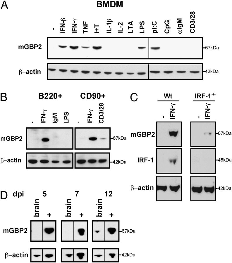
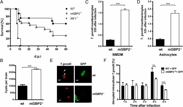
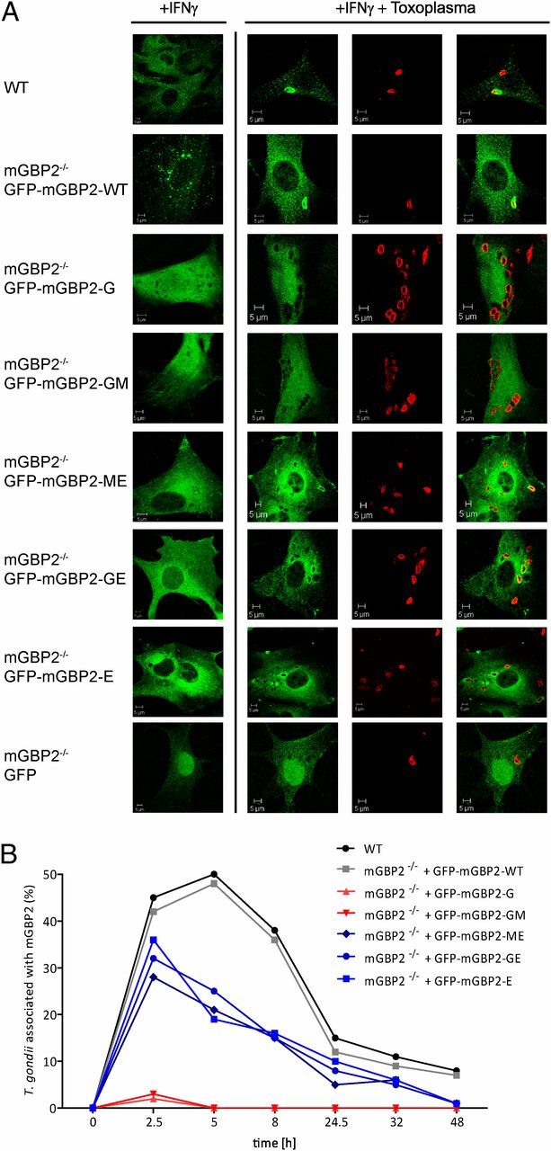
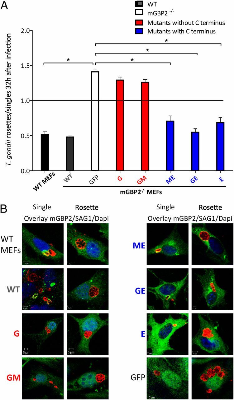
IFN-γ orchestrates the host response against intracellular pathogens. Members of the guanylate binding proteins (GBP) comprise the most abundant IFN-γ-induced transcriptional response. mGBPs are GTPases that are specifically up-regulated by IFN-γ, other proinflammatory cytokines, toll-like receptor agonists, as well as in response to Listeria monocytogenes and Toxoplasma gondii infection. mGBP2 localizes at the parasitophorous vacuole (PV) of T. gondii; however, the molecular function of mGBP2 and its domains in T. gondii infection is not known. Here, we show that mGBP2 is highly expressed in several cell types, including T and B cells after stimulation. We provide evidence that the C-terminal domain is sufficient and essential for recruitment to the T. gondii PV. Functionally, mGBP2 reduces T. gondii proliferation because mGBP2-deficient cells display defects in the replication control of T. gondii. Ultimately, mGBP2-deficient mice reveal a marked immune susceptibility to T. gondii. Taken together, mGBP2 is an essential immune effector molecule mediating antiparasitic resistance.
Conflict of interest statement
The authors declare no conflict of interest.
Figures
Similar articles
-
Essential Role of mGBP7 for Survival of Toxoplasma gondii Infection.mBio. 2020 Jan 21;11(1):e02993-19. doi: 10.1128/mBio.02993-19. mBio. 2020. PMID: 31964735 Free PMC article.
-
The GTPase activity of murine guanylate-binding protein 2 (mGBP2) controls the intracellular localization and recruitment to the parasitophorous vacuole of Toxoplasma gondii.J Biol Chem. 2012 Aug 10;287(33):27452-66. doi: 10.1074/jbc.M112.379636. Epub 2012 Jun 22. J Biol Chem. 2012. PMID: 22730319 Free PMC article.
-
mGBP2 engages Galectin-9 for immunity against Toxoplasma gondii.PLoS One. 2025 Jan 24;20(1):e0316209. doi: 10.1371/journal.pone.0316209. eCollection 2025. PLoS One. 2025. PMID: 39854420 Free PMC article.
-
T lymphocyte-dependent effector mechanisms of immunity to Toxoplasma gondii.Microbes Infect. 1999 Jul;1(9):699-708. doi: 10.1016/s1286-4579(99)80071-9. Microbes Infect. 1999. PMID: 10611747 Review.
-
Exposing Toxoplasma gondii hiding inside the vacuole: a role for GBPs, autophagy and host cell death.Curr Opin Microbiol. 2017 Dec;40:72-80. doi: 10.1016/j.mib.2017.10.021. Epub 2017 Nov 12. Curr Opin Microbiol. 2017. PMID: 29141239 Free PMC article. Review.
Cited by
-
NADPH Oxidase and Guanylate Binding Protein 5 Restrict Survival of Avirulent Type III Strains of Toxoplasma gondii in Naive Macrophages.mBio. 2018 Aug 28;9(4):e01393-18. doi: 10.1128/mBio.01393-18. mBio. 2018. PMID: 30154263 Free PMC article.
-
Innate immunity to Toxoplasma gondii infection.Nat Rev Immunol. 2014 Feb;14(2):109-21. doi: 10.1038/nri3598. Nat Rev Immunol. 2014. PMID: 24457485 Review.
-
Interferon-induced guanylate-binding proteins: Guardians of host defense in health and disease.J Exp Med. 2019 Mar 4;216(3):482-500. doi: 10.1084/jem.20182031. Epub 2019 Feb 12. J Exp Med. 2019. PMID: 30755454 Free PMC article. Review.
-
Caspase-11 activation requires lysis of pathogen-containing vacuoles by IFN-induced GTPases.Nature. 2014 May 15;509(7500):366-70. doi: 10.1038/nature13157. Epub 2014 Apr 16. Nature. 2014. PMID: 24739961
-
Murine Irgm Paralogs Regulate Nonredundant Functions To Execute Host Defense to Toxoplasma gondii.Infect Immun. 2021 Oct 15;89(11):e0020221. doi: 10.1128/IAI.00202-21. Epub 2021 Aug 2. Infect Immun. 2021. PMID: 34338548 Free PMC article.
References
-
- Huang S, et al. Immune response in mice that lack the interferon-gamma receptor. Science. 1993;259(5102):1742–1745. - PubMed
-
- Pfeffer K, et al. Mice deficient for the 55 kd tumor necrosis factor receptor are resistant to endotoxic shock, yet succumb to L. monocytogenes infection. Cell. 1993;73(3):457–467. - PubMed
-
- Endres R, et al. Listeriosis in p47(phox-/-) and TRp55-/- mice: protection despite absence of ROI and susceptibility despite presence of RNI. Immunity. 1997;7(3):419–432. - PubMed
-
- Boehm U, Klamp T, Groot M, Howard JC. Cellular responses to interferon-gamma. Annu Rev Immunol. 1997;15:749–795. - PubMed
-
- Martens S, Howard J. The interferon-inducible GTPases. Annu Rev Cell Dev Biol. 2006;22:559–589. - PubMed
Publication types
MeSH terms
Substances
LinkOut - more resources
Full Text Sources
Molecular Biology Databases
Research Materials
