The Yersinia virulence effector YopM binds caspase-1 to arrest inflammasome assembly and processing
- PMID: 23245324
- PMCID: PMC3703949
- DOI: 10.1016/j.chom.2012.10.020
The Yersinia virulence effector YopM binds caspase-1 to arrest inflammasome assembly and processing
Abstract
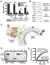
Inflammasome assembly activates caspase-1 and initiates the inflammatory cell death program pyroptosis, which is protective against numerous pathogens. Consequently, several pathogens, including the plague causing bacterium Yersinia pestis, avoid activating this pathway to enhance their virulence. However, bacterial molecules that directly modulate the inflammasome have yet to be identified. Examining the contribution of Yersinia type III secretion effectors to caspase-1 activation, we identified the leucine-rich repeat effector YopM as a potent antagonist of both caspase-1 activity and activation. YopM directly binds caspase-1, which both inhibits caspase-1 activity and sequesters it to block formation of the mature inflammasome. Caspase-1 activation antagonizes Yersinia survival in vivo, and consequently YopM inhibition of caspase-1 is required for Yersinia pathogenesis. Thus, a bacterium obstructs pyroptosis utilizing a direct mechanism of caspase-1 inhibition that is distinct from known viral or host inhibitors.
Copyright © 2012 Elsevier Inc. All rights reserved.
Figures


Comment in
-
YopM puts caspase-1 on ice.Cell Host Microbe. 2012 Dec 13;12(6):737-8. doi: 10.1016/j.chom.2012.11.006. Cell Host Microbe. 2012. PMID: 23245318 Free PMC article.
Similar articles
-
YopM puts caspase-1 on ice.Cell Host Microbe. 2012 Dec 13;12(6):737-8. doi: 10.1016/j.chom.2012.11.006. Cell Host Microbe. 2012. PMID: 23245318 Free PMC article.
-
Manipulation of Interleukin-1β and Interleukin-18 Production by Yersinia pestis Effectors YopJ and YopM and Redundant Impact on Virulence.J Biol Chem. 2016 May 6;291(19):9894-905. doi: 10.1074/jbc.M115.697698. Epub 2016 Feb 16. J Biol Chem. 2016. PMID: 26884330 Free PMC article.
-
The Yersinia pestis Effector YopM Inhibits Pyrin Inflammasome Activation.PLoS Pathog. 2016 Dec 2;12(12):e1006035. doi: 10.1371/journal.ppat.1006035. eCollection 2016 Dec. PLoS Pathog. 2016. PMID: 27911947 Free PMC article.
-
Current activities of the Yersinia effector protein YopM.Int J Med Microbiol. 2015 May;305(3):424-32. doi: 10.1016/j.ijmm.2015.03.009. Epub 2015 Apr 1. Int J Med Microbiol. 2015. PMID: 25865799 Review.
-
Activation and Evasion of Inflammasomes by Yersinia.Curr Top Microbiol Immunol. 2016;397:69-90. doi: 10.1007/978-3-319-41171-2_4. Curr Top Microbiol Immunol. 2016. PMID: 27460805 Review.
Cited by
-
Interleukin 1α and the inflammatory process.Nat Immunol. 2016 Jul 19;17(8):906-13. doi: 10.1038/ni.3503. Nat Immunol. 2016. PMID: 27434011 Free PMC article. Review.
-
Phosphorylation of caspases by a bacterial kinase inhibits host programmed cell death.Nat Commun. 2024 Sep 30;15(1):8464. doi: 10.1038/s41467-024-52817-1. Nat Commun. 2024. PMID: 39349471 Free PMC article.
-
Nigericin Promotes NLRP3-Independent Bacterial Killing in Macrophages.Front Immunol. 2019 Oct 1;10:2296. doi: 10.3389/fimmu.2019.02296. eCollection 2019. Front Immunol. 2019. PMID: 31632394 Free PMC article.
-
Programmed Cell Death in the Evolutionary Race against Bacterial Virulence Factors.Cold Spring Harb Perspect Biol. 2020 Feb 3;12(2):a036459. doi: 10.1101/cshperspect.a036459. Cold Spring Harb Perspect Biol. 2020. PMID: 31501197 Free PMC article. Review.
-
Subversion of GBP-mediated host defense by E3 ligases acquired during Yersinia pestis evolution.Nat Commun. 2022 Aug 4;13(1):4526. doi: 10.1038/s41467-022-32218-y. Nat Commun. 2022. PMID: 35927280 Free PMC article.
References
-
- Bauernfeind FG, Horvath G, Stutz A, Alnemri ES, MacDonald K, Speert D, Fernandes-Alnemri T, Wu J, Monks BG, Fitzgerald KA, et al. Cutting Edge: NF-kB Activating Pattern Recognition and Cytokine Receptors License NLRP3 Inflammasome Activation by Regulating NLRP3 Expression. J Immunol. 2009;183:787–791. - PMC - PubMed
Publication types
MeSH terms
Substances
Grants and funding
LinkOut - more resources
Full Text Sources
Other Literature Sources
Miscellaneous

