Distinct gene expression profiles of viral- and nonviral-associated merkel cell carcinoma revealed by transcriptome analysis
- PMID: 23223137
- PMCID: PMC3597750
- DOI: 10.1038/jid.2012.445
Distinct gene expression profiles of viral- and nonviral-associated merkel cell carcinoma revealed by transcriptome analysis
Abstract
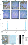
Merkel cell carcinoma (MCC) is an aggressive cutaneous neuroendocrine tumor with high mortality rates. Merkel cell polyomavirus (MCPyV), identified in the majority of MCCs, may drive tumorigenesis via viral T antigens. However, the mechanisms underlying pathogenesis in MCPyV-negative MCCs remain poorly understood. To nominate genes contributing to the pathogenesis of MCPyV-negative MCCs, we performed DNA microarray analysis on 30 MCCs. The MCPyV status of MCCs was determined by PCR for viral DNA and RNA. A total of 1,593 probe sets were differentially expressed between MCPyV-negative and MCPyV-positive MCCs, with significant differential expression defined as at least a 2-fold change in either direction and a P-value 0.05. MCPyV-negative tumors showed decreased RB1 expression, whereas MCPyV-positive tumors were enriched for immune response genes. Validation studies included immunohistochemistry demonstration of decreased RB protein expression in MCPyV-negative tumors and increased peritumoral CD8+ T lymphocytes surrounding MCPyV-positive tumors. In conclusion, our data suggest that loss of RB1 expression may have an important role in the tumorigenesis of MCPyV-negative MCCs. Functional and clinical validation studies are needed to determine whether this tumor-suppressor pathway represents an avenue for targeted therapy.
Conflict of interest statement
Conflict of Interest, The authors state no conflict of interest
Figures


Similar articles
-
Decreased H3K27me3 Expression Is Associated With Merkel Cell Polyomavirus-negative Merkel Cell Carcinoma, Especially Combined With Cutaneous Squamous Cell Carcinoma.Anticancer Res. 2019 Oct;39(10):5573-5579. doi: 10.21873/anticanres.13751. Anticancer Res. 2019. PMID: 31570452
-
Merkel cell polyomavirus infection, large T antigen, retinoblastoma protein and outcome in Merkel cell carcinoma.Clin Cancer Res. 2011 Jul 15;17(14):4806-13. doi: 10.1158/1078-0432.CCR-10-3363. Epub 2011 Jun 3. Clin Cancer Res. 2011. PMID: 21642382
-
LT and SOX9 expression are associated with gene sets that distinguish Merkel cell polyomavirus (MCPyV)-positive and MCPyV-negative Merkel cell carcinoma.Br J Dermatol. 2024 May 17;190(6):876-884. doi: 10.1093/bjd/ljae033. Br J Dermatol. 2024. PMID: 38261397
-
A review on the oncogenesis of Merkel cell carcinoma: Several subsets arise from different stages of differentiation of stem cell.Medicine (Baltimore). 2023 Apr 14;102(15):e33535. doi: 10.1097/MD.0000000000033535. Medicine (Baltimore). 2023. PMID: 37058042 Free PMC article. Review.
-
Merkel cell polyomavirus and non-Merkel cell carcinomas: guilty or circumstantial evidence?APMIS. 2020 Feb;128(2):104-120. doi: 10.1111/apm.13019. Epub 2020 Jan 28. APMIS. 2020. PMID: 31990105 Review.
Cited by
-
MiR-375 Regulation of LDHB Plays Distinct Roles in Polyomavirus-Positive and -Negative Merkel Cell Carcinoma.Cancers (Basel). 2018 Nov 14;10(11):443. doi: 10.3390/cancers10110443. Cancers (Basel). 2018. PMID: 30441870 Free PMC article.
-
Merkel Cell Carcinoma: An Immunotherapy Fairy-Tale?Front Oncol. 2021 Sep 23;11:739006. doi: 10.3389/fonc.2021.739006. eCollection 2021. Front Oncol. 2021. PMID: 34631574 Free PMC article. Review.
-
Activation of Oncogenic and Immune-Response Pathways Is Linked to Disease-Specific Survival in Merkel Cell Carcinoma.Cancers (Basel). 2022 Jul 23;14(15):3591. doi: 10.3390/cancers14153591. Cancers (Basel). 2022. PMID: 35892849 Free PMC article.
-
Response of Merkel cell polyomavirus-positive merkel cell carcinoma xenografts to a survivin inhibitor.PLoS One. 2013 Nov 18;8(11):e80543. doi: 10.1371/journal.pone.0080543. eCollection 2013. PLoS One. 2013. PMID: 24260412 Free PMC article.
-
Merkel cell polyomavirus small tumour antigen activates the p38 MAPK pathway to enhance cellular motility.Biochem J. 2020 Jul 31;477(14):2721-2733. doi: 10.1042/BCJ20200399. Biochem J. 2020. PMID: 32639530 Free PMC article.
References
-
- Becker JC. Merkel cell carcinoma. Ann Oncol. 2010;21(7):vii81–5. - PubMed
-
- Becker JC, Houben R, Ugurel S, Trefzer U, Pfohler C, Schrama D. MC polyomavirus is frequently present in Merkel cell carcinoma of European patients. J Invest Dermatol. 2009;129:248–50. - PubMed
-
- Ben-Arie N, Hassan BA, Bermingham NA, Malicki DM, Armstrong D, Matzuk M, et al. Functional conservation of atonal and Math1 in the CNS and PNS. Development. 2000;127:1039–48. - PubMed
-
- Benjamini Y, Hochberg Y. Controlling the false discovery rate: a practical and powerful approach to multiple testing. J R Stat Soc Ser B Stat Methodol. 1995;57:289–300.
Publication types
MeSH terms
Substances
Grants and funding
LinkOut - more resources
Full Text Sources
Other Literature Sources
Medical
Molecular Biology Databases
Research Materials
Miscellaneous

