. 2012 Dec 27;2(6):1475-84.
doi: 10.1016/j.celrep.2012.11.011. Epub 2012 Dec 6.

Deubiquitinases as a signaling target of oxidative stress

Affiliations

Deubiquitinases as a signaling target of oxidative stress

Xiomaris M Cotto-Rios et al. Cell Rep. .

Abstract

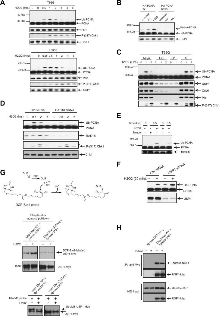
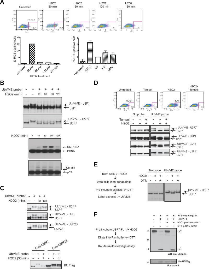
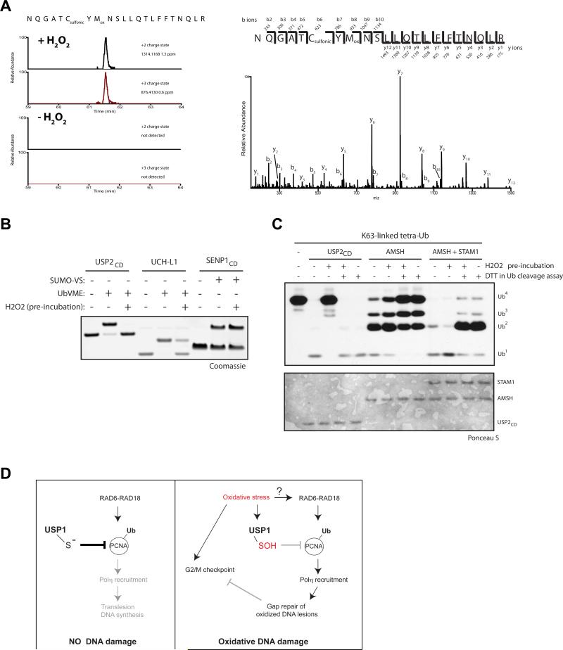
Deubiquitinating enzymes (DUBs) constitute a large family of cysteine proteases that have a broad impact on numerous biological and pathological processes, including the regulation of genomic stability. DUBs are often assembled onto multiprotein complexes to assist in their localization and substrate selection, yet it remains unclear how the enzymatic activity of DUBs is modulated by intracellular signals. Herein, we show that bursts of reactive oxygen species (ROS) reversibly inactivate DUBs through the oxidation of the catalytic cysteine residue. Importantly, USP1, a key regulator of genomic stability, is reversibly inactivated upon oxidative stress. This, in part, explains the rapid nature of PCNA monoubiquitination-dependent DNA damage tolerance in response to oxidative DNA damage in replicating cells. We propose that DUBs of the cysteine protease family act as ROS sensors in human cells and that ROS-mediated DUB inactivation is a critical mechanism for fine-tuning stress-activated signaling pathways.


Figures

Figure 1
Figure 1. Oxidation of USP1 is Dependent on its Catalytic Site Competency
(A) PCNA monoubiquitination is transiently induced by oxidative stress, while checkpoint activation is prolonged. T98G and U2OS cells were treated with H2O2 (final concentration 1mM) for the indicated amount of time. Samples were collected, lysed and analyzed by Western blot with the indicated antibodies. (B) Oxidative stress-induced PCNA monoubiquitination is dependent on an intact Lys 164 site, but independent of USP1 degradation. U2OS cells stably expressing HA-PCNA wild type or HA-PCNA K164R mutant were either left untreated or treated with 50 Joules/M2 UVC for 3hrs, or treated with 1 mM H2O2 for 30min. Samples were collected, lysed and analyzed by Western blot with the indicated antibodies. (C) Oxidative stress-induced PCNA monoubiquitination mainly occurs during S phase. T98G cells were synchronized in G0/G1 by serum-deprivation for 72 hrs. Cells were then replenished with fresh media and collected at the indicated time points. Cells were either left untreated or treated with a final concentration of 0.5 mM H2O2. Synchronized cells were treated at 0 hrs after release from serum deprivation for G0, after 4 hrs of serum deprivation release for G1 phase, or after 24 hrs of release for early S-phase. Samples were collected, lysed and analyzed by Western blot with the indicated antibodies. Asterisk indicates a non-specific, cross-reactive band when probing with anti-phospho Chk1 antibody (p317). (D) Depletion of Rad18 results in increased Chk1 activation. U2OS cells were transfected for 48 hrs with a control (Ctrl) siRNA or Rad18 siRNA. Cells were then treated with H2O2 (final concentration 0.5 mM) for the indicated amount of time. Samples were collected, lysed and analyzed by Western blot with the indicated antibodies. Asterisk indicates a non-specific, cross-reactive band. (E) The anti-oxidant activity of Tempol reduces the levels of PCNA monoubiquitination after oxidative stress. U2OS cells in the presence or absence of Tempol (3 mM) were treated with H2O2 (final concentration 0.3 mM) for the indicated amount of time. Samples were collected, lysed and analyzed by Western blot with the indicated antibodies. (F) Depletion of USP1 further elevates PCNA monoubiquitination in the presence of oxidative stress. U2OS cells were transfected for 48 hrs with a control (Ctrl) siRNA or USP1 siRNA. Cells were then treated with H2O2 (final concentration 0.3 mM) for 30 minutes. Samples were collected, lysed and analyzed by Western blot with the indicated antibodies. (G) Oxidation of USP1 catalytic Cys to the sulfenic acid intermediate can be captured by DCP-Bio1 probe. U2OS cells were transfected for 48 hrs with Usp1-Myc WT or (DDAA)-Myc mutant along with Xpress-UAF1. Cells were then treated with H2O2 (final concentration 0.3 mM) for 30 minutes. Extracts were made and divided for two separate reactions: labeling with DCP-Bio1 probe or with the UbVME DUB activity probe. Input represents 30% of extracts used for the DCP-Bio1 reaction. (H) Oxidative stress does not disrupt the binding between Usp1 and UAF1 in cells. U2OS cells were transfected with Xpress-UAF1 only or co-transfected with Usp1-Myc WT and Xpress-UAF1 and treated with H2O2 (final concentration 1 mM) for 30 minutes. Extracts were subjected to immunoprecipitation with anti-Myc antibody and probed with indicated antibodies.
Figure 2
Figure 2. Reversible Inhibition of DUBs by Oxidative Stress
(A) Temporal analysis of ROS levels inside cells after H2O2 treatment. T98G cells were treated with a final concentration of 1 mM H2O2 for the indicated amount of time (upper panel, left bottom panel). Cells were trypsinized, collected, and incubated with CM-H2DCFDA for 30 min at 37°C prior to FACS analysis. Additionally, T98G cells were either left untreated or treated with 0.2 mM H2O2 for 30 min, 50 Joules/M2 UVC for 30 min, 2 mM HU for 18 hrs or MMC for 18 hrs (right bottom panel) and trypsinized, collected, and incubated with CM-H2DCFDA for FACS analysis. (B) USP1 and USP7 are reversibly inactivated by oxidative stress and correlate with the ubiquitination status of their respective substrates. U2OS cells were left untreated or treated with a final concentration of 1 mM H2O2 for the indicated time-course. Extracts were made in parallel to assess DUB activity by the UbVME probe and by whole cell lysis to assess the ubiquitination status of PCNA and p53 proteins. (C) Ectopically expressed DUBs are similarly affected by oxidative stress as their endogenous counterparts. 293T cells were either transfected or not for 48 hrs with wildtype FLAG-USP7 or FLAG-USP28 expression constructs. Then cells were left untreated or treated with 1mM H2O2 for 30 min or with the indicated time-course. Samples were then collected and process for DUB activity assay as in (B), and probed with the indicated antibodies for Western blot. Additional bands that appear are likely different isoforms of USP28 (USP28 antibody). (D) Tempol can reverse the DUB catalytic inhibition by oxidative stress. U2OS cells were left untreated or treated with 0.3 mM H2O2 for 30 min in the absence or presence of Tempol. Cells were then trypsinized, collected, and incubated with CM-H2DCFDA as in (A). In parallel analysis, samples were collected and process for DUB activity assay as in (B). (E) ROS-inactivated USP7 in U2OS cells can be reversed after in vitro DTT treatment. U2OS cells were treated with 1 mM H2O2 for 30 min. Extracts were then pre-incubated on ice with 20 mM DTT (or not) prior to the labeling with UbVME as in (B). (F) Recombinant USP7 can be reversibly inhibited by ROS in vitro. His-tagged recombinant full-length USP7 is subjected to a ubiquitin cleavage assay of K48-linked tetra-ubiquitin chains in the presence or absence of H2O2 (1 mM) pre-incubation. DUB reaction was done in the presence or absence of 20 mM DTT.
Figure 3
Figure 3. The Metalloprotease AMSH is not susceptible to ROS-mediated catalytic inhibition
(A) Detecting the oxidation of the active site Cys of USP7 by mass spectrometry. (Left panel) Extracted ion chromatograms for the peptide harboring the catalytic Cys NQGATCYMNSLLQTLFFTNQLR in the H2O2 treated top panel and untreated sample (left panel). The m/z values for the doubly (monoisotopic m/z = 1314.1151) and the triply charged peptide (monoisotopic m/z = 876.4125) carrying a sulfonic acid modification on the Cys residue and an oxidation on Met are extracted with a mass window of 0.1 Da. In the H2O2 treated sample, the doubly and triply charged precursor of the sulfonic acid modified peptide with an oxidation on the Met residue could be detected (see also Right panel). The mass error is within 2 ppm of the theoretical value. In the untreated sample, there are no ions detected for the doubly or triply charged peptide carrying a sulfonic acid modification and a methionine oxidation. In addition, an m/z value for the peptide carrying just the sulfonic acid modification could not be detected (data not shown). (Right panel) MS/MS spectrum of the triply charged ion of peptide NQGATCYMNSLLQTLFFTNQLR carrying a sulfonic acid modification on the Cys residue and an oxidation on Met. Observed peptide bond cleavage is indicated in the sequence. The corresponding theoretical N-terminal (b-type ion) and C-terminal (y-type ions) ion series for the observed fragment ions is shown above and below the sequence, respectively. Note the mass error on the precursor ion and all fragment ions is better than 2 ppm. Neutral loss of water from y- and b- type ions is not indicated in the spectrum. (B) UCH-L1, but not SENP1, can be inhibited by ROS in vitro. Recombinant UCH-L1 and USP2CD (catalytic domain) were labeled by UbVME, while recombinant SENP1CD was labeled by SUMO1-VS. Samples were either pre-incubated with or without H2O2 (1 mM). Reactions were analyzed by Coomassie stained SDS-PAGE. (C) AMSH cleavage of K63-linked tetra-ubiquitin chains is unaffected by ROS. Recombinant USP2CD, AMSH, or AMSH plus STAM1 are subjected to ubiquitin cleavage assay of K63-linked tetra-ubiquitin chains in the presence or absence of H2O2 (1 mM) pre-incubation. The DUB reaction was done in the presence or absence of 20 mM DTT. Samples were then analyzed by Western blot with anti-ubiquitin antibody. (D) Model depicting how reversible oxidation of USP1 could modulate PCNA monoubiquitination in the presence of oxidative DNA damage. (Left panel) In the absence of DNA damage, USP1 associated with its catalytic co-factor, UAF1, is in the catalytically active state and can readily deubiquitinate or suppress monoubiquitinated PCNA to prevent aberrant recruitment of error-prone TLS polymerases. (Right panel) In response to oxidative stress or oxidative DNA damage, USP1 becomes transiently inactivated by direct oxidation of its catalytic Cys to the sulfenic acid intermediate. This renders USP1 unable to deubiquitinate PCNA, allowing for monoubiquitinated PCNA to engage with TLS polymerases that are responsible for DNA damage tolerance and/or gap repair. In the absence of oxidative DNA damage-induced checkpoint response, the cells become more reliant on the DNA damage tolerance pathway to ensure that the genome is fully replicated in a timely manner during S phase.

Similar articles

Cited by

References

    1. Andersen JK. Oxidative stress in neurodegeneration: cause or consequence? Nat Med. 2004;10(Suppl):S18–25. - PubMed
    1. Bienko M, Green CM, Crosetto N, Rudolf F, Zapart G, Coull B, Kannouche P, Wider G, Peter M, Lehmann AR, et al. Ubiquitin-binding domains in Y-family polymerases regulate translesion synthesis. Science. 2005;310:1821–1824. - PubMed
    1. Borodovsky A, Ovaa H, Kolli N, Gan-Erdene T, Wilkinson KD, Ploegh HL, Kessler BM. Chemistry-based functional proteomics reveals novel members of the deubiquitinating enzyme family. Chem Biol. 2002;9:1149–1159. - PubMed
    1. Bossis G, Melchior F. Regulation of SUMOylation by reversible oxidation of SUMO conjugating enzymes. Mol Cell. 2006;21:349–357. - PubMed
    1. Charles RL, Schröder E, May G, Free P, Gaffney PR, Wait R, Begum S, Heads RJ, Eaton P. Protein sulfenation as a redox sensor: proteomics studies using a novel biotinylated dimedone analogue. Mol Cell Proteomics. 2007;6:1473–1484. - PubMed

Publication types

LinkOut - more resources