. 2012;8(11):e1003059.
doi: 10.1371/journal.ppat.1003059. Epub 2012 Nov 29.

Species-specific inhibition of RIG-I ubiquitination and IFN induction by the influenza A virus NS1 protein

Affiliations

Species-specific inhibition of RIG-I ubiquitination and IFN induction by the influenza A virus NS1 protein

Ricardo Rajsbaum et al. PLoS Pathog. 2012.

Abstract

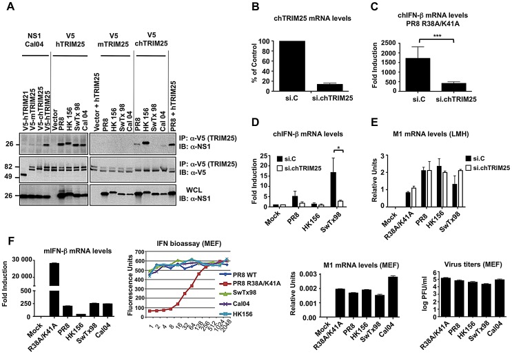
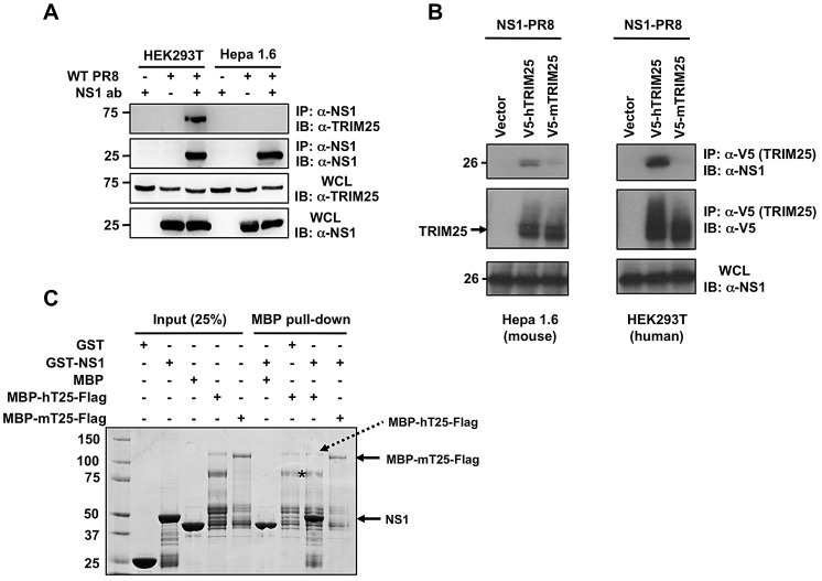
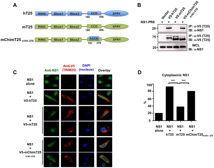
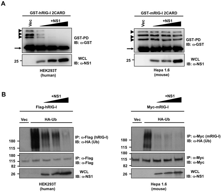
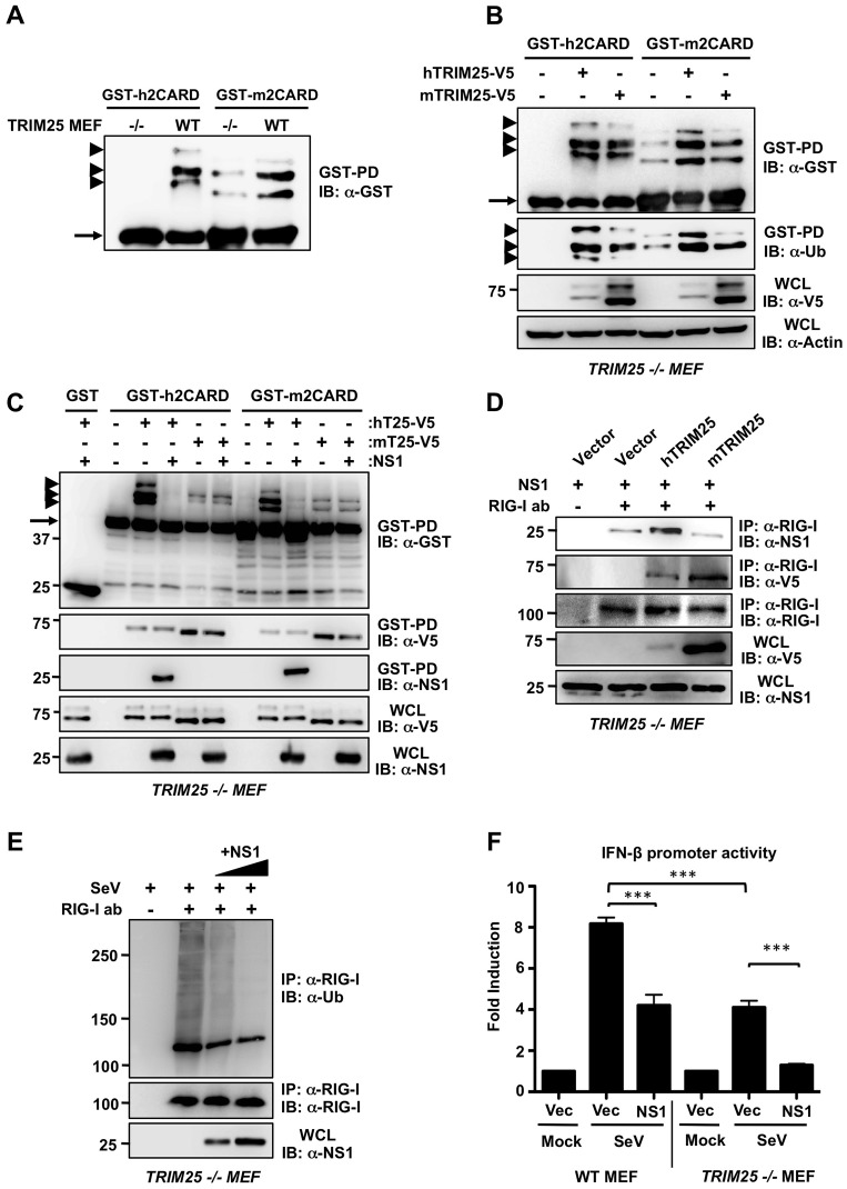
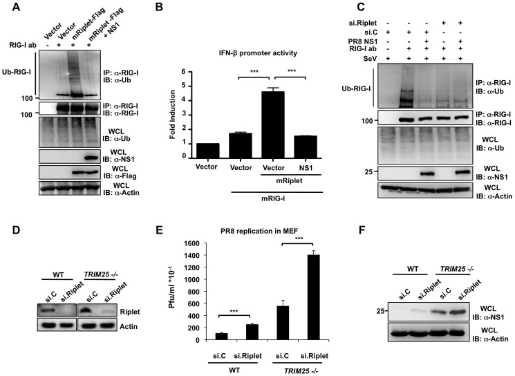
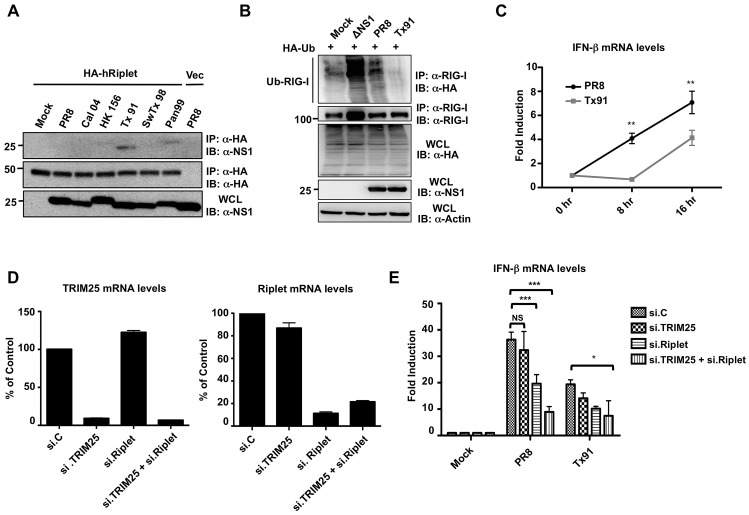
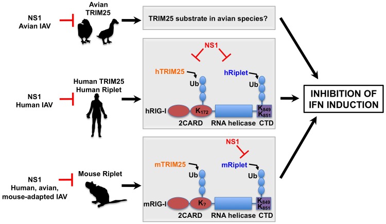
Influenza A viruses can adapt to new host species, leading to the emergence of novel pathogenic strains. There is evidence that highly pathogenic viruses encode for non-structural 1 (NS1) proteins that are more efficient in suppressing the host immune response. The NS1 protein inhibits type-I interferon (IFN) production partly by blocking the TRIM25 ubiquitin E3 ligase-mediated Lys63-linked ubiquitination of the viral RNA sensor RIG-I, required for its optimal downstream signaling. In order to understand possible mechanisms of viral adaptation and host tropism, we examined the ability of NS1 encoded by human (Cal04), avian (HK156), swine (SwTx98) and mouse-adapted (PR8) influenza viruses to interact with TRIM25 orthologues from mammalian and avian species. Using co-immunoprecipitation assays we show that human TRIM25 binds to all tested NS1 proteins, whereas the chicken TRIM25 ortholog binds preferentially to the NS1 from the avian virus. Strikingly, none of the NS1 proteins were able to bind mouse TRIM25. Since NS1 can inhibit IFN production in mouse, we tested the impact of TRIM25 and NS1 on RIG-I ubiquitination in mouse cells. While NS1 efficiently suppressed human TRIM25-dependent ubiquitination of RIG-I 2CARD, NS1 inhibited the ubiquitination of full-length mouse RIG-I in a mouse TRIM25-independent manner. Therefore, we tested if the ubiquitin E3 ligase Riplet, which has also been shown to ubiquitinate RIG-I, interacts with NS1. We found that NS1 binds mouse Riplet and inhibits its activity to induce IFN-β in murine cells. Furthermore, NS1 proteins of human but not swine or avian viruses were able to interact with human Riplet, thereby suppressing RIG-I ubiquitination. In conclusion, our results indicate that influenza NS1 protein targets TRIM25 and Riplet ubiquitin E3 ligases in a species-specific manner for the inhibition of RIG-I ubiquitination and antiviral IFN production.


Conflict of interest statement

The authors have declared that no competing interests exist.

Figures

Figure 1
Figure 1. Influenza A Virus NS1 interacts with TRIM25 in a species-specific manner.
(A) HEK293T cells were transfected with V5-tagged mouse TRIM25 (V5-mTRIM25), human TRIM25 (V5-hTRIM25) or chicken TRIM25 (V5-chTRIM25) together with NS1-A/California/04/09 (Cal04) (left), or NS1-PR8, NS1-A/Hong Kong/156 (HK156), NS1-A/Swine Texas/98 (SwTx98), or NS1-Cal04 (right panel). Empty vector and V5-tagged human TRIM21 were used as negative controls. Whole-cell lysates (WCLs) of HEK293T cells were subjected to immunoprecipitation (IP) using anti-V5 antibody, followed by immunoblotting (IB) with anti-NS1. (B–E) TRIM25 is important for virus-induced IFN-β production in chicken cells. Chicken LMH cells were transiently transfected with non-silencing control siRNA (si.C), or with a siRNA specific for chicken TRIM25 (si.chTRIM25). At 40 h posttransfection, cells were infected with recombinant influenza viruses expressing the NS1 RNA-binding mutant R38A/K41A (C), or NS1 from PR8, SwTx98, or HK156 (D) at an MOI of 0.1 for 24 h. Knockdown was confirmed by determining chTRIM25 mRNA levels (B). Furthermore, the IFN-β (C and D) and viral M1 (E) mRNA levels were assessed by qPCR and the values were normalized to chicken GAPDH. (F) IFN induction and viral replication in murine (MEF) cells. 12 hours postinfection, IFN-β (far left panel) and viral M1 (right panel) mRNA levels were assessed by qPCR from MEFs infected with the different NS1 recombinant viruses at an MOI of 2. Values were normalized to mouse actin. IFN protein was quantified by VSV-GFP bioassay on L929 cells treated with 2-fold dilutions of post-influenza virus supernatants from MEF cells (left panel). Relative fluorescence values reported on the y-axis represent the levels of VSV-GFP replication. High IFN concentrations in the supernatants correspond to low levels of VSV-GFP replication (low fluorescence values). Each sample was assayed in triplicate, and results are representative of two independent experiments. Supernatants were assayed for progeny virus yields 12 h postinfection in standard plaque titrations (far right panel). Virus yields are depicted in PFU/ml. *p<0.05; ***p<0.001.
Figure 2
Figure 2. Influenza A Virus NS1 does not interact with mouse TRIM25.
(A) WCLs of human (HEK293T) or mouse (Hepa1.6) cells, that had been mock-treated or infected with PR8 virus at MOI 2 for 18 h, were subjected to IP with or without anti-NS1 antibody, followed by IB with anti-TRIM25. Expression of NS1 and endogenous TRIM25 was determined by IB with anti-NS1 or anti-TRIM25 antibody. (B) Mouse Hepa1.6 or human HEK293T cells were transfected with empty vector, V5-tagged mouse TRIM25 (V5-mTRIM25), or V5-tagged human TRIM25 (V5-hTRIM25), together with NS1-PR8. At 30 h posttransfection, WCLs were prepared and subjected to IP with anti-V5 antibody, followed by immunoblotting with anti-NS1 or anti-V5. (C) In vitro binding of NS1 with TRIM25. Maltose-binding protein (MBP), GST, and the recombinant fusion proteins MBP-human TRIM25-Flag (MBP-hTRIM25-Flag), MBP-mouse TRIM25-Flag (MBP-mTRIM25-Flag), and GST-NS1 (PR8) were purified from bacteria. Purified MBP-hTRIM25-Flag or MBP-mTRIM25-Flag was incubated with GST-NS1. As controls for binding, purified MBP was incubated with GST-NS1, and MBP-hTRIM25-Flag was incubated with GST. Protein complexes were then precipitated with Amylose agarose gel, and precipitates resolved by SDS-PAGE, followed by Coomassie staining. Asterisk indicates the main degradation product of MBP-hTRIM25-Flag.
Figure 3
Figure 3. Chimeric mouse TRIM25 containing the CCD of human TRIM25 recovers NS1 binding.
(A) Schematic representations of human TRIM25 (hT25), mouse TRIM25 (mT25) and the mouse/human TRIM25 chimera (mChimT25h191–379). Numbers indicate amino acid. (B) WCLs of HEK293T cells, that had been transfected with empty vector, V5-hTRIM25, V5-mTRIM25 or V5-mChimT25h191–379 together with NS1-PR8, were subjected to IP with anti-V5 antibody, followed by IB with anti-NS1. (C) Localization of NS1-PR8 in HeLa cells determined by confocal microscopy. At 30 h posttransfection with NS1-PR8 alone, NS1-PR8 together with V5-hTRIM25, V5-mTRIM25, or V5-mChimT25h191–379, HeLa cells were stained with anti-NS1 (green), anti-V5 (red) and DAPI (nucleus, blue). (D) Quantification of cytoplasmic NS1 localization from (C). From three independent experiments, 50 cells with expression of NS1 alone or NS1 together with hTRIM25, mTRIM25 or mChimT25h191–379, respectively, were counted and the percentage of cells with cytoplasmic NS1 is shown, ***p<0.001 by Fisher's exact test.
Figure 4
Figure 4. NS1 inhibits the ubiquitination of full-length RIG-I but not that of RIG-I 2CARD in murine cells.
(A) Human HEK293T (left) or mouse Hepa 1.6 (right) cells were transfected with human GST-RIG-I 2CARD (GST-hRIG-I 2CARD) (left) or mouse GST-RIG-I 2CARD (GST-mRIG-I 2CARD) (right) and increasing amounts of NS1-PR8 (left and right). At 40 h posttransfection, WCLs were subjected to GST-pulldown (GST-PD), followed by IB with anti-GST antibody. Arrow heads indicate ubiquitinated bands. Arrow indicates non-ubiquitinated 2CARD. (B) Human HEK293T (left) or mouse Hepa1.6 (right) cells were transfected with Flag-tagged human RIG-I (Flag-hRIG-I) or Myc-tagged mouse RIG-I (Myc-mRIG-I) respectively, together with empty vector or HA-ubiquitin and increasing amounts of NS1-PR8. At 24 h posttransfection, cells were infected with SeV (10 HA units/ml) for 10 h. WCLs were subjected to IP with anti-Flag (left) or anti-Myc (right), followed by IB with the indicated antibodies.
Figure 5
Figure 5. NS1-mediated suppression of RIG-I ubiquitination and IFN induction in mouse is TRIM25-independent.
(A) Ubiquitination of the RIG-I CARDs in WT and TRIM25 −/− MEF cells. WT and TRIM25 −/− MEFs were transfected with human GST-RIG-I 2CARD (GST-h2CARD) or mouse GST-RIG-I 2CARD (GST-m2CARD). At 40 h posttransfection, WCLs were subjected to GST-pulldown (GST-PD), followed by IB with anti-GST antibody. Arrow heads indicate ubiquitinated bands. Arrow indicates non-ubiquitinated 2CARD. (B) Ubiquitination of the RIG-I CARDs in TRIM25 −/− cells reconstituted with mouse or human TRIM25. At 40 h posttransfection with human or mouse GST-RIG-I 2CARD together with empty vector, V5-tagged hTRIM25 or mTRIM25, WCLs of TRIM25 −/− MEFs were subjected to GST-PD, followed by IB with anti-GST or anti-ubiquitin (Ub) antibody. Expression of TRIM25 proteins was determined by IB with anti-V5 antibody. Arrow heads indicate ubiquitinated bands. Arrow indicates non-ubiquitinated 2CARD. (C) NS1 inhibits the RIG-I CARD ubiquitination induced by human TRIM25 but not by mouse TRIM25. TRIM25 −/− MEFs were transfected with GST, human GST-RIG-I 2CARD (GST-h2CARD) or mouse GST-RIG-I 2CARD (GST-m2CARD) together with vector, hTRIM25-V5 or mTRIM25-V5 with or without NS1. At 40 h posttransfection, WCLs were subjected to GST-PD, followed by IB with anti-GST for detecting RIG-I CARD ubiquitination, anti-V5 for detecting TRIM25 binding, or anti-NS1 antibody for detecting NS1 binding. Arrow heads indicate ubiquitinated bands. Arrow indicates non-ubiquitinated 2CARD. (D) NS1 interacts with mouse RIG-I in a TRIM25-independent manner. TRIM25 −/− MEFs were transfected with empty vector, V5-hTRIM25, or V5-mTRIM25 together with NS1-PR8. WCLs were subjected to IP with anti-RIG-I antibody, followed by IB with anti-NS1, anti-V5, or anti-RIG-I antibody. Expression of NS1 and TRIM25 proteins in the WCLs was determined by anti-NS1 and anti-V5 antibody, respectively. (E) NS1 inhibits endogenous mouse RIG-I ubiquitination in a TRIM25-independent manner. TRIM25 −/− MEFs were transfected with increasing amounts of NS1-PR8. At 32 h posttransfection, cells were infected with SeV (50 HA units/ml) for 10 h. WCLs were subjected to IP with anti-RIG-I antibody, followed by IB with anti-Ub or anti-RIG-I antibody. (F) NS1 inhibits IFN-β induction in murine cells in a TRIM25-independent manner. WT and TRIM25 −/− MEFs were transfected with an IFN-β luciferase construct together with empty vector or NS1. At 24 h posttransfection, cells were either mock-treated, or infected with SeV (10 HA units/ml) for 16 h. Samples were then subjected to a dual luciferase assay. Data represent the mean ± SD (n = 3).
Figure 6
Figure 6. Influenza A Virus NS1 interacts with mouse Riplet.
(A) At 30 h posttransfection with Myc-tagged mouse Riplet (Myc-mRiplet) together with NS1-PR8, HEK293T cells were mock-treated or infected with SeV (10 HA units/ml) for 10 h. V5-tagged hTRIM25 was co-transfected as positive control. WCLs were subjected to IP with anti-Myc (Riplet) or anti-V5 (TRIM25), followed by IB with anti-NS1 antibody. (B) Hepa1.6 cells were transfected with Flag-tagged mouse Riplet. At 30 h posttransfection, cells were either mock-treated or infected with recombinant A/PR/8/34 virus at an MOI of 2. 18 h later, WCLs were subjected to IP with anti-NS1 antibody, followed by immunoblotting using the indicated antibodies. (C) Localization of NS1-PR8 and mouse Riplet in HeLa cells determined by confocal microscopy. At 30 h posttransfection with Myc-mRiplet alone, NS1-PR8 alone, or NS1 together with Myc-mRiplet, HeLa cells were stained with anti-Myc (green), anti-NS1 (red), and DAPI (nucleus, blue). Fifty cells from three independent experiments were counted and the percentage of cells with cytoplasmic NS1 is shown, ***p<0.001 by Fisher's exact test. (D and E) Hepa1.6 cells were transfected with Flag-tagged mouse Riplet. At 30 h posttransfection, cells were either mock-treated or infected with recombinant A/PR/8/34 virus expressing NS1 PR8, Cal04, HK156, Tx91, SwTx98, or Pan99 at an MOI of 2 (D), or R38A/K41A or E96A/E97A NS1 mutant at an MOI of 4 (E). 18 h later, WCLs were subjected to IP with anti-NS1 antibody, followed by immunoblotting using the indicated antibodies.
Figure 7
Figure 7. NS1 inhibits the Riplet-dependent RIG-I ubiquitination and IFN induction in murine cells.
(A) Mouse Hepa 1.6 cells were transfected with vector or Flag-tagged mRiplet with or without NS1-PR8. WCLs were subjected to IP with RIG-I antibody, followed by IB with anti-Ub or anti-RIG-I antibody. Expression of mRiplet, NS1, and ubiquitin (Ub) was determined in the WCLs by IB with anti-Flag, anti-NS1 or anti-Ub antibody. (B) Mouse Hepa1.6 cells were transfected with IFN-β luciferase reporter plasmid together with empty vector or mRIG-I together with or without mRiplet and NS1-PR8. At 24 h posttransfection, cells were lysed and subjected to luciferase assay. Data shown is representative of 3 independent experiments and depicted is the mean ± SD (n = 3). (C) Influenza NS1 protein specifically inhibits the Riplet-dependent ubiquitination of mouse RIG-I. Hepa1.6 cells were transiently transfected with non-silencing control siRNA (si.C), or with a siRNA specific for mouse Riplet (si.Riplet) together with empty vector or NS1-PR8. At 24 h posttransfection, cells were infected with SeV (50 HA units/ml) for 22 h. WCLs were used for IP with anti-RIG-I antibody, followed by IB with anti-Ub or anti-RIG-I antibody. (D–F) Knockdown of endogenous Riplet in mouse embryonic fibroblasts enhances influenza A virus replication. WT or TRIM25 −/− MEFs were transfected with non-silencing control siRNA (si.C), or with a siRNA specific for mouse Riplet (si.Riplet). At 30 h posttransfection, cells were infected with recombinant A/PR/8/34 WT virus (MOI 0.1). Knockdown of endogenous Riplet was confirmed by RT-PCR (D). Supernatants were assayed for progeny virus yields 24 h postinfection in standard plaque titrations (E). Virus yields are depicted in Pfu/ml. The results of three independent experiments are shown. Furthermore, viral NS1 protein expression was determined in the WCLs of infected cells (F).
Figure 8
Figure 8. NS1 proteins from human influenza strains bind and inhibit human Riplet.
(A) HEK293T cells were transfected with empty vector or HA-tagged human Riplet (HA-hRiplet). At 30 h posttransfection, cells were either mock-treated, or infected with the indicated recombinant A/PR/8/34 viruses at an MOI of 2. 18 h later, WCLs were subjected to IP with anti-HA antibody, followed by immunoblotting using the indicated antibodies. (B) Tx91 recombinant virus suppresses the endogenous RIG-I ubiquitination more potently than PR8 virus. HEK293T cells, that had been transfected with HA-tagged ubiquitin, were either mock-treated, or infected with ΔNS1 PR8, PR8 WT, or Tx91-NS1 recombinant virus at an MOI of 2 for 18 h. WCLs were subjected to IP with anti-RIG-I antibody, followed by IB with anti-HA or anti-RIG-I antibody. Expression of HA-ubiquitin, viral NS1, and Actin was further determined in the WCLs. (C) A549 cells were infected with PR8-NS1 or Tx91-NS1 recombinant virus at an MOI of 0.1. Cells were collected at the indicated time points and IFN-β mRNA was measured by qPCR. (D and E) A549 cells were transiently transfected with non-silencing control siRNA (si.C), or with siRNA specific for TRIM25 (si.TRIM25), Riplet (si.Riplet), or both. At 40 h posttransfection, cells were infected with PR8 WT or Tx91 recombinant virus at an MOI of 2 for 30 h. The mRNA levels of TRIM25 and Riplet were measured by qPCR for analyzing their knockdown efficiency (D). Furthermore, IFN-β mRNA levels were assessed by qPCR (E). Results are triplicates from 3 independent experiments. NS; statistically non-significant.
Figure 9
Figure 9. Proposed model of the species-specific inhibition of RIG-I by influenza A virus NS1 protein.
The NS1 proteins of avian influenza viruses bind to chicken TRIM25, thereby suppressing IFN induction. The substrate of TRIM25 in avian cells has yet to be determined. The NS1 proteins from human influenza viruses bind to and inhibit both human TRIM25 and human Riplet, thereby suppressing the ubiquitination of the RIG-I CARD and CTD. The NS1 proteins from avian, human and mouse-adapted influenza viruses block Riplet but not TRIM25 for the inhibition of RIG-I signaling in mouse cells.

Similar articles

Cited by

References

    1. Johnson NP, Mueller J (2002) Updating the accounts: global mortality of the 1918–1920 “Spanish” influenza pandemic. Bull Hist Med 76: 105–115. - PubMed
    1. Molinari NA, Ortega-Sanchez IR, Messonnier ML, Thompson WW, Wortley PM, et al. (2007) The annual impact of seasonal influenza in the US: measuring disease burden and costs. Vaccine 25: 5086–5096. - PubMed
    1. Taubenberger JK, Kash JC (2010) Influenza virus evolution, host adaptation, and pandemic formation. Cell Host Microbe 7: 440–451. - PMC - PubMed
    1. Basler CF, Aguilar PV (2008) Progress in identifying virulence determinants of the 1918 H1N1 and the Southeast Asian H5N1 influenza A viruses. Antiviral Res 79: 166–178. - PMC - PubMed
    1. Herfst S, Schrauwen EJ, Linster M, Chutinimitkul S, de Wit E, et al. (2012) Airborne transmission of influenza A/H5N1 virus between ferrets. Science 336: 1534–1541. - PMC - PubMed

Publication types

MeSH terms