. 2013 Feb;195(4):672-81.
doi: 10.1128/JB.02032-12. Epub 2012 Nov 30.

Protein export by the mycobacterial SecA2 system is determined by the preprotein mature domain

Affiliations

Protein export by the mycobacterial SecA2 system is determined by the preprotein mature domain

Meghan E Feltcher et al. J Bacteriol. 2013 Feb.

Abstract

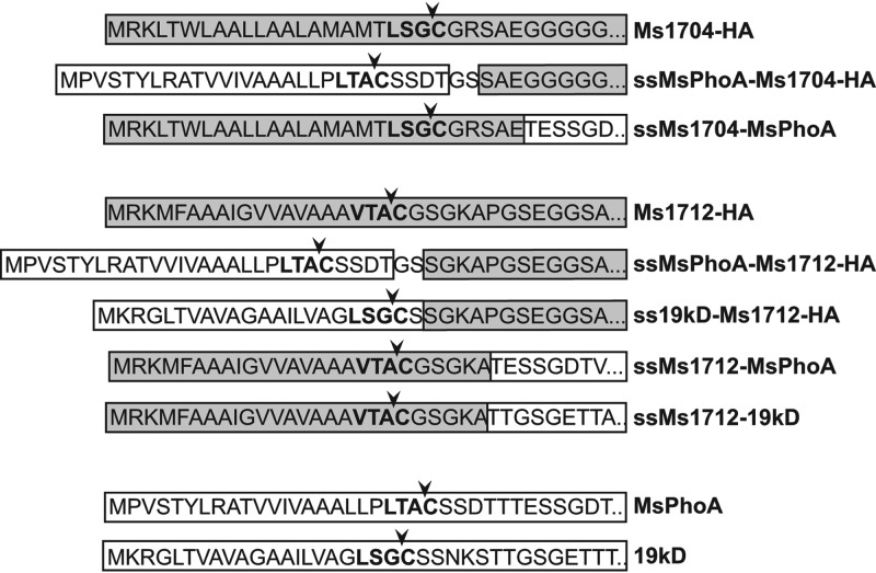
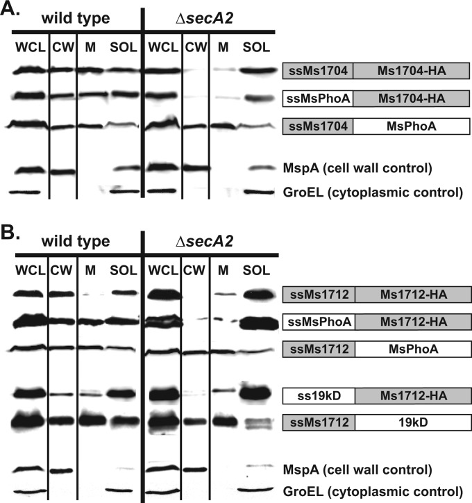
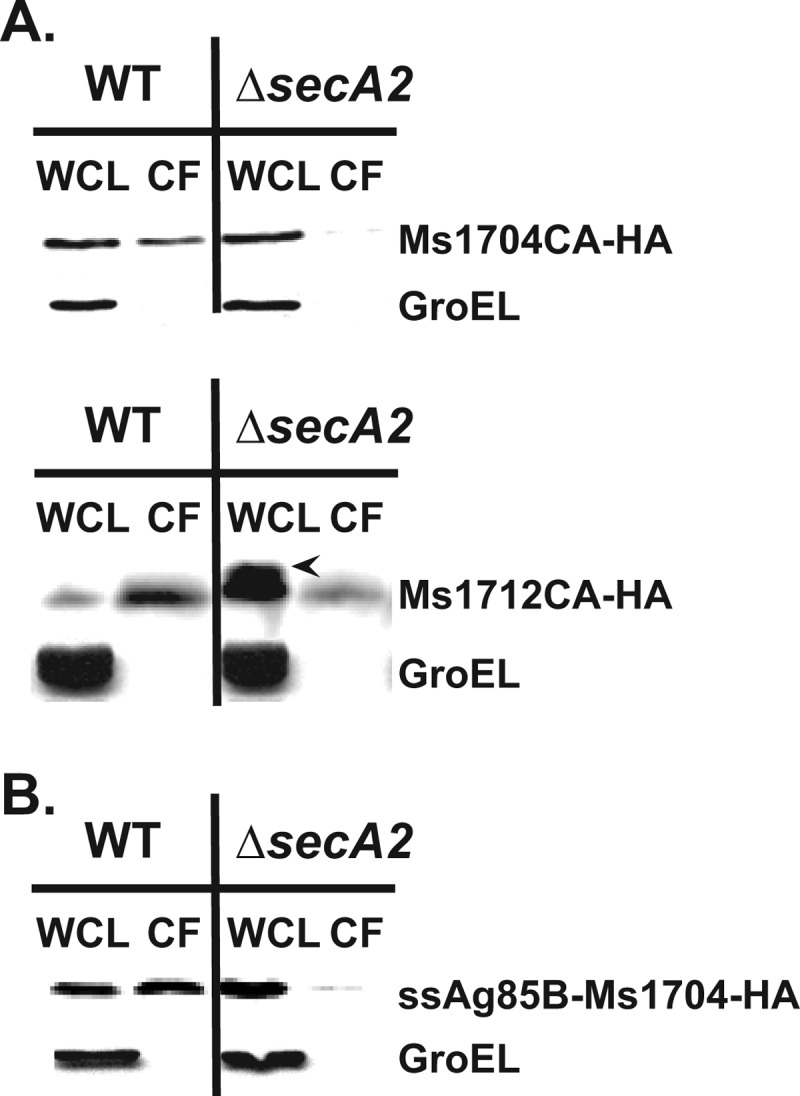
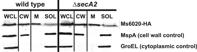
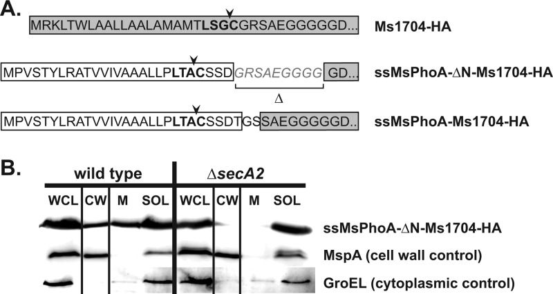
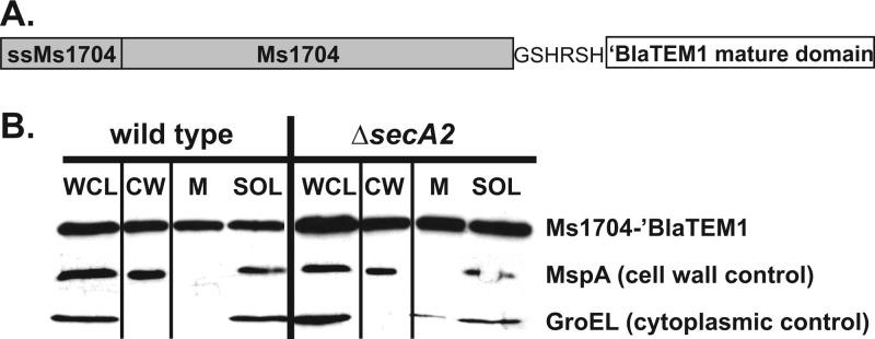
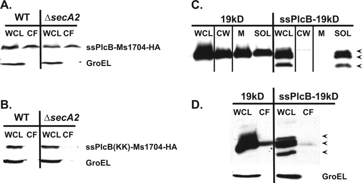
At the core of the bacterial general secretion (Sec) pathway is the SecA ATPase, which powers translocation of unfolded preproteins containing Sec signal sequences through the SecYEG membrane channel. Mycobacteria have two nonredundant SecA homologs: SecA1 and SecA2. While the essential SecA1 handles "housekeeping" export, the nonessential SecA2 exports a subset of proteins and is required for Mycobacterium tuberculosis virulence. Currently, it is not understood how SecA2 contributes to Sec export in mycobacteria. In this study, we focused on identifying the features of two SecA2 substrates that target them to SecA2 for export, the Ms1704 and Ms1712 lipoproteins of the model organism Mycobacterium smegmatis. We found that the mature domains of Ms1704 and Ms1712, not the N-terminal signal sequences, confer SecA2-dependent export. We also demonstrated that the lipid modification and the extreme N terminus of the mature protein do not impart the requirement for SecA2 in export. We further showed that the Ms1704 mature domain can be efficiently exported by the twin-arginine translocation (Tat) pathway. Because the Tat system exports only folded proteins, this result implies that SecA2 substrates can fold in the cytoplasm and suggests a putative role of SecA2 in enabling export of such proteins. Thus, the mycobacterial SecA2 system may represent another way that bacteria solve the problem of exporting proteins that can fold in the cytoplasm.


Figures

Fig 1
Fig 1
Schematic of signal sequence chimeras. The signal sequence regions of chimeras generated between M. smegmatis SecA2 substrates and SecA2-independent lipoproteins are shown. Amino acid sequences from either SecA2 substrate (Ms1704 or Ms1712) are highlighted in gray, while sequences from SecA2-independent proteins (PhoA or 19-kDa protein) are boxed in white. The predicted lipobox in each signal sequence is highlighted in bold, and the predicted cleavage site adjacent to the invariant cysteine (site of lipid modification) at the +1 position of the mature domain is noted with an arrow. Amino acids introduced from cloning are unboxed.
Fig 2
Fig 2
The mature domains of Ms1704 and Ms1712 require SecA2 for export to the cell wall. (A) Equalized whole-cell lysates (WCL) generated from wild-type and ΔsecA2 mutant M. smegmatis cells expressing either Ms1704-HA, ssPhoA-Ms1704-HA, or ssMs1704-PhoA were subjected to ultracentrifugation to generate subcellular fractions. Fractions were separated by SDS-PAGE, and proteins were detected with either anti-HA or anti-PhoA antibody. The total amount of cell wall (CW), membrane (M), and soluble (SOL) fractions shown is equivalent to the amount of WCL loaded. Native MspA and GroEL were detected as cell wall and cytoplasmic controls, respectively. (B) Wild-type or ΔsecA2 mutant M. smegmatis cells expressing either Ms1712-HA, ssPhoA-Ms1712-HA, ssMs1712-PhoA, ss19-kDa-Ms1712-HA, or ssMs1712-19-kDa were fractionated, and material was separated by SDS-PAGE. Immunoblotting was performed as described in panel A with the addition of the anti-19-kDa antibody when appropriate. The experiment shown is representative of three independent experiments.
Fig 3
Fig 3
SecA2-dependent export occurs regardless of lipidation. (A) Culture filtrate (CF) proteins from M. smegmatis cells expressing either Ms1704(CA)-HA or Ms1712(CA)-HA proteins were analyzed by SDS-PAGE and immunoblotting using an anti-HA antibody. Whole-cell lysate (WCL) and CF material loaded were obtained from an equal number of wild-type (WT) or ΔsecA2 M. smegmatis cells for each experiment. Native GroEL protein was detected as a loading and cell lysis control. Accumulation of presumptive precursor Ms1712(CA)-HA, containing an uncleaved signal sequence, in the WCL of the ΔsecA2 mutant is noted with an arrowhead. (B) Immunoblot of WCL and CF proteins from wild-type and ΔsecA2 M. smegmatis cells expressing the nonlipidated ssAg85B-Ms1704-HA chimera. The experiments shown are representative of three independent experiments.
Fig 4
Fig 4
Ms6020-HA is exported to the cell wall independent of SecA2. Equalized whole-cell lysates (WCL) generated from wild-type and ΔsecA2 M. smegmatis cells expressing HA-tagged Ms6020 were subjected to ultracentrifugation to generate subcellular fractions. Ms6020-HA was detected with an anti-HA antibody while native MspA and GroEL were detected as cell wall and cytoplasmic controls, respectively. The total amount of cell wall (CW), membrane (M), and soluble (SOL) material shown is equivalent to the amount of WCL loaded. Shown here is a representative of three independent experiments.
Fig 5
Fig 5
The extreme N terminus of the Ms1704 mature domain is not required for SecA2-mediated export. (A) The first 9 amino acids (italics) were removed from the Ms1704 mature domain, adjacent to the signal peptide cleavage site (arrowhead), to assess the contribution of this region to SecA2-mediated export. This extreme N terminus truncation was created in the ssPhoA-Ms1704-HA signal sequence chimera, where sequence derived from M. smegmatis PhoA is boxed in white and sequence from Ms1704 is boxed in gray. (B) Equalized whole-cell lysates (WCL) generated from wild-type and ΔsecA2 M. smegmatis cells expressing this ssPhoA-ΔN-Ms1704-HA protein were subjected to ultracentrifugation to generate subcellular fractions. Fractions were separated by SDS-PAGE, and proteins were detected by an anti-HA antibody. The total amount of cell wall (CW), membrane (M), and soluble (SOL) fractions shown is equivalent to the amount of WCL loaded. Native MspA and GroEL were detected as cell wall and cytoplasmic controls, respectively. Shown here is a representative of three independent experiments.
Fig 6
Fig 6
An Ms1704-′BlaTEM1 fusion is exported to the cell wall independently of SecA2. (A) The structure of the Ms1704-′BlaTEM1 fusion protein is shown. The signal peptide and entire Ms1704 mature domain constitute the N terminus of this fusion and are depicted in the gray rectangle. The E. coli ′BlaTEM1 (lacking its native signal sequence) was fused to the C terminus of Ms1704. Amino acids introduced from cloning are unboxed. (B) Equalized whole-cell lysates (WCL) generated from wild-type and ΔsecA2 M. smegmatis cells expressing Ms1704-′BlaTEM1 were subjected to ultracentrifugation to generate subcellular fractions. The Ms1704-′BlaTEM1 fusion was detected with an anti-BlaTEM1 antibody while native MspA and GroEL were detected as cell wall and cytoplasmic controls, respectively. The total amount of cell wall (CW), membrane (M), and soluble (SOL) fractions shown is equivalent to the amount of WCL loaded. Shown is a representative of two independent experiments.
Fig 7
Fig 7
Ms1704 is compatible with export by the twin-arginine translocation (Tat) pathway. (A) Concentrated culture filtrate (CF) proteins from M. smegmatis cells expressing ssPlcB-Ms1704-HA were analyzed by SDS-PAGE and immunoblotting using an anti-HA antibody. Whole-cell lysate (WCL) and CF material loaded were obtained from an equal number of wild-type and ΔsecA2 M. smegmatis cells for each experiment. Native GroEL was detected as a loading and cell lysis control. (B) In the signal sequence of the ssPlcB(KK)-Ms1704-HA chimera, the twin arginine residues were changed to lysines to abolish targeting to the Tat machinery. Secretion of this chimera was analyzed as described in panel A. (C) Export of the 19-kDa protein containing its native Sec signal sequence and the ssPlcB-19-kDa chimera were analyzed by SDS-PAGE and immunoblotting using an anti-19-kDa antibody. Equalized whole-cell lysates (WCL) generated from wild-type M. smegmatis cells expressing each protein were subjected to ultracentrifugation to generate cell wall (CW), membrane (M), and soluble (SOL) fractions. (D) WCL and culture filtrate (CF) proteins from wild-type M. smegmatis cells expressing either protein were also analyzed by immunoblotting. Wild-type 19-kDa protein is readily detected in both CW and CF fractions while the ssPlcB-19-kDa chimera is not exported. The ssPlcB-19-kDa chimera also exhibits three forms in the WCL, SOL, and CF fractions (indicated by arrowheads in panels C and D), likely representing precursor protein containing an uncleaved signal sequence and cytoplasmic degradation products. All immunoblots are representative of at least two independent experiments.
Fig 8
Fig 8
Models for mycobacterial SecA2 export. (A) The feature(s) that make a preprotein dependent on SecA2 for export is not contained in the N-terminal signal sequence. Instead, the mature domain of select proteins, such as Ms1704 and Ms1712, impart the requirement for SecA2. Lipidation of the mature domain is not a factor. (B) SecA2 is required for the export of certain proteins, likely through the canonical SecYEG channel. One possibility is that glycosylation or another posttranslational modification in the mature domain (depicted as attached circles) prevents export by the canonical Sec pathway and/or directs preproteins to SecA2 for export (1). A second possibility is that the mature domain contains an amino acid sequence that directs the preprotein to SecA2 and/or away from SecA1 (depicted as a thick black oval) (2). A third possibility is that the defining feature of SecA2 substrates is a tendency to fold in the cytoplasm (3). Our data are consistent with this last possibility, where SecA2 would then function in maintaining an unfolded preprotein conformation prior to or during export. The signal sequence of preproteins is depicted by the oval with diagonal lines.

Similar articles

Cited by

References

    1. Palmer T, Berks BC. 2012. The twin-arginine translocation (Tat) protein export pathway. Nat. Rev. Microbiol. 10:483–496 - PubMed
    1. Lycklama AN, Driessen AJ. 2012. The bacterial Sec-translocase: structure and mechanism. Philos. Trans. R. Soc. Lond. B Biol. Sci. 367:1016–1028 - PMC - PubMed
    1. Brundage L, Hendrick JP, Schiebel E, Driessen AJ, Wickner W. 1990. The purified E. coli integral membrane protein SecY/E is sufficient for reconstitution of SecA-dependent precursor protein translocation. Cell 62:649–657 - PubMed
    1. von Heijne G. 1990. The signal peptide. J. Membr. Biol. 115:195–201 - PubMed
    1. Auclair SM, Moses JP, Musial-Siwek M, Kendall DA, Oliver DB, Mukerji I. 2010. Mapping of the signal peptide-binding domain of Escherichia coli SecA using Forster resonance energy transfer. Biochemistry 49:782–792 - PMC - PubMed

Publication types

MeSH terms

LinkOut - more resources