Bioprinted amniotic fluid-derived stem cells accelerate healing of large skin wounds
- PMID: 23197691
- PMCID: PMC3659666
- DOI: 10.5966/sctm.2012-0088
Bioprinted amniotic fluid-derived stem cells accelerate healing of large skin wounds
Abstract
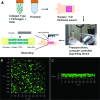
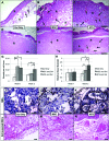
Stem cells obtained from amniotic fluid show high proliferative capacity in culture and multilineage differentiation potential. Because of the lack of significant immunogenicity and the ability of the amniotic fluid-derived stem (AFS) cells to modulate the inflammatory response, we investigated whether they could augment wound healing in a mouse model of skin regeneration. We used bioprinting technology to treat full-thickness skin wounds in nu/nu mice. AFS cells and bone marrow-derived mesenchymal stem cells (MSCs) were resuspended in fibrin-collagen gel and "printed" over the wound site. At days 0, 7, and 14, AFS cell- and MSC-driven wound closure and re-epithelialization were significantly greater than closure and re-epithelialization in wounds treated by fibrin-collagen gel only. Histological examination showed increased microvessel density and capillary diameters in the AFS cell-treated wounds compared with the MSC-treated wounds, whereas the skin treated only with gel showed the lowest amount of microvessels. However, tracking of fluorescently labeled AFS cells and MSCs revealed that the cells remained transiently and did not permanently integrate in the tissue. These observations suggest that the increased wound closure rates and angiogenesis may be due to delivery of secreted trophic factors, rather than direct cell-cell interactions. Accordingly, we performed proteomic analysis, which showed that AFS cells secreted a number of growth factors at concentrations higher than those of MSCs. In parallel, we showed that AFS cell-conditioned media induced endothelial cell migration in vitro. Taken together, our results indicate that bioprinting AFS cells could be an effective treatment for large-scale wounds and burns.
Figures


Similar articles
-
Mesenchymal stem cells enhance wound healing through differentiation and angiogenesis.Stem Cells. 2007 Oct;25(10):2648-59. doi: 10.1634/stemcells.2007-0226. Epub 2007 Jul 5. Stem Cells. 2007. PMID: 17615264
-
[Effects of human adipose-derived mesenchymal stem cells and platelet-rich plasma on healing of wounds with full-thickness skin defects in mice].Zhonghua Shao Shang Za Zhi. 2018 Dec 20;34(12):887-894. doi: 10.3760/cma.j.issn.1009-2587.2018.12.013. Zhonghua Shao Shang Za Zhi. 2018. PMID: 30585053 Chinese.
-
Comparative study on characterization and wound healing potential of goat (Capra hircus) mesenchymal stem cells derived from fetal origin amniotic fluid and adult bone marrow.Res Vet Sci. 2017 Jun;112:81-88. doi: 10.1016/j.rvsc.2016.12.009. Epub 2017 Jan 3. Res Vet Sci. 2017. PMID: 28135618
-
The Role of MSC in Wound Healing, Scarring and Regeneration.Cells. 2021 Jul 8;10(7):1729. doi: 10.3390/cells10071729. Cells. 2021. PMID: 34359898 Free PMC article. Review.
-
Mesenchymal stem cells and cutaneous wound healing: novel methods to increase cell delivery and therapeutic efficacy.Stem Cell Res Ther. 2016 Mar 9;7:37. doi: 10.1186/s13287-016-0303-6. Stem Cell Res Ther. 2016. PMID: 26960535 Free PMC article. Review.
Cited by
-
Emerging Biofabrication Techniques: A Review on Natural Polymers for Biomedical Applications.Polymers (Basel). 2021 Apr 8;13(8):1209. doi: 10.3390/polym13081209. Polymers (Basel). 2021. PMID: 33918049 Free PMC article. Review.
-
Skin microbiome considerations for long haul space flights.Front Cell Dev Biol. 2022 Sep 8;10:956432. doi: 10.3389/fcell.2022.956432. eCollection 2022. Front Cell Dev Biol. 2022. PMID: 36158225 Free PMC article.
-
Intravital three-dimensional bioprinting.Nat Biomed Eng. 2020 Sep;4(9):901-915. doi: 10.1038/s41551-020-0568-z. Epub 2020 Jun 22. Nat Biomed Eng. 2020. PMID: 32572195
-
Three-Dimensional Bioprinting: A Comprehensive Review for Applications in Tissue Engineering and Regenerative Medicine.Bioengineering (Basel). 2024 Jul 31;11(8):777. doi: 10.3390/bioengineering11080777. Bioengineering (Basel). 2024. PMID: 39199735 Free PMC article. Review.
-
History of burns: The past, present and the future.Burns Trauma. 2014 Oct 25;2(4):169-80. doi: 10.4103/2321-3868.143620. eCollection 2014. Burns Trauma. 2014. PMID: 27574647 Free PMC article.
References
-
- Cherry DK, Hing E, Woodwell DA, et al. National Ambulatory Medical Care Survey: 2006 summary. Natl Health Stat Report. 2008:1–39. - PubMed
-
- Pitts SR, Niska RW, Xu J, et al. National Hospital Ambulatory Medical Care Survey: 2006 emergency department summary. Natl Health Stat Report. 2008:1–38. - PubMed
-
- Miller SF, Bessey P, Lentz CW, et al. National burn repository 2007 report: A synopsis of the 2007 call for data. J Burn Care Res. 2008;29:862–870. discussion 871. - PubMed
-
- Peck MD. Epidemiology of burns throughout the world. Part I: Distribution and risk factors. Burns. 2011;37:1087–1100. - PubMed
Publication types
MeSH terms
Substances
Grants and funding
LinkOut - more resources
Full Text Sources
Other Literature Sources
Medical
Research Materials

