. 2012;7(11):e49112.
doi: 10.1371/journal.pone.0049112. Epub 2012 Nov 20.

GSK3beta-mediated Drp1 phosphorylation induced elongated mitochondrial morphology against oxidative stress

Affiliations

GSK3beta-mediated Drp1 phosphorylation induced elongated mitochondrial morphology against oxidative stress

Chia-Hua Chou et al. PLoS One. 2012.

Abstract

Multiple phosphorylation sites of Drp1 have been characterized for their functional importance. However, the functional consequence of GSK3beta-mediated phosphorylation of Drp1 remains unclear. In this report, we pinpointed 11 Serine/Threonine sites spanning from residue 634~736 of the GED domain and robustly confirmed Drp1 Ser693 as a novel GSK3beta phosphorylation site. Our results suggest that GSK3beta-mediated phosphorylation at Ser693 does cause a dramatic decrease of GTPase activity; in contrast, GSK3beta-mediated phosphorylation at Ser693 appears not to affect Drp1 inter-/intra-molecular interactions. After identifying Ser693 as a GSK3beta phosphorylation site, we also determined that K679 is crucial for GSK3beta-binding, which strongly suggests that Drp1 is a novel substrate for GSK3beta. Thereafter, we found that overexpressed S693D, but not S693A mutant, caused an elongated mitochondrial morphology which is similar to that of K38A, S637D and K679A mutants. Interestedly, using H89 and LiCl to inhibit PKA and GSK3beta signaling, respectively, it appears that a portion of the elongated mitochondria switched to a fragmented phenotype. In investigating the biofunctionality of phosphorylation sites within the GED domain, cells overexpressing Drp1 S693D and S637D, but not S693A, showed an acquired resistance to H(2)O(2)-induced mitochondrial fragmentation and ensuing apoptosis, which affected cytochrome c, capase-3, -7, and PARP, but not LC3B, Atg-5, Beclin-1 and Bcl2 expressions. These results also showed that the S693D group is more effective in protecting both non-neuronal and neuronal cells from apoptotic death than the S637D group. Altogether, our data suggest that GSK3beta-mediated phosphorylation at Ser693 of Drp1 may be associated with mitochondrial elongation via down-regulating apoptosis, but not autophagy upon H(2)O(2) insult.


Conflict of interest statement

Competing Interests: The authors have declared that no competing interests exist.

Figures

Figure 1
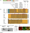
Figure 1. Interaction and co-localization of Drp1/Hydn IV with GSK3beta.
(A) Schematic diagram showing the domain structure of Drp1 as previously described and relative positions of truncated Drp1 constructs used in this study. Yeast two-hybrid assays showing interactions of various Drp1 constructs as bait (pAS2-1) with GSK3beta as prey (pACT-2) as indicated. Blue (B) and white (W) assay was applied for reporter gene beta-galactosidase expression. Strength of interaction was assayed by beta-galactosidase and HIS3 induction as described previously by Hong et al. . (B) Specific binding region of Drp1 with GSK3beta is highly conserved. Multiple alignment of Drp 1 GED domain (colored in orange) was performed using ClustalW2 by which seven Drp1 homologues and human Dynamin 1 were aligned. Note the very high conservation in the GSK3 binding domain (BD, indicated by red bar) in Drp1 but not Dynamin 1 and Ser or Lys residues at positions corresponding to human Drp1 S637, K679, and S693 as indicated by red or white rectangles. Compared to human Drp1, the percentage of amino acid identity corresponding to each BD in GED is listed in front of the sequence. The prospective phosphorylated Ser/Thr residues are indicated by “O”. (C) Total cell lysates were prepared from pEGFP C1-Drp1, pEGPC1-GSK3beta overexpressed 293 cells after transient expression for 24 hours, and subjected to co-immunoprecipitation (IP) assays with anti-GSK3 (left panel), anti-Drp1 (right panel), respectively. The anti-GFP antibody was used to detect the exogenous expressed GFP-tagged proteins in Western blotting (as indicated) which has been described in Materials and Methods. Data are representative of three independent experiments. The IP-Drp1 and IP-GSK3 represents the co-precipitated protein respects to antibodies used (D) The binding fashion of Drp1634–690 and Drp1444–736 fragment with GSK3beta was confirmed by far-Western blotting. Recombinant His-tagged fusion protein, either Drp1 full-length or truncated fragments of Drp1, was separated on 12% SDS polyacrylamide gel. Proteins were transferred into PVDF and incubated with HeLa cell lysate containing FLAG-tagged GSK3beta. Arrowhead indicates binding. (E) Co-localization study was performed by immunocytochemistry staining with confocal fluorescent microscopy as described in Materials and Methods. At 24 hours, 293 cells were stained with anti-GSK3beta antibody to detect endogenous GSK3beta (red, frame a). Frame b is the localization of endogenous Drp1 (green). Frame c is the merged image of the Drp1 and anti- GSK3beta stained cells. Bar = 5 µm.
Figure 2
Figure 2. Drp1 is phosphorylated by GSK3beta at Ser693.
Drp1 is phosphorylated by GSK3beta. (A) Purified GSK3beta was used in an in vitro kinase assay to detect whether and which truncated His-tagged Drp1 proteins can act as a substrate to be phosphorylated by GSK3beta. The upper panel is a coomassie blue stained blot showing the His-tagged Drp1 proteins were successfully expressed. The lower panel indicates GSK3beta autophosphorylation (arrow) and phosphorylation of truncated His-Drp1 (empty arrow). (B) In vitro kinase assay to determine the GSK3beta phosphorylation site using site-directed mutagenesis technique. Numbers indicate the mutated residue within the Drp1634–690 fragment. The upper panel is a coomassie blue stained blot showing that His-tagged Drp1634–690 wild-type and mutants were expressed. The lower panel indicates the phosphorylation level with respect to His-tagged Drp1634–690 wild-type and different mutants. (C) Numbers indicate the mutated residue within the Drp1691–736 fragment. The upper panel is a coomassie blue stained blot showing that His-tagged Drp1691–736 wild-type and mutants were expressed. The lower panel indicates the phosphorylation level with respect to His-tagged Drp1691–736 wild-type and different mutants. (D) Both S693A and S693D Drp1 mutants were confirmed to lose their GSK3beta-mediated phosphorylation in vitro. The upper panel is the coomassie blue staining of His-tagged mutated Drp1 fragments. Numbers indicate the mutated residue within the Drp1691–736 fragment. The lower panel indicates the phosphorylation of mutated His-tagged Drp1. (E) The specificity of GSK3beta-mediated phosphorylation was tested on Drp1691–736 fragments. GSK3beta inhibitor, GSKIP, was added in kinase assay with different doses and the effect on the phosphorylation of Drp1691–736 is shown. The coomassie blue image of GSKIP protein as loading/input control was shown in the lower panel.
Figure 3
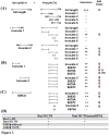
Figure 3. Yeast two-hybrid assay identifying Drp1 inter-/intra- interaction and residue responsible for GSK3beta-Drp1 binding.
The design of the yeast two-hybrid assays showing interactions of various bait (pAS2-1) or prey (pACT2) constructs is indicated. The strength of interaction was assayed as described in Methods. beta-galactosidase and HIS3 induction was quantified as described in Materials and Methods. The result of the interaction was shown by “+” or “−”. (A) The inter-molecular interaction of Drp1 was tested by using full-length Drp1 as bait and various truncated Drp1 as prey to verify their interacting ability. The intra-molecular interaction of Drp1 was tested by using truncate 1 or 2 of Drp1 as bait and various truncated Drp1 as prey to verify their interacting ability. (B) Drp1 truncated fragments with point-mutation were tested to verify their interaction with Drp1 truncate 1 or 2. Some of our results were inconsistent with previous reports , and are indicated as “☆”and “☆☆”, respectively (C). Some Drp1 truncate 2 mutants were tested for their possible interaction with GSK3beta. (D) Matrix of yeast two-hybrid assays showing interactions of various Drp1 N- and C-terminal deletion fragments, GSK3beta wt and GSK3beta V276G mutant in bait constructs, tested against C-terminal Drp1 prey constructs with or without K679A mutation as indicated. The result of interaction was shown as “+” or “−”.
Figure 4
Figure 4. GTPase hydrolysis activity of Drp1mutants.
A GTPase hydrolysis activity assay was performed followed the procedures published by Ingerman and Nunnari . E. coli were transformed by Drp1 mutant plasmids and 15 µg of cell lysate was applied to the assay. O.D. 340 was measured to reflect the constant of the NADH. The slope reflects the consumption of NADH. The data are representative of three independent experiments and are shown as mean values ± SD.
Figure 5
Figure 5. Mitochondrial dynamics of HeLa cells with Drp1 wt and mutants.
(A) HeLa cells were transfected with GFP-tagged Drp1 wt or other mutants for 24 hours. Mitochondrial morphology was observed by confocal fluorescent microscope with Mitotracker dye. Cell nuclei were counter-stained by DAPI. Insets are magnifications of the Mitotracker signal at the indicated areas. Inset 1 represents the non-transfected cells, and inset 2 indicates the transfected cells. Indications (white arrows) represent typical elongated mitochondria morphology. (B) Statistical result of mitochondrial morphology. After 24 hours, over 100 transfected cells were categorized into 3 groups depending on mitochondrial morphology. *p<0.05, **p<0.01, ***p<0.001.
Figure 6
Figure 6. Mitochondrial dynamics of HeLa cells with Drp1 wt and mutants and were treated with LiCl and H89.
(A) HeLa cells were transfected with GFP-tagged Drp1wt or other mutants for 24 hours. Then cells were treated with 10 mM LiCl and 10 µM H89 for another 24 hours. Mitochondrial morphology of HeLa cells was observed by staining with Mitotracker under confocal microscopy. Cell nuclei were counter-stained by using DAPI. Insets are magnifications of the Mitotracker signal at the indicated areas. Inset 1 represents the non-transfected cells, and inset 2 indicates the transfected cells. Indications (white arrows) represent typical elongated mitochondria morphology. (B) Statistical result of mitochondrial morphology. After 24 hours treated with inhibitors (upper: LiCl, lower: H89), over 100 transfected cells were categorized into 3 groups depending on mitochondrial morphology. *p<0.05, **p<0.01, ***p<0.001.
Figure 7
Figure 7. Mitochondrial dynamics of HeLa cells with Drp1 wt and mutants treated with H2O2.
(A) HeLa cells were transfected with GFP-tagged Drp1wt or other mutants for 24 hours. Then cells were treated with 500 µM H2O2 for another 24 hours. Mitochondrial morphology of HeLa cells was observed by staining with Mitotracker under confocal microscopy. Cell nuclei were counter-stained by using DAPI. Insets are magnifications of the Mitotracker signal at the indicated areas. Inset 1 represents the non-transfected cells, and inset 2 indicates the transfected cells. Indications (white arrows) represent typical elongated mitochondria morphology. (B) Statistical results demonstrated mitochondrial morphology of HeLa cells with or without Drp1 expression under H2O2 treatment for 24 hours; over 100 transfected cells were categorized into 3 groups depending on mitochondrial morphology. **p<0.001.
Figure 8
Figure 8. Overexpressed Drp1 S693D can protect against H2O2-induced mitochondrial fragmentation and ensuing apoptosis but does not induce autophagy.
SH-SY5Y cells were transfected with GFP alone, GFP-tagged Drp-1 wild-type and other mutants for 24 hours. Then cells were treated with 500 µM H2O2 for another 24 hours, and were lysed and detected by Western blotting using anti-cytochrome c, cleaved-caspase 3, cleaved-caspase 7, cleaved-PARP, -Bcl2, -LC3, p62, -Beclin 1 and -Atg5 antibody, respectively. beta-actin served as a protein loading control. The data are representative of three independent experiments.
Figure 9
Figure 9. Model represents both GSK3β- and PKA-mediated Drp1 phosphorylation induction of mitochondrial elongation which subsequently causes acquired resistance to H2O2-induced apoptosis rather than inducing autophagy.
Mitochondria dynamics regulate the GTPase hydrolysis activity of proteins (Drp1, Opa1, Mfn1 and 2) resulting in mitochondrial fission or fusion. In this model, two Drp1 phosphorylation sites could serve a regulatory function, including phosphorylation by PKA/AKAP1 on Ser637 or by GSK3β on Ser693 (as shown in this study), leading to diminished mitochondrial fission resulting in mitochondrial elongation. GSK3β might be recruited to mitochondria through AKAP220 and be dephosphorylated by PP1, 2, and 3 . Such mitochondrial morphological changes could also result in cell fate determination. Mitochondrial fission is involved in the initiation of apoptosis, whereas mitochondrial fusion may induce autophagy. Both phosphorylation events occurring at S637 and S693 cause elongated mitochondrial morphology and lead to acquired resistance to H2O2-induced mitochondrial fragmentation and ensuing apoptosis via down-regulating cytochrome c release, capase-3, -7 and PARP activations rather than inducing autophagy.

Similar articles

Cited by

References

    1. Cross DA, Alessi DR, Cohen P, Andjelkovich M, Hemmings BA (1995) Inhibition of glycogen synthase kinase-3 by insulin mediated by protein kinase B. Nature 378: 785–789. - PubMed
    1. Jope RS, Johnson GV (2004) The glamour and gloom of glycogen synthase kinase-3. Trends Biochem Sci 29: 95–102. - PubMed
    1. Cho J, Rameshwar P, Sadoshima J (2009) Distinct roles of glycogen synthase kinase (GSK)-3alpha and GSK-3beta in mediating cardiomyocyte differentiation in murine bone marrow-derived mesenchymal stem cells. J Biol Chem 284: 36647–36658. - PMC - PubMed
    1. Hooper C, Killick R, Lovestone S (2008) The GSK3 hypothesis of Alzheimer's disease. J Neurochem 104: 1433–1439. - PMC - PubMed
    1. Hong YR, Chen CH, Cheng DS, Howng SL, Chow CC (1998) Human dynamin-like protein interacts with the glycogen synthase kinase 3beta. Biochem Biophys Res Commun 249: 697–703. - PubMed

Publication types

MeSH terms

Grants and funding

This study was sponsored by National Science Council-98-2320-B-037-024-MY3; National Science Council -98-2321-B-037-061; National Science Council-99-2321-B-037-001; National Health Research Intitute-EX100-9809SI (Taiwan); National Sun Yet-San University-Kaohsiung Medical University Joint Research Project-101-020 to Yi-Ren Hong and National Science Council-100-2314-B-037-010 (Taiwan) to Shen-Long Howng. The funders had no role in study design, data collection and analysis, decision to publish, or preparation of the manuscript.