. 2012 Nov 21;32(47):16560-73.
doi: 10.1523/JNEUROSCI.1787-12.2012.

An early onset progressive motor neuron disorder in Scyl1-deficient mice is associated with mislocalization of TDP-43

Affiliations

An early onset progressive motor neuron disorder in Scyl1-deficient mice is associated with mislocalization of TDP-43

Stephane Pelletier et al. J Neurosci. .

Abstract

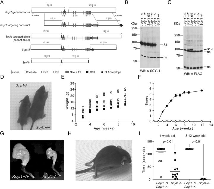
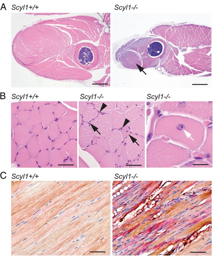
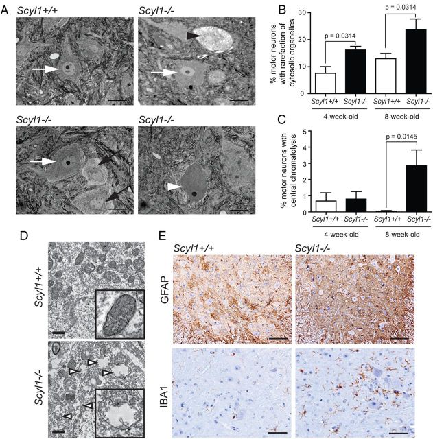
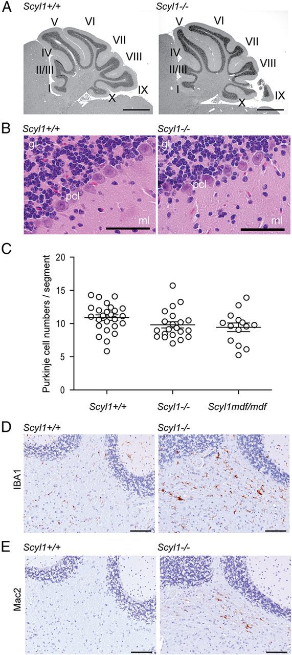
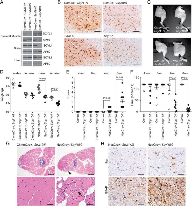
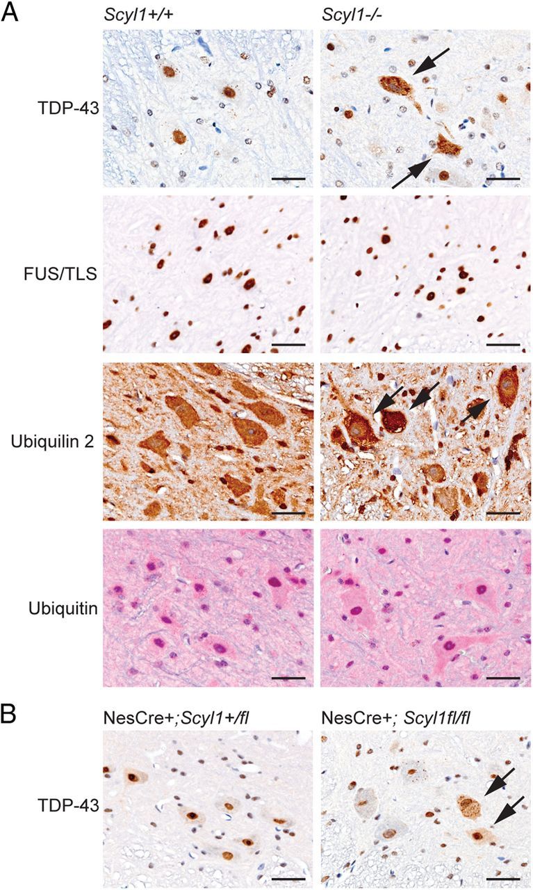
The molecular and cellular bases of motor neuron diseases (MNDs) are still poorly understood. The diseases are mostly sporadic, with ~10% of cases being familial. In most cases of familial motor neuronopathy, the disease is caused by either gain-of-adverse-effect mutations or partial loss-of-function mutations in ubiquitously expressed genes that serve essential cellular functions. Here we show that deletion of Scyl1, an evolutionarily conserved and ubiquitously expressed gene encoding the COPI-associated protein pseudokinase SCYL1, causes an early onset progressive MND with characteristic features of amyotrophic lateral sclerosis (ALS). Skeletal muscles of Scyl1(-/-) mice displayed neurogenic atrophy, fiber type switching, and disuse atrophy. Peripheral nerves showed axonal degeneration. Loss of lower motor neurons (LMNs) and large-caliber axons was conspicuous in Scyl1(-/-) animals. Signs of neuroinflammation were seen throughout the CNS, most notably in the ventral horn of the spinal cord. Neural-specific, but not skeletal muscle-specific, deletion of Scyl1 was sufficient to cause motor dysfunction, indicating that SCYL1 acts in a neural cell-autonomous manner to prevent LMN degeneration and motor functions. Remarkably, deletion of Scyl1 resulted in the mislocalization and accumulation of TDP-43 (TAR DNA-binding protein of 43 kDa) and ubiquilin 2 into cytoplasmic inclusions within LMNs, features characteristic of most familial and sporadic forms of ALS. Together, our results identify SCYL1 as a key regulator of motor neuron survival, and Scyl1(-/-) mice share pathological features with many human neurodegenerative conditions.


Figures

Figure 1.
Figure 1.
SCYL1 deficiency causes growth defects and early onset progressive motor deficits. A, Schematic representation of the Scyl1 locus, the targeting vector, and predicted Scyl1 mutated locus. After Cre- or Flp-mediated recombination in ES cells, Scyl1− and Scyl1fl loci were generated. Following Cre-mediated recombination, only exon 1 remained. Exons are indicated with gray bars. Black bars represent XhoI sites. Black triangles indicate loxP and Frt sites. Gray box indicates the Neo–TK cassette. Diphtheria toxin cassette is illustrated by a black arrow. The black circle labeled with an f indicates the position of the FLAG epitope. B, C, SCYL1 and FLAG-tagged SCYL1 expression in the cerebrum of Scyl1+/+, Scyl1+/, Scyl1+/fl, Scyl1fl/fl, and Scyl1−/− mice. Cerebrum extracts obtained from adult Scyl1+/+, Scyl1+/−, Scyl1+/fl, Scyl1fl/fl, and Scyl1−/− mice were resolved by SDS-PAGE and analyzed by Western blotting, using antibodies against Scyl1 (α-SCYL1) (B) or the FLAG epitope (C). Note the absence of SCYL1 (S1) in Scyl1-deficient mice. Also note the expression of FLAG-tagged SCYL1 (S1-F) in Scyl1+/fl and Scyl1fl/fl mice. Nonspecific (ns) bands served as loading control. D, Representative photograph of 6-week-old Scyl1+/+ and Scyl1−/− male littermates illustrating the size differences between Scyl1+/+ and Scyl1−/− mice. E, Growth curve of male (circle) and female (square) Scyl1+/+ (white) and Scyl1−/− (black) mice from 3 to 9 weeks of age. Scyl1−/− mice did not recover and stayed smaller than their wild-type littermates. Values are expressed as the mean ± SEM. Males: Scyl1+/+, n = 9; Scyl1−/−, n = 5; females: Scyl1+/+, n = 6; Scyl1−/−, n = 13. F, Disease progression in Scyl1-deficient mice. The clinical progression of defects in mice was assessed by an objective grading system (for details, see Materials and Methods). Values are expressed as the mean ± SEM. Scyl1+/+, n = 13; Scyl1−/−, n = 13. G, Representative photograph of hindlimbs obtained from 12-week-old Scyl1+/+ and Scyl1−/− males showing massive muscle wasting in Scyl1−/− mice. H, Representative photograph of an end-stage Scyl1−/− mouse. The hindlimbs of the Scyl1−/− mouse are atrophied and dorsally contracted. In some mice, flexion of the hindlimb digits is still possible. Also note the flattening of the pelvis. At this stage of the disease, the affected mice move by the power of their front legs. I, Motor defects in Scyl1-deficient mice. The inverted grid test was performed on 4-week-old and 8- to 12-week-old Scyl1+/+ and Scyl1−/− mice to assess their motor functions for up to 120 s as described in Materials and Methods. Four-week-old Scyl1+/+, n = 22; 4-week-old Scyl1−/−, n = 11; 8- to 12-week-old Scyl1+/+, n = 10; 8- to 12-week-old Scyl1−/−, n = 10.
Figure 2.
Figure 2.
Myopathological abnormalities in Scyl1-deficient mice. A, Representative micrographs of H&E-stained sections of the anterior muscles in the rear legs of 12-week-old Scyl1+/+ and Scyl1−/− mice. Note the massive muscle wasting in the quadriceps femoris of Scyl1−/− mice (arrow). Scale bar, 1 mm. B, Higher magnification of H&E-stained sections of quadriceps femoris muscle fibers obtained from Scyl1+/+ and Scyl1−/− mice. Lesions in Scyl1−/− muscles include fibers of different sizes, angulated (atrophied) fibers surrounded by rounded fibers, group atrophy (arrows), nuclear clumps (arrowheads), and centrally localized nuclei (white arrow). Scale bars: left, middle, 50 μm; right, 25 μm. C, Representative micrographs of longitudinal sections of quadriceps femoris from 12-week-old Scyl1+/+ and Scyl1−/− mice illustrating fiber type switching from type II (fast, brown) to type I (slow, red) in Scyl1−/− muscles. Scale bar, 50 μm.
Figure 3.
Figure 3.
Loss of large-diameter axons in peripheral nerves of Scyl1-deficient mice. A, B, Representative micrograph of toluidine blue-stained semithin sections of the sciatic nerve obtained from 3.5-week-old (A) and 8-week-old (B) Scyl1+/+ and Scyl1−/− mice. Scale bar, 20 μm. C, D, Histogram of the frequency of myelinated axons of the sciatic nerve in function of their axonal diameter in 3.5-week-old (C) or 8-week-old (D) mice. A total of 5882 axons from three 8-week-old Scyl1+/+ mice, 4911 axons from three 8-week-old Scyl1−/− mice, 4050 axons from three 3.5-week-old Scyl1+/+, and 2820 axons from three 3.5-week-old Scyl1−/− mice were evaluated. Note the significant reduction in large-caliber axons (≥5 μm) and increased frequency of small-caliber axons (<1–2 μm) in 8-week-old Scyl1-deficient mice. Values are expressed as the mean ± SEM. *p < 0.05. E, Myelinated axon counts in the sciatic nerve of 3.5- and 8-week-old Scyl1+/+ and Scyl1−/− mice. Values are expressed as the mean ± SEM; n = 3 each. F, Segmental demyelination in the sciatic nerve of Scyl1−/− mice. H&E-stained longitudinal sections of sciatic nerves obtained from 4- and 8-week-old Scyl1+/+ (left) and Scyl1−/− male mice. Arrows indicates myelin ovoids with myelin debris. Scale bar, 50 μm.
Figure 4.
Figure 4.
Degenerating motor neurons in the ventral horn of Scyl1−/− mice. A, Representative micrographs of toluidine blue-stained lumbar ventral horn motor neurons of Scyl1+/+ (top left) and Scyl1−/− (top right and bottom panels) mice. Morphological changes in motor neurons of the lumbar spinal ventral horn included rarefaction of cytosolic organelles, enlarged cytoplasmic vacuoles, and central chromatolysis. White arrows indicate healthy motor neurons. Black arrows point to motor neurons with rarefaction of cytosolic organelles. Black arrowhead points to a motor neuron with enlarged vacuoles. White arrowhead points to a motor neuron showing central chromatolysis. Scale bar, 20 μm. B, Quantification of lumbar ventral horn motor neurons showing rarefaction of cytosolic organelles (including motor neurons with enlarged vacuoles) in 4- and 8-week-old Scyl1+/+ and Scyl1−/− mice. Values are expressed as the mean ± SEM. A minimum of 23 images from three different mice per genotype and age group were analyzed (see Materials and Methods). C, Quantification of chromatolytic lumbar ventral horn motor neurons in 4- and 8-week-old Scyl1+/+ and Scyl1−/− mice. Values are expressed as the mean ± SEM. A minimum of 23 images from three different mice per genotype and age group were analyzed (see Materials and Methods). D, Mitochondrial swelling in motor neurons of Scyl1-deficient mice. In motor neurons of Scyl1+/+ mice, mitochondria of the perikaryon appear normal (top panel and inset). In select LMNs of Scyl1−/− mice, mitochondria are swollen, with disrupted cristae (bottom panel and inset). Mitochondrial swelling was observed in 75 (4.9%) of the 1549 mitochondria analyzed from Scyl1+/+ motor neurons versus 247 (16.7%) of the 1479 mitochondria analyzed in Scyl1−/− mice. Scale bar, 1 μm. E, Neuroinflammation in the spinal ventral horn of Scyl1-deficient mice. Immunohistochemistry using antibodies against Iba1 and GFAP on spinal ventral horn sections obtained from 8-week-old Scyl1+/+ and Scyl1−/− mice. Note the increased Iba1 and GFAP staining in the ventral horn of Scyl1-deficient mice. Scale bars: top row, 100 μm; bottom row, 50 μm.
Figure 5.
Figure 5.
Histological examination of the layered structures of Scyl1+/+ and Scyl1−/− cerebellum. A, Representative micrographs of H&E-stained sagittal sections of cerebellums obtained from 17-week-old Scyl1+/+ and Scyl1−/− mice. No gross deformation in the cerebellar structures was observed in Scyl1−/− mice. I–X, Cerebellar lobules. Scale bar, 1 mm. B, Representative micrographs of H&E-stained sections of 8-week-old Scyl1+/+ and Scyl1−/− mice showing the layered structures of the cerebellum. The layered structures of the cerebellum of Scyl1−/− were undistinguishable from Scyl1+/+ mice. gl, Granular layer; pcl, Purkinje cell layer; ml, molecular layer. Scale bar, 50 μm. C, Purkinje cell numbers in Scyl1+/+ and Scyl1−/− mice. Purkinje cells numbers were determined as described in Materials and Methods. For Purkinje cell counts, a total of 25 segments from three Scyl1+/+, 21 segments from three Scyl1−/−, and 14 segments from three Scyl1mdf/mdf male mice of 17 weeks of age were counted. Values are expressed as mean ± SEM. p > 0.05. D, E, Microglial activation in the white track of the cerebellar peduncle. Immunohistochemistry using antibodies against Iba1 (D) and Mac2 (E) on cerebellar sections of 8-week-old Scyl1+/+ (left) and Scyl1−/− (right) mice. Note the increased Iba1 and Mac2 staining in the cerebellar peduncle of Scyl1-deficient mice. Scale bar, 100 μm.
Figure 6.
Figure 6.
Neural- and skeletal muscle-specific deletion of Scyl1. A, Western blot analysis of brain, liver, and skeletal muscle extracts obtained from CkmmCre+;Scyl1+/fl, CkmmCre+;Scyl1fl/fl, NesCre+;Scyl1+/fl, and NesCre+;Scyl1fl/fl mice using antibodies against SCYL1 or AP50 as loading control. Note the selective absence of SCYL1 in the brain of NesCre+;Scyl1fl/fl mice and in the skeletal muscle of CkmmCre+;Scyl1fl/fl mice. B, Immunohistochemical staining of spinal cord cross-sections from NesCre+;Scyl1+/fl and NesCre+;Scyl1fl/fl, Scyl1+/+ and Scyl1−/− mice, using an anti-SCYL1 antibody. In NesCre+;Scyl1fl/fl sections, >90% of neuronal cells are deficient in SCYL1 expression. Note the absence of SCYL1 in all neurons of spinal sections obtained from Scyl1−/− mice compared with Scyl1+/+ mice. A nonspecific nuclear staining is detected by using this antibody. This nuclear staining was also seen in Scyl1−/− mouse embryonic fibroblasts (data not shown) and in sections obtained from Scyl1−/− mice. Scale bar, 200 μm. C, Hindlimb morphology of neural and skeletal muscle mutants of Scyl1. Representative photographs of hindlimbs obtained from 12-week-old CkmmCre+;Scyl1+/fl, CkmmCre+;Scyl1fl/fl, NesCre+;Scyl1+/fl, and NesCre+;Scyl1fl/fl mice. D, Body weight of 8-week-old male (circles) and female (squares) CkmmCre+;Scyl1+/fl (males, n = 5; females, n = 4), CkmmCre+;Scyl1fl/fl (males, n = 4; females, n = 4), NesCre+;Scyl1+/fl (males, n = 5; females, n = 3), and NesCre+;Scyl1fl/fl (males, n = 5; females, n = 6) mice. Values are expressed as the mean ± SEM. E, Early onset progressive motor deficit in neural-specific but not muscle-specific mutant of Scyl1. Score of 4- and 8-week-old CkmmCre+;Scyl1+/fl [4-week-old (wo) animals, n = 18; 8-week-old animals, n = 8], CkmmCre+;Scyl1fl/fl (4-week-old animals, n = 12; 8-week-old animals, n = 7), NesCre+;Scyl1+/fl (4-week-old animals, n = 10; 8-week-old animals, n = 15), and NesCre+; Scyl1fl/fl (4-week-old animals, n = 12; 8-week-old animals, n = 19) mice. Values are expressed as the mean ± SEM. F, Motor defects in tissue-specific mutants of Scyl1. The inverted grid test was performed on 4- and 8-week-old CkmmCre+;Scyl1+/fl (4-week-old animals, n = 25; 8-week-old animals, n = 12), CkmmCre+;Scyl1fl/fl (4-week-old animals, n = 8; 8-week-old animals, n = 7), NesCre+;Scyl1+/fl (4-week-old animals, n = 10; 8-week-old animals, n = 15), and NesCre+;Scyl1fl/fl (4-week-old animals, n = 12; 8-week-old animals, n = 9) mice to assess their motor functions as described in Materials and Methods. Values are expressed as the mean ± SEM. G, Myopathy in neural-specific but not in muscle-specific mutants of Scyl1. Representative micrographs of skeletal muscles (quadriceps femoris) from CkmmCre+;Scyl1fl/fl (n = 3) mice and NesCre+;Scyl1fl/fl (n = 3) mice. Lesions in NesCre+;Scyl1fl/fl muscles include fibers of different size, angulated (atrophied) fibers surrounded by rounded fibers, group atrophy, nuclear clumps (white arrow), and centrally localized nuclei (black arrow). Scale bars: top row, 1 mm; bottom row, 50 μm. H, Neuroinflammation in the spinal ventral horn of NesCre+;Scyl1fl/fl mice. Representative immunohistochemical staining using antibodies against Iba1 (top row) and GFAP (bottom row) on spinal ventral horn sections obtained from 8-week-old control NesCre+;Scyl1+/fl (n = 3) and NesCre+;Scyl1fl/fl (n = 3) mice. Note the increased Iba1 and GFAP staining in the ventral horn of NesCre+;Scyl1fl/fl mice compared with control animals. Scale bar, 50 μm.
Figure 7.
Figure 7.
Nuclear to cytoplasmic relocalization of TDP-43 in Scyl1-deficient motor neurons. A, Immunohistochemistry using antibodies against TDP-43, FUS/TLS, ubiquilin 2, and ubiquitin on spinal ventral horn sections obtained from 8- to 12-week-old Scyl1+/+ and Scyl1−/− mice. Note the relocalization of TDP-43 (arrows) but not FUS/TLS from the nucleus to cytoplasmic inclusions in large motor neurons of Scyl1−/− mice. Also note the cytoplasmic accumulation of ubiquilin 2 (arrows) but not ubiquitin in select motor neurons of the spinal ventral horn of Scyl1-deficient mice. Data shown are representative of three mice for each genotype. Scale bars, 25 μm. B, Immunohistochemistry using antibodies against TDP-43 on spinal ventral horn sections obtained from 8- to 12-week-old NesCre+;Scyl1+/fl and NesCre+;Scyl1fl/fl mice. Note the relocalization of TDP-43 (arrows) in large motor neurons of NesCre+;Scyl1fl/fl mice. Data shown are representative of three mice for each genotype. Scale bars, 25 μm.

Similar articles

Cited by

References

    1. Appenzeller-Herzog C, Hauri HP. The ER-Golgi intermediate compartment (ERGIC): in search of its identity and function. J Cell Sci. 2006;119:2173–2183. - PubMed
    1. Baloh RH. TDP-43: the relationship between protein aggregation and neurodegeneration in amyotrophic lateral sclerosis and frontotemporal lobar degeneration. FEBS J. 2011;278:3539–3549. - PMC - PubMed
    1. Banks GT, Kuta A, Isaacs AM, Fisher EM. TDP-43 is a culprit in human neurodegeneration, and not just an innocent bystander. Mamm Genome. 2008;19:299–305. - PMC - PubMed
    1. Blot S, Poirier C, Dreyfus PA. The mouse mutation muscle deficient (mdf) is characterized by a progressive motoneuron disease. J Neuropathol Exp Neurol. 1995;54:812–825. - PubMed
    1. Buratti E, Baralle FE. Characterization and functional implications of the RNA binding properties of nuclear factor TDP-43, a novel splicing regulator of CFTR exon 9. J Biol Chem. 2001;276:36337–36343. - PubMed

Publication types

MeSH terms