. 2012 Dec;2(12):1100-8.
doi: 10.1158/2159-8290.CD-12-0206. Epub 2012 Nov 21.

MicroRNAs reprogram normal fibroblasts into cancer-associated fibroblasts in ovarian cancer

Affiliations

MicroRNAs reprogram normal fibroblasts into cancer-associated fibroblasts in ovarian cancer

Anirban K Mitra et al. Cancer Discov. 2012 Dec.

Abstract

Cancer-associated fibroblasts (CAF) are a major constituent of the tumor stroma, but little is known about how cancer cells transform normal fibroblasts into CAFs. microRNAs (miRNA) are small noncoding RNA molecules that negatively regulate gene expression at a posttranscriptional level. Although it is clearly established that miRNAs are deregulated in human cancers, it is not known whether miRNA expression in resident fibroblasts is affected by their interaction with cancer cells. We found that in ovarian CAFs, miR-31 and miR-214 were downregulated, whereas miR-155 was upregulated when compared with normal or tumor-adjacent fibroblasts. Mimicking this deregulation by transfecting miRNAs and miRNA inhibitors induced a functional conversion of normal fibroblasts into CAFs, and the reverse experiment resulted in the reversion of CAFs into normal fibroblasts. The miRNA-reprogrammed normal fibroblasts and patient-derived CAFs shared a large number of upregulated genes highly enriched in chemokines, which are known to be important for CAF function. The most highly upregulated chemokine, CCL5, (C-C motif ligand 5) was found to be a direct target of miR-214. These results indicate that ovarian cancer cells reprogram fibroblasts to become CAFs through the action of miRNAs. Targeting these miRNAs in stromal cells could have therapeutic benefit.

Significance: The mechanism by which quiescent fibroblasts are converted into CAFs is unclear. The present study identifies a set of 3 miRNAs that reprogram normal fibroblasts to CAFs. These miRNAs may represent novel therapeutic targets in the tumor microenvironment.


Figures

Figure 1
Figure 1
Identification of miRNAs that are both markers and regulators of cancer associated fibroblasts. A, Left: Isolation of cancer associated fibroblasts (CAFs) and tumor adjacent normal fibroblasts (NOFs) from partially tumor-transformed omentum of patients with serous ovarian cancer (OvCa). Right: Generation of induced CAFs by co-culturing fluorescently labeled NOFs from the omentum of non-cancer patients with OvCa cell lines. B, List of miRNAs upregulated or downregulated. miRNAs were sorted according to the p-value of correlation. C, Migration of NOFs triple transfected with anti-miR-31, anti-miR-214, and pre-miR-155 (to generate miR-CAFs) and CAFs triple transfected with pre-miR-214, pre-miR-31 and anti-miR-155 (to generate NOFs) compared to matching scrambled controls. D-E, Tumor cell invasion and colony formation of HeyA8 (D) and SKOV3ip1 (E) cells in the presence of triple transfected NOFs/CAFs. Data are shown as fold change normalized to NOFs transfected with matching scrambled controls. Error bars represent s.d. of three independent experiments. One-tailed t-tests were performed. F, Subcutaneous tumor growth in mice (10 mice/group) injected with either HeyA8 cells expressing luciferase alone, or HeyA8 cells co-injected with immortalized NOFs transiently expressing either scrambled control (scr) or anti-miR-31, anti-miR-214, and pre-miR-155, or with immortalized CAFs. Luciferase activity was quantified after 14 days and normalized to HeyA8 cells alone and the fold change in radiance plotted. One-way ANOVA was performed comparing all groups to HeyA8+scrambled control NOFs (scr). * p<0.05. G, Immunohistochemistry (Ki-67 staining for proliferation, F4/80 staining for macrophages, H&E) of tumors isolated from mice injected with either HeyA8 cells + NOFs transiently expressing scrambled control (scr) or miR-31, miR-214, and anti-miR-155 to induce CAFs (miR-CAFs). Ki-67 staining was quantified. Inset: mouse spleen as positive control for F4/80 staining. Scale bar=50 μm.
Figure 2
Figure 2
Chemokines are the most highly upregulated genes in CAFs, induced CAFs (iCAFs) and miR-CAFs. A, Comparison of genes upregulated (green) and downregulated (red) in patient-derived CAFs (compared to matched aNOFs), induced CAFs derived from a 7 day co-culture of NOFs with HeyA8 cells (compared to matched NOFs) and miR-CAFs 2 days after triple transfection (compared to matched NOFs). B, Ranked list of genes found to be up- (green) and down-regulated (red) in CAFs, induced CAFs and miR-CAFs from all OvCa patients. C, qRT-PCR to assess upregulation of the three highest upregulated chemokines for each array analysis. Error bars represent s.d. of a triplicate experiment.
Figure 3
Figure 3
Identification of CCL5 as a miR-214 target secreted by cancer associated fibroblasts. A, The culture medium of NOFs triple transfected with anti-miR-214, anti-miR-31, and pre-miR-155 (to generate miR-CAFs) was analyzed three days after transfection using a custom chemokine array designed to detect ten cytokines. Shown is one of two independent analyses which gave similar results. B, Schematic of the miR-214 and miR-31 seed matches in the human CCL5 3′-UTR and mutated binding sites introduced into luciferase 3′-UTR constructs. C, Detection of the cytokine CCL5 by ELISA in culture supernatant of NOFs transfected with the indicated anti-miRs or pre-miRs, thereby creating miR-CAFs. D, 293T cells were co-transfected with the CCL5 3′-UTR (wild type (wt), miR-214 mutant, miR-31 mutant) and either pre-miR-31 or pre-miR-214. Changes in repression of luciferase activity are shown. E, Changes in luciferase activity in NOFs or CAFs transfected with the different CCL5 3′-UTR constructs. Error bars represent s.d. of triplicate experiments. One tailed t-tests were performed.
Figure 4
Figure 4
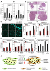
CCL5 is critical for the activity of CAFs and miR-CAFs. A, HeyA8 cells expressing luciferase were co-injected orthotopically into the mouse ovary with either NOFs triple-transfected with anti-miR-31, anti-miR-214, and pre-miR-155 (miR-CAF) or with equivalent scrambled controls (NOF). Mice were injected with either control IgG or a neutralizing anti-CCL5 antibody (αCCL5). Tumor growth was monitored by bioluminescence using Xenogen IVIS Spectrum and the fold change in radiance plotted. One-way ANOVA was performed comparing all groups to HeyA8+miR-CAFs treated with IgG for the same time point (* p<0.05). B, HeyA8 cells expressing luciferase were co-injected orthotopically with either NOFs or CAFs. Mice were injected twice with either control IgG or a neutralizing anti-CCL5 antibody (αCCL5). Tumor growth was monitored by bioluminescence using Xenogen IVIS Spectrum and the fold change in radiance plotted. Number of mice per group is indicated at the base of the columns. One-way ANOVA was performed comparing all groups to HeyA8+CAFs treated with IgG for the same time point (* p<0.05). C, H&E staining of ovaries from IgG injected mice shown in A (left two panels), and B (right two panels). Scale bar = 1500 μm. Insets show close up of tumor tissues (labeled by box), scale bar in insets = 200 μm. D, Plug-homing assay. Matrigel plugs were embedded with NOFs transfected with anti-miR-31 and anti-miR-214, scrambled control (scr) or no cells (/) and placed equidistant in the same culture dish and overlaid with GFP expressing HeyA8 cells. Half of the wells were treated with an IgG control antibody and half were treated with a neutralizing anti-CCL5 antibody (αCCL5). Homing of OvCa cells to the matrigel plugs was imaged (left) and quantified (right). E, Quantification of the homing assay shown in D, performed with GFP expressing SKOV3ip1 cells. F, Matrigel plugs were embedded with iCAFs (NOFs co-cultured with HeyA8), corresponding parental NOFs (Ctr) or no cells (/) and placed equidistant in the same culture dish and overlaid with GFP expressing HeyA8 cells. Half of the wells were treated with an IgG control antibody and half were treated with a neutralizing anti-CCL5 antibody (αCCL5). Homing of OvCa cells to the matrigel plugs was imaged and quantified. G, Matrigel plugs were embedded with NOFs transfected with anti-miR-31 and anti-miR-214, scrambled control (scr) or no cells (/) and placed equidistant in the same culture dish and overlaid with GFP expressing HeyA8 cells. Wells were treated with the CCR1/3 inhibitor J113863 (CCR1/3 Inh) or DMSO control (Ctr). Homing of cancer cells to the matrigel plugs was imaged and quantified. For E-G standard errors are shown for three independent experiments using fibroblasts from three to four different patients. One tailed t-tests were performed. H, Homing assay shown in G, performed with GFP expressing SKOV3ip1 cells. Standard errors are shown for three independent experiments. One tailed t-tests were performed. I, Co-invasion of HeyA8 with NOFs transfected with human CCL5 and treated with a CCL5 blocking antibody (NOF-CCL5+αCCL5) or IgG control (NOF-CCL5) were compared to co-invasion of HeyA8 with NOFs transfected with vector control (NOF-vect). Co-invasion of HeyA8 with CAFs treated with a CCL5 blocking antibody (CAF + αCCL5) or IgG control (CAF). All standard errors represent al least three independent experiments. One tailed t-tests were performed. J, Schematic of miRNA induced reprograming of NOFs to CAFs in ovarian cancer. Cancer cells induce a change in expression of miR-214, miR-31 and miR-155 in NOFs resulting in reprogramming them into CAFs which then promotes tumor growth and invasion through increased secretion of cytokines (e.g.) CCL5.

Comment in

Similar articles

Cited by

References

    1. Hanahan D, Coussens LM. Accessories to the crime: Functions of cell recruited to the tumor microenvironment. Cancer Cell. 2012;21:309–22. - PubMed
    1. Barkan D, Green JE, Chambers AF. Extracellular matrix: A gatekeeper in the transition from dormancy to metastatic growth. Eur J Cancer. 2010;46:1181–8. - PMC - PubMed
    1. Schauer IG, Sood AK, Mok S, Liu J. Cancer-associated fibroblasts and their putative role in potentiating the initiation and development of epithelial ovarian cancer. Neoplasia. 2011;13:393–405. - PMC - PubMed
    1. Orimo A, Gupta P, Sgroi D, Arenzana-Seisdedos F, Delaunay T, Naeem R, et al. Stromal fibroblasts present in invasive human breast carcinomas promote tumor growth and angiogenesis throught elevated SDF-1/CXCL 12 secretion. Cell. 2005;121:335–48. - PubMed
    1. Ambros V. The functions of animal microRNAs. Nature. 2004;431:350–5. - PubMed

Publication types

Associated data