InlA promotes dissemination of Listeria monocytogenes to the mesenteric lymph nodes during food borne infection of mice
- PMID: 23166492
- PMCID: PMC3499570
- DOI: 10.1371/journal.ppat.1003015
InlA promotes dissemination of Listeria monocytogenes to the mesenteric lymph nodes during food borne infection of mice
Abstract
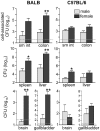
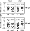
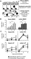
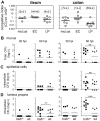
Intestinal Listeria monocytogenes infection is not efficient in mice and this has been attributed to a low affinity interaction between the bacterial surface protein InlA and E-cadherin on murine intestinal epithelial cells. Previous studies using either transgenic mice expressing human E-cadherin or mouse-adapted L. monocytogenes expressing a modified InlA protein (InlA(m)) with high affinity for murine E-cadherin showed increased efficiency of intragastric infection. However, the large inocula used in these studies disseminated to the spleen and liver rapidly, resulting in a lethal systemic infection that made it difficult to define the natural course of intestinal infection. We describe here a novel mouse model of oral listeriosis that closely mimics all phases of human disease: (1) ingestion of contaminated food, (2) a distinct period of time during which L. monocytogenes colonize only the intestines, (3) varying degrees of systemic spread in susceptible vs. resistant mice, and (4) late stage spread to the brain. Using this natural feeding model, we showed that the type of food, the time of day when feeding occurred, and mouse gender each affected susceptibility to L. monocytogenes infection. Co-infection studies using L. monocytogenes strains that expressed either a high affinity ligand for E-cadherin (InlA(m)), a low affinity ligand (wild type InlA from Lm EGDe), or no InlA (ΔinlA) showed that InlA was not required to establish intestinal infection in mice. However, expression of InlA(m) significantly increased bacterial persistence in the underlying lamina propria and greatly enhanced dissemination to the mesenteric lymph nodes. Thus, these studies revealed a previously uncharacterized role for InlA in facilitating systemic spread via the lymphatic system after invasion of the gut mucosa.
Conflict of interest statement
The authors have declared that no competing interests exist.
Figures


Similar articles
-
Mucosal CD8 T Cell Responses Are Shaped by Batf3-DC After Foodborne Listeria monocytogenes Infection.Front Immunol. 2020 Sep 11;11:575967. doi: 10.3389/fimmu.2020.575967. eCollection 2020. Front Immunol. 2020. PMID: 33042159 Free PMC article.
-
Functional genomic studies of the intestinal response to a foodborne enteropathogen in a humanized gnotobiotic mouse model.J Biol Chem. 2007 May 18;282(20):15065-72. doi: 10.1074/jbc.M610926200. Epub 2007 Mar 27. J Biol Chem. 2007. PMID: 17389602
-
Listeria adhesion protein orchestrates caveolae-mediated apical junctional remodeling of epithelial barrier for Listeria monocytogenes translocation.mBio. 2024 Mar 13;15(3):e0282123. doi: 10.1128/mbio.02821-23. Epub 2024 Feb 20. mBio. 2024. PMID: 38376160 Free PMC article.
-
Crossing the Intestinal Barrier via Listeria Adhesion Protein and Internalin A.Trends Microbiol. 2019 May;27(5):408-425. doi: 10.1016/j.tim.2018.12.007. Epub 2019 Jan 17. Trends Microbiol. 2019. PMID: 30661918 Review.
-
The mucosal phase of Listeria infection.Immunobiology. 1999 Dec;201(2):164-77. doi: 10.1016/S0171-2985(99)80056-4. Immunobiology. 1999. PMID: 10631565 Review.
Cited by
-
Fecal transplantation does not transfer either susceptibility or resistance to food borne listeriosis in C57BL/6 and BALB/c/By mice.F1000Res. 2013 Aug 20;2:177. doi: 10.12688/f1000research.2-177.v1. eCollection 2013. F1000Res. 2013. PMID: 24555086 Free PMC article.
-
Contributions of a LysR Transcriptional Regulator to Listeria monocytogenes Virulence and Identification of Its Regulons.J Bacteriol. 2020 Apr 27;202(10):e00087-20. doi: 10.1128/JB.00087-20. Print 2020 Apr 27. J Bacteriol. 2020. PMID: 32179628 Free PMC article.
-
A genome-wide screen in macrophages identifies PTEN as required for myeloid restriction of Listeria monocytogenes infection.PLoS Pathog. 2023 May 22;19(5):e1011058. doi: 10.1371/journal.ppat.1011058. eCollection 2023 May. PLoS Pathog. 2023. PMID: 37216395 Free PMC article.
-
CD169+ macrophages orchestrate innate immune responses by regulating bacterial localization in the spleen.Sci Immunol. 2017 Oct 6;2(16):eaah5520. doi: 10.1126/sciimmunol.aah5520. Sci Immunol. 2017. PMID: 28986418 Free PMC article.
-
Differentiation of distinct long-lived memory CD4 T cells in intestinal tissues after oral Listeria monocytogenes infection.Mucosal Immunol. 2017 Mar;10(2):520-530. doi: 10.1038/mi.2016.66. Epub 2016 Jul 27. Mucosal Immunol. 2017. PMID: 27461178 Free PMC article.
References
-
- Bartt R (2000) Listeria and atypical presentations of Listeria in the central nervous system. Semin Neurol 20: 361–373. - PubMed
-
- Munoz P, Rojas L, Bunsow E, Saez E, Sanchez-Cambronero L, et al. (2011) Listeriosis: An emerging public health problem especially among the elderly. J Infect 64: 19–33. - PubMed
-
- Ooi ST, Lorber B (2005) Gastroenteritis due to Listeria monocytogenes . Clin Infect Dis 40: 1327–1332. - PubMed
Publication types
MeSH terms
Substances
Grants and funding
LinkOut - more resources
Full Text Sources
Other Literature Sources
Medical

