. 2013 Jan;14(1):52-60.
doi: 10.1038/ni.2474. Epub 2012 Nov 18.

Nitric oxide controls the immunopathology of tuberculosis by inhibiting NLRP3 inflammasome-dependent processing of IL-1β

Affiliations

Nitric oxide controls the immunopathology of tuberculosis by inhibiting NLRP3 inflammasome-dependent processing of IL-1β

Bibhuti B Mishra et al. Nat Immunol. 2013 Jan.

Abstract

Interleukin 1 (IL-1) is an important mediator of innate immunity but can also promote inflammatory tissue damage. During chronic infections such as tuberculosis, the beneficial antimicrobial role of IL-1 must be balanced with the need to prevent immunopathology. By exogenously controlling the replication of Mycobacterium tuberculosis in vivo, we obviated the requirement for antimicrobial immunity and discovered that both IL-1 production and infection-induced immunopathology were suppressed by lymphocyte-derived interferon-γ (IFN-γ). This effect was mediated by nitric oxide (NO), which we found specifically inhibited assembly of the NLRP3 inflammasome via thiol nitrosylation. Our data indicate that the NO produced as a result of adaptive immunity is indispensable in modulating the destructive innate inflammatory responses elicited during persistent infections.


Figures

Figure 1
Figure 1. Lymphocytes, IFN-γ, and iNOS modulate TB immunopathology
a. Paired groups of mutant and wild type C57BL/6 mice were infected with Mtb, and lung histopathology was assessed by hematoxylin/eosin staining on the indicated day post-infection (dpi). b. Lesions in C57BL/6 mice were histocytic, whereas the inflammatory lesions of immunocompromised mice were pyogranulomatous and contained a larger number of neutrophils (insets). c. Bacterial burdens in the lungs of the indicated mice were determined by plating organ homogenates. CFU: colony forming unit. d. Relative cytokine levels were determined by ELISA in lung lysates generated at the same time points as in panel “a”. Data are presented as the % abundance of cytokine in knockout relative to wild type animals. Statistics are provided for cytokines that were significantly increased in all three mutant strains. P values were calculated by two-way ANOVA. e. IL-1β concentrations were determined by ELISA in the lung lysates shown in panel “d”. P values were determined by student’s t-test. Data are representative of two independent experiments. f. Immunoblot analysis of pro-IL-1β (p31) and mature-IL-1β p17 in lung lysates. Numbers indicate individual animals. “*” indicates a nonspecific band. Right panel shows densitometry analysis (ratio of processed to unprocessed IL-1β). Throughout, error bars represent standard deviation (sd). Each group contained 3-5 animals.
Figure 2
Figure 2. Nitric oxide’s anti-inflammatory activities are independent of its anti-mycobacterial function
a. The streptomycin-dependent Mtb (strain 18b) infection model. Mice were infected with Mtb 18b and administered streptomycin for two weeks. At the indicated time points after streptomycin withdrawal, disease progression was assessed in lungs (for intratracheal) or spleens (for intravenous). b. Organs of C57BL/6 and iNos−/− mice harbored similar numbers of viable Mtb throughout the infection. c. Representative histopathology of lungs (day 36) and spleen (day 40) of the indicated mouse strains. d. Body weight of intratracheally-infected mice at day 36, relative to day 0. Mean ± sd of two independent experiments, P value was determined by student’s t-test. e. Neutrophil and T cell counts in bronchoalveolar lavage (BAL) and dissociated lung tissue (Lung) were quantified by flow cytometry. P values were determined by two-way ANOVA. f. Myeloperoxidase activity per milligram of protein in lung lysates was quantified. P value was determined by student’s t-test. g. Relative cytokine levels in lung homogenates were determined by ELISA (presented as in Fig. 1d). P values were determined by two-way ANOVA. h. IL-1β in lung lysates was assessed by immunoblot. Data are representative of 2 independent experiments. All bar graphs depict the mean ± sd of 3-5 mice.
Figure. 3
Figure. 3. IL-1R1 and ASC promote the enhanced neutrophil recruitment observed upon iNOS inhibition
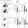
a. C57BL/6 and Il1r1−/− mice were infected intratracheally with Mtb 18b and treated with or without aminoguanidine hemisulfate (AGHS) throughout the infection. Seven weeks after streptomycin withdrawal (62 days post-infection), neutrophils (CD11b+, Gr1hi cells) were quantified in the BAL by flow cytometry and in the lung lysates by assaying myeloperoxidase activity per milligram of total protein. Representative flow cytometry plots of AGHS treated mice are shown. b. Asc−/− and C57BL/6 control mice were infected with Mtb 18b. Four weeks after streptomycin withdrawal, neutrophil numbers and myeloperoxidase activity in the dissociated lung tissues are shown as in panel “a”. Bars represent the mean ± sd of 4-6 mice, P values were calculated by two-way ANOVA; “ns” indicates P > 0.05.
Figure 4
Figure 4. IFN-γ activation of macrophages inhibits IL-1β processing
a. 24 hours before Mtb infection (multiplicity of infection, MOI, =10), bone marrow derived macrophages (BMDMs) were primed with/without LPS and stimulated with/without IFN-γ. IL-1β and TNF were measured in the supernatants at the indicated time points post-infection. b. The indicated genotypes of BMDMs were treated as shown, and the IL-1β concentration in the supernatant was measured 24 hours later by ELISA. c. At 24 hours post infection, the amount of pro-IL-1β (p31) in cell lysates and mature-IL-1β (p17) in supernatants (SN) were assessed by immunoblotting. Cell lysate samples were normalized for total protein content. d. BMDMs were primed with the indicated TLR stimulant (LPS: TLR4, Pam3CSK4: TLR2, poly (I:C): TLR3) and infected as in panel “a”. IL-1β and IL-1α levels in the supernatant were measured 24 hours later by ELISA. Throughout the figure, bars represent mean ± sd of at least 3 replicates. P values were calculated by two-way ANOVA. Data are representative of three (b) to six (a, c-d) independent experiments.
Figure 5
Figure 5. IFN-γ specifically inhibits NLRP3 inflammasome activation
a and b. BMDMs primed with LPS±IFN-γ were treated with 10μM Nigericin for 1hr to stimulate the NLRP3 inflammasome or transfected with poly(dA:dT) to stimulate the AIM2 inflammasome. Cytokines were measured by ELISA (a) or immunoblotting (b). P values were calculated by two-way ANOVA. c. Different intracellular pathogens trigger a distinct combination NLRP3 and NLRC4 inflammasomes. BMDMs of the indicated genotypes were infected with M. marinum (MOI=10) or S. typhimurium (MOI=25), and the IL-1β found in the supernatants after 6hrs (S.typhimurium) and 24hrs (M.marinum) was measured by ELISA. P values were calculated by student’s t-test (two tailed). d. LPS-primed BMDMs were treated with or without IFN-γ before infection with M.marinum and S.typhimurium. IL-1β and TNF were measured in the culture supernatants as in panel “c”. To determine statistical significance, the IL-1β levels were first normalized to the amount of TNF produced, and then compared using a student’s t-test (two tailed). “ns” indicates P > 0.05. The same supernatant samples were probed by western blotting for the presence of mature IL-1β (p17). Throughout the figure, bars represent the mean ± sd of at least 3 replicates. Data are representative of two (a-c) to four (d) independent experiments.
Figure 6
Figure 6. Nitric oxide is necessary for IFN-γ-mediated suppression of IL-1β processing
a and b. BMDMs were primed with LPS or Pam3CSK4 ± IFN-γ and infected with Mtb (MOI=10). 24 hours later, cell lysates and supernatants were analyzed by ELISA for IL-1β (a) or immunoblotting for IL-1β and caspase-1 (b). Pro-forms in lysates and mature products in supernatants are indicated. c. Pam3CSK4-primed BMDMs were treated with aminoguanidine hemisulfate (AGHS) before Mtb infection. Cytokines were measured in culture supernatants after 24 hrs. d and e. Macrophages were primed with Pam3CSK4 and treated with IFN-γ or NO donors, S-nitrosoglutathione (GSNO) or S-nitroso-N-acetyl-DL-penicillamine (SNAP), before Mtb infection. Secretion of IL-1β and TNF was measured by ELISA. IL-1β and caspase-1 processing in cell lysates and supernatants was assessed by immunoblotting. f. BMDMs from C57BL/6 and iNos−/− mice were co-cultured in the indicated combinations (1:1 ratio), primed with Pam3CSK4 ± IFN-γ, and infected with Mtb. 24 hours later, IL-1β secretion was measured by ELISA. One-way or two-way ANOVA was applied to calculate P values, as appropriate. “ns” indicates P > 0.05. Bars represent the mean ± sd of at least 3 replicates. Data are representative of two (c-f) to five (a and b) independent experiments.
Figure 7
Figure 7. IFN-γ-induced nitric oxide posttranslationally inhibits the assembly and activation of the NLRP3 inflammasome via thiol nitrosylation.
a. Inflammasomes were reconstituted in 293T cells by transfection. Cells were treated ± SNAP, and IL-1β processing was assessed by immunoblotting. b. BMDMs were primed with LPS ± IFN-γ or LPS ± SNAP, and stimulated with nigericin or poly (dA:dT). ASC oligomerization was assayed by cross-linking inflammasome components, followed by immunoblotting. Predicted mono- and multi-meric forms are indicated. “Lysate” indicates unfractionated lysates. c. BMDMs were treated with dehydroascorbate concomitantly with Pam3CSK4 ± IFN-γ. NO production and IL-1β release were assessed at 16 hours (upper panels). Pro- and mature forms of IL-1β and caspase-1 were assessed by immunoblotting (lower panel). Data are representative of at least three independent experiments. P values were determined by student’s t-test. d. BMDMs were subjected to the indicated treatments. 16 hours later, S-nitrosylated cysteines were biotinylated, and the corresponding proteins were purified using streptavidin. Total S-nitrosylated species or the indicated proteins were detected in the biotinylated or total lysate fractions by immunoblotting. e. Independent 293T cultures were transfected with NLRP3 or caspase-1 expression plasmids, treated ± SNAP, and lysed after 16 hours (lysate 1). Separate cultures were transfected to produce the other three components of the complex (lysate 2). Lysates 1 and 2 were mixed and IL-1β processing was assessed by immunoblotting.

Comment in

  • Inflammasome: Turning on and off NLRP3.
    Leavy O. Leavy O. Nat Rev Immunol. 2013 Jan;13(1):1. doi: 10.1038/nri3366. Epub 2012 Nov 30. Nat Rev Immunol. 2013. PMID: 23197112 No abstract available.
  • Just say NO to NLRP3.
    Rayamajhi M, Miao EA. Rayamajhi M, et al. Nat Immunol. 2013 Jan;14(1):12-4. doi: 10.1038/ni.2493. Nat Immunol. 2013. PMID: 23238751 No abstract available.

Similar articles

Cited by

References

    1. Lamkanfi M, Dixit VM. Inflammasomes: guardians of cytosolic sanctity. Immunol Rev. 2009;227:95–105. - PubMed
    1. Vance RE, Isberg RR, Portnoy DA. Patterns of pathogenesis: discrimination of pathogenic and nonpathogenic microbes by the innate immune system. Cell Host Microbe. 2009;6:10–21. - PMC - PubMed
    1. Dinarello CA. Immunological and inflammatory functions of the interleukin-1 family. Annu Rev Immunol. 2009;27:519–550. - PubMed
    1. Dinarello CA. Role of interleukin-1 in infectious diseases. Immunol Rev. 1992;127:119–146. - PubMed
    1. Hise AG, et al. An essential role for the NLRP3 inflammasome in host defense against the human fungal pathogen Candida albicans. Cell Host Microbe. 2009;5:487–497. - PMC - PubMed

Publication types

MeSH terms