. 2012 Dec;30(12):1217-24.
doi: 10.1038/nbt.2434. Epub 2012 Nov 18.

Microparticles bearing encephalitogenic peptides induce T-cell tolerance and ameliorate experimental autoimmune encephalomyelitis

Affiliations

Microparticles bearing encephalitogenic peptides induce T-cell tolerance and ameliorate experimental autoimmune encephalomyelitis

Daniel R Getts et al. Nat Biotechnol. 2012 Dec.

Abstract

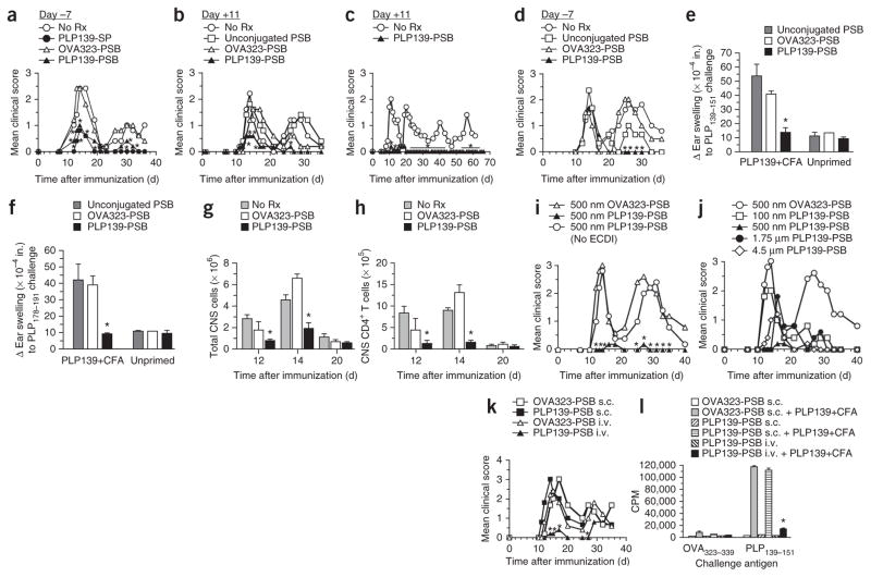
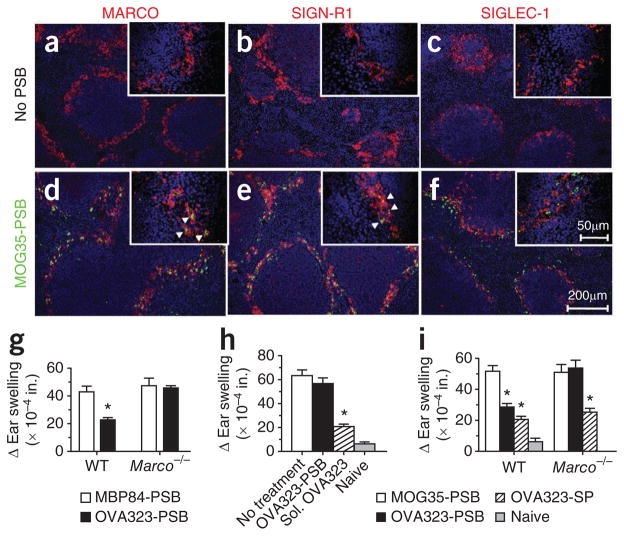
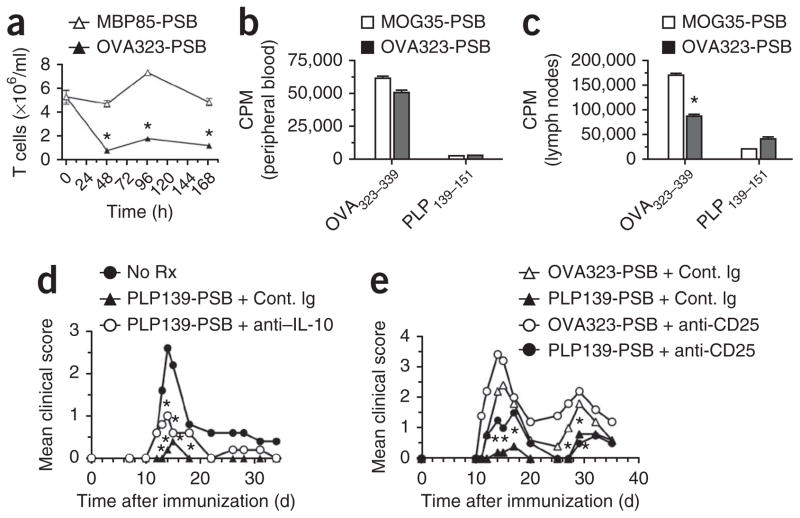
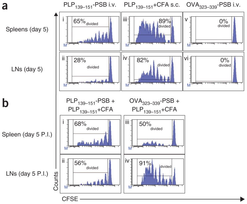
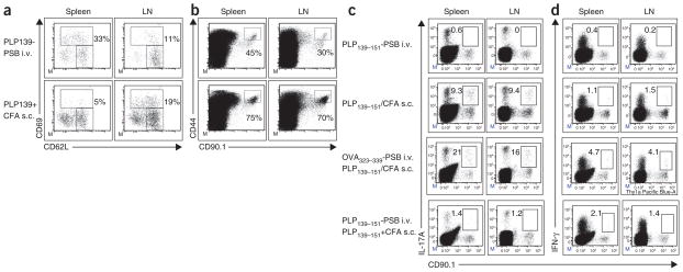
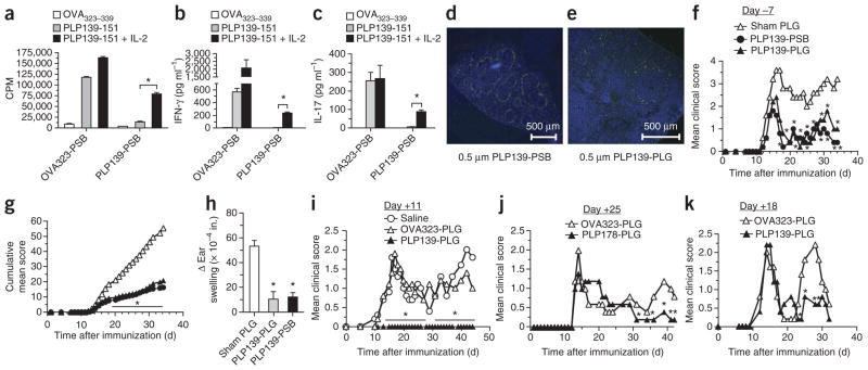
Aberrant T-cell activation underlies many autoimmune disorders, yet most attempts to induce T-cell tolerance have failed. Building on previous strategies for tolerance induction that exploited natural mechanisms for clearing apoptotic debris, we show that antigen-decorated microparticles (500-nm diameter) induce long-term T-cell tolerance in mice with relapsing experimental autoimmune encephalomyelitis. Specifically, intravenous infusion of either polystyrene or biodegradable poly(lactide-co-glycolide) microparticles bearing encephalitogenic peptides prevents the onset and modifies the course of the disease. These beneficial effects require microparticle uptake by marginal zone macrophages expressing the scavenger receptor MARCO and are mediated in part by the activity of regulatory T cells, abortive T-cell activation and T-cell anergy. Together these data highlight the potential for using microparticles to target natural apoptotic clearance pathways to inactivate pathogenic T cells and halt the disease process in autoimmunity.


Conflict of interest statement

COMPETING FINANCIAL INTERESTS

The authors declare no competing financial interests.

Figures

Figure 1
Figure 1
Antigen-coupled polystyrene microparticles are effective for inducing tolerance for the prevention and treatment of EAE. (a) Mean clinical score of SJL/J mice injected i.v. with 500-nm carboxylated PSB coupled to PLP139–151 (PLP139-PSB) or OVA323–339 (OVA323-PSB) 7 d before initiation of EAE by s.c. immunization with PLP139–151 plus CFA. A separate group was tolerized with PLP139–151-SP (PLP139-SP). No Rx, no treatment. (b,c) Mean clinical score of mice that received PLP139–151-PSB, OVA323–339-PSB or unconjugated PSB at the onset of hindlimb paralysis (11 d after priming); disease symptoms were scored for a total of 35 and 66 d, respectively. (d) Mean clinical score of mice injected i.v. with 500-nm carboxylated PSB coupled to PLP139–151, OVA323–339 or nothing 7 d before induction of EAE with PLP178–191. (e,f) Ear swelling, as a measure of DTH, 24 h after ear challenge with the priming PLP139–151 epitope (e) or the PLP178–191 spread epitope (f) at 36 d after priming in selected representative mice from the PLP139–151 plus CFA (PLP139+CFA)-primed groups in a (OVA323–339-PSB, PLP139–151-PSB and no particles). Additional mice included in this analysis received doses of PSB i.v. but were not primed for EAE. Responses to a control OVA323–339 peptide were subtracted from each measure of ear swelling. (g,h) The number of CD45hi cells (g) and CD3+CD4+ T cells (h) determined by flow cytometry at the onset of disease (day 12), peak of disease (day 14) and remission (day 20) in the brains and spinal cords of SJL/J mice injected i.v. with 500-nm carboxylated PSB coupled with PLP139–151, OVA323–339 or nothing 7 d before EAE priming with PLP139–151 plus CFA. (i) Mean clinical score in SJL/J mice treated with i.v. injection of 500-nm carboxylated PSB incubated with PLP139–151 in the presence or absence of ECDI or treated with 500-nm OVA323–339-PSB 7 d before priming with PLP139–151 plus CFA. (j) Mean clinical score in SJL/J mice treated i.v. with PSB of varying diameters (100 nm, 500 nm, 1.75 μm or 4.5 μm) coupled to PLP139–151 or treated with 500-nm OVA323–339-PSB 7 d before priming with PLP139–151 plus CFA. (k) Mean clinical score in SJL/J mice treated with 500-nM PLP139–151-PSB or OVA323–339-PSB in the lateral tail vein (i.v.) or on the flank (s.c.) 7 d before priming with PLP139–151 plus CFA. (l) In vitro proliferative responses to stimulation with the PLP139–151 priming epitope or a control peptide (OVA323–339) determined by [3H]-thymidine uptake in spleens and lymph nodes collected from a subset of the mice in k. CPM, counts per minute. All experiments consisted of 5–10 mice per group and are representative of 2–4 repeats. *P ≤ 0.05 (ANOVA) for the differences in mean clinical scores, DTH responses, numbers of CNS-infiltrating cells or proliferative responses compared to the responses in groups tolerized to the appropriate irrelevant control peptide. Error bars, s.e.m.
Figure 2
Figure 2
MARCO has a crucial role in tolerance induction using antigen-coupled microparticles. (af) MARCO (a,d, red), SIGN-R1 (b,e, red), SIGLEC-1 (c,f, red) and 4,6-diamidino-2-phenylindole (DAPI, blue) staining in dissected and snap-frozen spleens from mice infused with PSB (no PSB) or FITC-labeled MOG35–55-PSB (MOG35-PSB, green). Arrowheads indicate phagocytized PSB. (g) Ear swelling 24 h after ear challenge with OVA323–339 or an irrelevant peptide (PLP129–151) in WT or Marco−/− BALB/c mice injected i.v. with OVA323–339-PSB (OVA323-PSB) or control MBP84–104-PSB (MBP84-PSB) 7 d before immunization with OVA323–339 plus CFA. Ear challenge, as a measure of DTH, was performed 8 d after immunization. (h,i) Ear swelling as described for g in WT (i) and Marco−/− (h,i) BALB/c mice treated i.v. with OVA323–339-PSB (h,i), soluble OVA323–339 (sol. OVA323) (h), MOG35–55-PSB (i) or OVA323–339-SP (i) 7–8 d before immunization with OVA323–339 plus CFA. All experiments consisted of 5–10 mice per group and are representative of at least 2–4 separate experiments. *P ≤ 0.05 (ANOVA) for differences in mean clinical scores and DTH responses compared to the responses in groups tolerized to the appropriate irrelevant control peptide. Error bars, s.e.m.
Figure 3
Figure 3
Response of antigen-specific T cells to tolerance induction with Ag-PSB. (a) T cell content 48, 39 and 168 h after treatment in female DO11.10 OVA323–339-specific TCR transgenic mice treated i.v. with 500-nm carboxylated PSB coupled to the cognate peptide (OVA323–339, OVA323-PSB) or an irrelevant peptide (MBP85–99, MBP85-PSB). (b,c) Proliferation, measured by [3H]-thymidine uptake, in 3 × 10⁵ T cells magnetically purified from peripheral blood (b) and peripheral lymph nodes (c) of DO11.10 mice treated i.v. with 500-nm carboxylated PSB coupled to the cognate peptide (OVA323–339) or an irrelevant peptide (MOG35–55, MOG35-PSB) and re-stimulated 48 h later with 1 mg ml−1 cognate OVA323–339 peptide or PLP139–151 in vitro. (d,e) Mean clinical score in naive SJL/J mice treated with control immunoglobulin (Cont. Ig) or anti–IL-10 (JES5-16E3; 200 μg intraperitoneally (i.p.)) (d) or control immunoglobulin or anti-CD25 (PC61; 500 μg i.p.) (e) 1 d before and 1 d after treatment with either OVA323–339-PSB or PLP139–151-PSB (PLP139-PSB); 7 d after tolerization, mice were primed for EAE with PLP139–151 plus CFA. Data are representative of three separate experiments. Error bars, s.e.m. *P ≤ 0.05 (Student’s t test) for the differences in T cell numbers, CPM and mean clinical scores compared to the responses in groups tolerized to the appropriate irrelevant control peptide.
Figure 4
Figure 4
Antigen-specific T cells undergo suboptimal proliferation in response to Ag-PSB. (a) Results from naive SJL/J (CD90.2+) recipient mice exposed i.v. to PLP139–151-PSB (i,ii), s.c. to PLP139–151 plus CFA (iii,iv) or i.v. to OVA323–339-PSB (v,vi) 48 h after being transferred with naive CD90.1+ PLP139–151-specific 5B6 TCR transgenic T cells sorted from donor lymph nodes and labeled with CFSE. Five days after these treatments (P.I.), spleens and lymph nodes (LNs) were collected, and the percentage of diving CD90.1+ T cells was assessed by measuring CFSE dilution using flow cytometry. (b) Flow cytometric analyses of CFSE dilution at 5 d after priming in mice additionally treated with PLP139–151-PSB (i,ii) or OVA323–339-PSB (iii,iv) 5 d after the initial treatments in a and then primed with PLP139–151 plus CFA. Three separate mice were analyzed in each group with representative plots shown. Data shown are representative of three separate experiments. Percentages in graphs reflect the percent of T cells that have divided.
Figure 5
Figure 5
Antigen-specific T cells are abortively activated after Ag-PSB encounter but do not synthesize IL-17A and IFN-γ after direct in vivo exposure to Ag-PSB or after subsequent immunogenic stimulation. (a,b) Flow cytometric analyses of T-cell activation markers CD62L, CD69 and CD44 5 d after treatment in spleens and lymph nodes (LNs) from naive CD90.2+ SJL/J recipients after being transferred with CFSE-labeled naive CD90.1+ 5B6 TCR transgenic T cells and then treated i.v. with PLP139–151-PSB (PLP139-PSB) or primed s.c. with PLP139–151 plus CFA. Transgenic T cells were identified by CD90.1 and CFSE signals. (c,d) Naive CD90.1+ 5B6 TCR transgenic T cells were CFSE labeled and transferred to naive CD90.2+ SJL/J recipients that were then treated i.v. with PLP139–151-PSB or primed s.c. with PLP139–151 plus CFA. Five days after treatment, levels of intracellular IL-17A (c) and IFN-gamma (d) were determined. Where indicated, SJL/J recipients of naive CFSE-labeled 5B6 TCR transgenic T cells were treated i.v. with OVA323–339-PSB or PLP139–151-PSB and primed with PLP139–151 plus CFA 5 d later. Data shown are representative of three independent experiments.
Figure 6
Figure 6
Short-term tolerance induced by i.v. treatment with Ag-PSB is caused primarily by anergy induction. (ac) SJL/J mice were treated i.v. with OVA323–339-PSB (OVA323-PSB) or PLP139–151-PSB (PLP139-PSB) 7 d before s.c. priming with PLP139–151 plus CFA. Eight days after priming, the proliferation of spleen and lymph nodes in response to stimulation with the priming antigen (PLP139–151) or a control antigen (OVA323–339) in the presence of absence of 200 U ml-1 of exogenous IL-2 was measured (a). Supernatants were collected for measurements of secreted IFN-γ (b) and IL-17A (c). Results are representative of 3–4 separate experiments. (d,e) SJL/J mice were treated with 500-nm FITC-PSB (d) or 500-nm biodegradable FITC-PLG microparticles (e) coupled with PLP139–151. Twelve hours later, frozen spleen sections were prepared from a subset of mice, and these sections were counterstained with DAPI (blue). (fi) On day –7 (fh) or day +11 (i) relative to PLP139–151 plus CFA priming, mice were injected with 500-nm PLP139–151-PSB or PLP139–151-PLG (PLP139-PLG) microparticles and monitored for development of clinical disease by assessing mean clinical score (f,i) and cumulative mean clinical score (g) over time. (h) At the conclusion of the experiment, the mice from f were ear challenged with PLP139–151, and DTH responses were determined. (j,k) SJL/J mice were tolerized with 500-nm PLP178–191-PLG (PLP178-PLG) or OVA323–339-PLG (OVA323-PLG) microparticles on day +25 after PLP139–151 plus CFA priming (j) or with 500-nm PLP139–151-PLG or OVA323–339-PLG microparticles on day +18 after PLP178–191 plus CFA priming (k) and monitored for clinical disease. Error bars, s.e.m. *P ≤ 0.01 (ANOVA) for the differences in proliferation, mean clinical scores and DTH responses compared to groups tolerized to sham PLG particles. Data shown are representative of 2–3 separate experiments of 5–7 mice per group.

Comment in

Similar articles

Cited by

References

    1. Christen U, von Herrath MG. Initiation of autoimmunity. Curr Opin Immunol. 2004;16:759–767. - PubMed
    1. Chatenoud L, Bluestone JA. CD3-specific antibodies: a portal to the treatment of autoimmunity. Nat Rev Immunol. 2007;7:622–632. - PubMed
    1. Kohm AP, Turley DM, Miller SD. Targeting the TCR: T-cell receptor and peptide-specific tolerance-based strategies for restoring self-tolerance in CNS autoimmune disease. Int Rev Immunol. 2005;24:361–392. - PubMed
    1. Miller SD, Turley DM, Podojil JR. Antigen-specific tolerance strategies for the prevention and treatment of autoimmune disease. Nat Rev Immunol. 2007;7:665–677. - PubMed
    1. Herold KC, et al. Treatment of patients with new onset Type 1 diabetes with a single course of anti-CD3 mAb Teplizumab preserves insulin production for up to 5 years. Clin Immunol. 2009;132:166–173. - PMC - PubMed

Publication types

MeSH terms