. 2012 Nov 13;2(11):e183.
doi: 10.1038/tp.2012.109.

Early accumulation of intracellular fibrillar oligomers and late congophilic amyloid angiopathy in mice expressing the Osaka intra-Aβ APP mutation

Affiliations

Early accumulation of intracellular fibrillar oligomers and late congophilic amyloid angiopathy in mice expressing the Osaka intra-Aβ APP mutation

L Kulic et al. Transl Psychiatry. .

Abstract

Pathogenic amyloid-β peptide precursor (APP) mutations clustered around position 693 of APP-position 22 of the Aβ sequence--are commonly associated with congophilic amyloid angiopathy (CAA) and intracerebral hemorrhages. In contrast, the Osaka (E693Δ) intra-Aβ APP mutation shows a recessive pattern of inheritance that leads to AD-like dementia despite low brain amyloid on in vivo positron emission tomography imaging. Here, we investigated the effects of the Osaka APP mutation on Aβ accumulation and deposition in vivo using a newly generated APP transgenic mouse model (E22ΔAβ) expressing the Osaka mutation together with the Swedish (K670N/M671L) double mutation. E22ΔAβ mice exhibited reduced α-processing of APP and early accumulation of intraneuronal fibrillar Aβ oligomers associated with cognitive deficits. In line with our in vitro findings that recombinant E22Δ-mutated Aβ peptides form amyloid fibrils, aged E22ΔAβ mice showed extracellular CAA deposits in leptomeningeal cerebellar and cortical vessels. In vitro results from thioflavin T aggregation assays with recombinant Aβ peptides revealed a yet unknown antiamyloidogenic property of the E693Δ mutation in the heterozygous state and an inhibitory effect of E22Δ Aβ42 on E22Δ Aβ40 fibrillogenesis. Moreover, E22Δ Aβ42 showed a unique aggregation kinetics lacking exponential fibril growth and poor seeding effects on wild-type Aβ aggregation. These results provide a possible explanation for the recessive trait of inheritance of the Osaka APP mutation and the apparent lack of amyloid deposition in E693Δ mutation carriers.


Figures
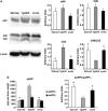
Figure 1
Figure 1
Intra-amyloid β (Aβ) mutations decrease α-cleavage in vivo. Transgene expression and Aβ peptide precursor (APP) processing were compared in three mouse models of Alzhemier's disease (AD). (a) Western blot analysis of cortical SDS extracts from 3-month-old E22ΔAβ mice in comparison with age-matched Tg2576 and arcAβ mice. Full-length APP and APP C-terminal fragments (C99 and C83) were detected with a C-terminal-specific APP antibody and values normalized to β-actin. Quantification reveals significant reductions in α-stub (C83) levels in both transgenic mouse lines bearing the intra-Aβ mutation. This results in an increased C99/C83 ratio in the E22ΔAβ and arcAβ mice as compared with the Tg2576 mice. *P<0.05 E22ΔAβ and arcAβ vs Tg2576 (Mann–Whitney U-test). n=4 per group. (b) MSD analysis of sAPPα and sAPPβ levels in cortical samples extracted with Tris buffer containing 2% SDS. In accordance with the results of the APP CTF analysis, sAPPβ levels were not significantly different among the three APP transgenic mouse lines, whereas sAPPα was reduced almost threefold in the E22ΔAβ and arcAβ mice as compared with the Tg2576 mice. *P=0.001 E22ΔAβ and arcAβ vs Tg2576 (Mann–Whitney U-test). n=6–8 per group.
Figure 2
Figure 2
Accumulation of detergent-insoluble amyloid β (Aβ) at different ages in the three Alzhemier's disease (AD) mouse models. MesoScale Discovery (MSD) analysis of Aβ38 (black column), Aβ40 (gray column) and Aβ42 (white column) in serially extracted cortical samples (Tris buffer, Tris buffer containing 1% Triton, Tris buffer containing 2% sodium dodecyl sulfate (SDS) and formic acid (FA)). (a) In E22ΔAβ mice most of the Aβ can be detected in the SDS-soluble protein fraction up to the age of 15 months, whereas Tris buffer-soluble, Triton-soluble and FA-soluble Aβ levels are comparatively low. Only at 24 months of age is detergent-insoluble Aβ detected. (b) In contrast, Tg2576 mice show higher soluble Aβ levels in Tris and Triton fractions and a prominent age-dependent Aβ accumulation in the detergent-insoluble FA fraction at 15 months. (c) The 3-month-old arcAβ mice show intermediate Tris- and Triton-soluble Aβ levels (as compared with age-matched E22ΔAβ and Tg2576 mice) and accumulation of Aβ in the FA fraction already at the age of 6 months. n=6–9 per group.
Figure 3
Figure 3
Lack of amyloid deposition in 15-month-old E22ΔAβ mice. (ad) Immunostaining with antibody 6E10 reveals a complete absence of extracellular amyloid deposits in 15-month-old E22ΔAβ mice (a: cortex; b: hippocampus). In contrast, arcAβ mice show a massive extracellular amyloid deposition at that age (c: cortex; d: hippocampus). Scale bars: 250 μm (ad). Aβ, amyloid β.
Figure 4
Figure 4
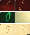
Aged E22ΔAβ mice develop congophilic amyloid angiopathy (CAA) at 24 months. (a+b) Immunostaining of amyloid-laden leptomeningeal vessels with polyclonal anti-amyloid antibody directed against human pan-Aβ in the cortex (a) and cerebellum (b) of a 24-month-old E22ΔAβ mouse. (c+d) Vascular amyloid deposits are thioflavin S (c) and Congo red positive (d). Note the classical apple-green birefringence of Congo red-stained vessels under polarized light (d). (e+f) Immunostainings with Aβ40-specific monoclonal antibody BA27 (e) and Aβ42-specific monoclonal antibody BC05 (f) reveal that Aβ40 is the dominant Aβ species deposited in the vessel walls. Scale bars: 125 μm (a+b) and 40 μm (cf). Aβ, amyloid β.
Figure 5
Figure 5
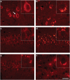
Intraneuronal amyloid β (Aβ) consists of fibrillar oligomers bearing a ‘toxic turn' conformation. (ad) Immunostaining with polyclonal OC antibody directed against fibrillar Aβ oligomers reveals age-dependent accumulation of intracellular oligomeric deposits in cortical (a and b) and CA1 hippocampal neurons (c and d). Note the temporal change of intraneuronal oligomers from rather diffuse and smaller dot-like aggregates at 3 months of age (a and c) to bigger and more compact deposits at 15 months of age (b and d). (e and f) Conformation-dependent monoclonal antibody 11A1 directed against the turn between positions 22 and 23 of the Aβ sequence stains hippocampal (e) and cortical (f) intraneuronal aggregates (representative immunostaining in a 15-month-old E22ΔAβ mouse). Scale bar: 40 μm (af).
Figure 6
Figure 6
Early cognitive deficits in E22ΔAβ mice. (a) Cognitive assessment in the Y-maze demonstrates a significant decrease in the percentage spatial alternation rate for E22ΔAβ mice as compared with their wild-type (wt) littermates at 3, 6 and 9 months of age (P<0.05 for all comparisons; Student's t-test). (b) Assessment of Barnes maze spatial learning at the age of 3 months reveals that both wild-type and E22ΔAβ transgenic mice show successful learning over the 4 days of training. However, E22ΔAβ mice, as compared with wild-type littermates, show significantly higher latencies to escape on day 3 (P=0.01) and day 4 (P=0.03), with similar but nonsignificant trends on day 1 (P=0.096) and day 2 (P=0.095) (analysis of variance (ANOVA), followed by post hoc Fisher's least significant difference (LSD) analysis). (c and d) Qualitative assessment of Barnes maze search strategies reveals a decrease in spatial and relative increase in serial search strategies in 3-month-old E22ΔAβ mice (d) in comparison to wild-type control mice (c). n=22–24 per group. Aβ, amyloid β.
Figure 7
Figure 7
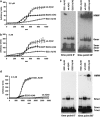
Unique aggregation kinetics of recombinant E22ΔAβ peptides in vitro. Fibril formation is measured via the absolute increase in thioflavin T fluorescence at 482 nm during aggregation. Aggregation of amyloid β (Aβ) peptides (identical total monomer concentrations: 2.5 μℳ in a+c, 5 μℳ in b) at pH 7.4 and 37 °C is initiated by a 10-fold dilution of Aβ stock solutions in 10 mℳ NaOH with buffer. Average curves of three independent experiments are shown (±s.e.m.). (a and b) Thioflavin T analysis of E22G (Arctic) Aβ42 and wild-type (wt) Aβ42 reveals that a typical aggregation kinetics with a lag phase, which is shorter for E22G Aβ42 than wild-type Aβ42, is followed by an exponential growth phase that eventually reaches a plateau of saturated β-sheet formation. In contrast, no obvious lag phase and no exponential growth phase is observed for E22Δ Aβ42 at 2.5 μℳ (a) and—more obvious—at 5 μℳ (b). Instead, E22Δ Aβ42 is characterized by slow non-exponential fibril growth; however, it forms detergent-insoluble high molecular weight (HMW) aggregates immediately after reconstitution at pH 7.4 as revealed by western blotting of the recombinant peptide preparations that were used for the thioflavin T analysis at time point 0 and time point 60 of the assay (c). (d) In contrast to E22Δ Aβ42, E22Δ Aβ40—similar to E22G Aβ40—shows an aggregation kinetics characterized by a lag phase, an exponential growth phase and a plateau phase. Note the unusually high thioflavin T fluorescence values for E22Δ Aβ40 in contrast to E22G and wild-type Aβ40, and the remarkably shorter lag phases of the E22-mutated Aβ40 variants in comparison to the wild-type peptide. (e) In contrast to the amyloid β (Aβ42) analysis, western blot analysis of the Aβ40 peptide variants reveals no immediate formation of detergent-insoluble HMW aggregates at time point 0 min. Only the E22G mutant develops prominent HMW aggregates after 60 min.
Figure 8
Figure 8
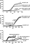
Anti-amyloidogenic property of E22ΔAβ peptides in mixtures of different Aβ peptide variants. (a and b) Co-aggregation of E22Δ Aβ42 with wild-type (wt) Aβ42 (a) or E22Δ Aβ40 with wild-type Aβ40 (b) leads to a significant extension of the lag phase and a delay of the growth phase in comparison with the aggregation of the respective Aβ peptide variants alone (see Figure 7) (total monomer concentrations for each peptide variant: 2.5 μℳ). In contrast, co-aggregation of 2.5 μℳ E22G (Arctic) Aβ42 with 2.5 μℳ wild-type Aβ42 (a) results in a similar aggregation curve as with 2.5 or 5 μℳ E22G Aβ42 alone (see Figure 7). Co-aggregation of E22G Aβ40 with wild-type Aβ40 (b) only slightly delays β-sheet formation in comparison with E22G Aβ40 alone. (c) Co-aggregation of E22Δ Aβ40 with E22Δ Aβ42 in a physiological 9:1 ratio (total monomer end concentration in solution: 2.5 μℳ) leads to a strong inhibition of E22Δ Aβ40 aggregation. Note the dramatic delay of the growth phase and remarkable loss of absolute thioflavin T fluorescence in comparison to E22Δ Aβ40 alone (Figure 7). In contrast, 9:1 mixtures of E22G Aβ40 and E22G Aβ42 result in an aggregation curve very similar to E22G Aβ40 alone. Average curves of three independent experiments are shown (±s.e.m.).

Similar articles

Cited by

References

    1. Thal DR, Del Tredici K, Braak H. Neurodegeneration in normal brain aging and disease. Sci Aging Knowledge Environ. 2004;2004:pe26. - PubMed
    1. Thal DR, Griffin WS, Braak H. Parenchymal and vascular Abeta-deposition and its effects on the degeneration of neurons and cognition in Alzheimer's disease. J Cell Mol Med. 2008;12:1848–1862. - PMC - PubMed
    1. Hardy J, Selkoe DJ. The amyloid hypothesis of Alzheimer's disease: progress and problems on the road to therapeutics. Science. 2002;297:353–356. - PubMed
    1. Tanzi RE, Bertram L. Twenty years of the Alzheimer's disease amyloid hypothesis: a genetic perspective. Cell. 2005;120:545–555. - PubMed
    1. Citron M, Oltersdorf T, Haass C, McConlogue L, Hung AY, Seubert P, et al. Mutation of the beta-amyloid precursor protein in familial Alzheimer's disease increases beta-protein production. Nature. 1992;360:672–674. - PubMed

Publication types

Substances